Review
. 2012:2012:949535.
doi: 10.1100/2012/949535. Epub 2012 Aug 1.

Beyond the definitions of the phenotypic complications of sickle cell disease: an update on management

Affiliations
Review

Beyond the definitions of the phenotypic complications of sickle cell disease: an update on management

Samir K Ballas et al. ScientificWorldJournal. 2012.

Erratum in

  • ScientificWorldJournal. 2013;2013:861251

Abstract

The sickle hemoglobin is an abnormal hemoglobin due to point mutation (GAG → GTG) in exon 1 of the β globin gene resulting in the substitution of glutamic acid by valine at position 6 of the β globin polypeptide chain. Although the molecular lesion is a single-point mutation, the sickle gene is pleiotropic in nature causing multiple phenotypic expressions that constitute the various complications of sickle cell disease in general and sickle cell anemia in particular. The disease itself is chronic in nature but many of its complications are acute such as the recurrent acute painful crises (its hallmark), acute chest syndrome, and priapism. These complications vary considerably among patients, in the same patient with time, among countries and with age and sex. To date, there is no well-established consensus among providers on the management of the complications of sickle cell disease due in part to lack of evidence and in part to differences in the experience of providers. It is the aim of this paper to review available current approaches to manage the major complications of sickle cell disease. We hope that this will establish another preliminary forum among providers that may eventually lead the way to better outcomes.


Figures

Figure 1
Figure 1
(A) Ultrawide field fundus photograph of the right eye shows a black sunburst lesion (arrow), a flat, round, black patch along the superior arcade temporally. (B) Ultrawide field fluorescein angiogram of the right eye in the arteriovenous phase demonstrates the staining of the sunburst lesion (arrow).
Figure 2
Figure 2
(A) Fundus photograph of new vascular formations characteristic of sickle cell disease called sea fan formations (arrow), which tend to occur in the temporal periphery. (B) Fluorescein angiogram in the arteriovenous phase shows peripheral nonperfusion and sea fan neovascularization at the border of vascularized and nonvascularized retina (arrow indicates the largest sea fan). (C) Late phase of the angiogram shows fluorescein leakage from sea fans as the dye study progresses.
Figure 3
Figure 3
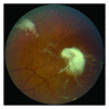
Color fundus photo shows a white autoinfarcted sea fan in the midperipheral retina with white, fibrous tissue remnants adherent to the cortical vitreous (top). Also note white, occluded retinal vessels.
Figure 4
Figure 4
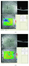
(a) Optical coherence tomography image of the right eye with a cross-section of the macula that demonstrates thinning of the inner layers (arrows), in the temporal aspect of the macula, whereas the nasal side of the macula (right) is normal in thickness. Thickness map below demonstrates the area of thinning, outlined by the blue color and shown by the arrow. (b) Optical coherence tomography image of the left eye shows a cross-section of the macula that demonstrates thinning of the inner layers, as indicated by the arrows, in the temporal aspect of the macula. Thickness map below demonstrates the two areas of thinning outlined by the blue color and shown by the arrows.
Figure 5
Figure 5
(A and B) Color fundus photographs of both eyes show white, occluded vessels (arrows) in the periphery bilaterally. (C and D) Ultrawide field fluorescein angiograms of the right and the left eye demonstrate the initial vascular remodeling at the junction of the perfused central, and nonperfused peripheral retina (arrows) includes arteriovenous (AV) anastomoses (black arrow head) and hairpin loops (solid white arrow head).
Figure 6
Figure 6
(A) Ultrawide field photograph of the left eye shows the sea fans with overlying preretinal hemorrhage in the superotemporal periphery as shown by the arrow. (B and C) Fluorescein angiogram delineates the nonperfused peripheral retina in the arteriovenous phase with 2 sea fans that leak as the study progresses. (D) Ultrawide field fundus image of the left eye demonstrates the guided scatter laser photocoagulation (asterisk) applied to the areas of nonperfusion noted on the fluorescein angiogram.

References

    1. Perumbeti A, Malik P. Genetic correction of sickle cell anemia and β-thalassemia: progress and new perspective. The Scientific World Journal. 2010;10:644–654. - PMC - PubMed
    1. Steinberg MH. Sickle cell anemia, the first molecular disease: overview of molecular etiology, pathophysiology, and therapeutic approaches. The Scientific World Journal. 2008;8:1295–1324. - PMC - PubMed
    1. Steinberg MH. Genetic etiologies for phenotypic diversity in sickle cell anemia. The Scientific World Journal. 2009;9:46–67. - PMC - PubMed
    1. Ballas SK, Lieff S, Benjamin LJ, et al. Definitions of the phenotypic manifestations of sickle cell disease. American Journal of Hematology. 2010;85(1):6–13. - PMC - PubMed
    1. Newaskar M, Hardy KA, Morris CR. Asthma in sickle cell disease. The Scientific World Journal. 2011;11:1138–1152. - PMC - PubMed

Publication types

MeSH terms