. 2010;2(2):12-25.
doi: 10.2174/1876504101002010012.

Near-Infrared Fluorescence Imaging in Humans with Indocyanine Green: A Review and Update

Affiliations

Near-Infrared Fluorescence Imaging in Humans with Indocyanine Green: A Review and Update

Milton V Marshall et al. Open Surg Oncol J. 2010.

Abstract

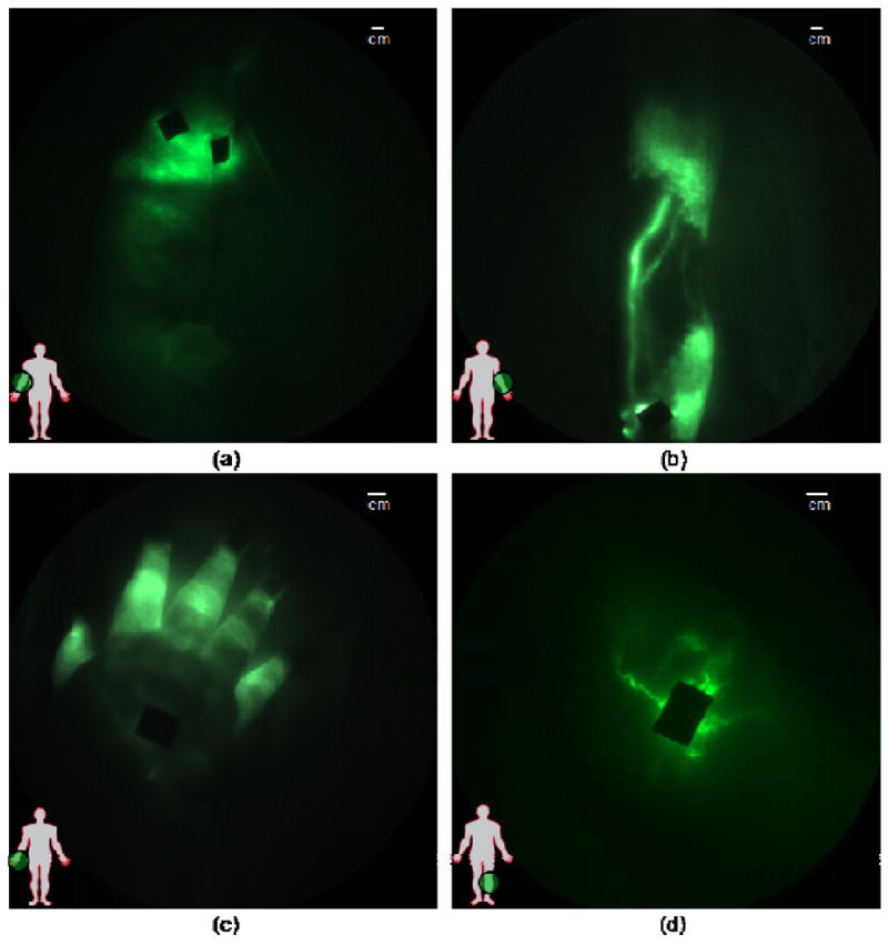
Near-infrared (NIR) fluorescence imaging clinical studies have been reported in the literature with six different devices that employ various doses of indocyanine green (ICG) as a non-specific contrast agent. To date, clinical applications range from (i) angiography, intraoperative assessment of vessel patency, and tumor/metastasis delineation following intravenous administration of ICG, and (ii) imaging lymphatic architecture and function following subcutaneous and intradermal ICG administration. In the latter case, NIR fluorescence imaging may enable new discoveries associated with lymphatic function due to (i) a unique niche that is not met by any other conventional imaging technology and (ii) its exquisite sensitivity enabling high spatial and temporal resolution. Herein, we (i) review the basics of clinical NIR fluorescence imaging, (ii) survey the literature on clinical application of investigational devices using ICG fluorescent contrast, (iii) provide an update of non-invasive dynamic lymphatic imaging conducted with our FDPM device, and finally, (iv) comment on the future NIR fluorescence imaging for non-invasive and intraoperative use given recent demonstrations showing capabilities for imaging following microdose administration of contrast agent.


Figures

Fig. (1)
Fig. (1)
(A) Fluorescent image taken following NIR excitation at 785 nm and collection of fluorescence at 835 nm showing a mouse containing no fluorophore and a vial containing 50 nM of NIR fluorophore, IRDye800CW. (B) Fluorescent image taken following red excitation at 690 nm and collection of fluorescence at 710 nm showing a mouse containing no fluorophore and a vial containing 50 nM of Cy5. The images show dramatically more autofluorescence that creates elevated background at red (690 nm) than at NIR (785 nm) excitation wavelengths. Reproduced with permission from Adams, et al. [15].
Fig. (2)
Fig. (2)
NIR fluorescent images of the normal left and right arms of a woman who underwent cancer surgery and left ALND three decades previous to the date of imaging. The subject has no lymphedema, but NIR fluorescent images show (A) unusual lymphatic vasculature on the left hand, and (B) normal lymphatic vasculature on her right hand. NIR fluorescent images of her (C) medial right arm shows normal functioning lymphatic vessels (See online supplemental video #1). NIR fluorescent images show drainage for both (D) left and (E) right axilla (See online supplemental videos #2 and #3).
Fig. (2)
Fig. (2)
NIR fluorescent images of the normal left and right arms of a woman who underwent cancer surgery and left ALND three decades previous to the date of imaging. The subject has no lymphedema, but NIR fluorescent images show (A) unusual lymphatic vasculature on the left hand, and (B) normal lymphatic vasculature on her right hand. NIR fluorescent images of her (C) medial right arm shows normal functioning lymphatic vessels (See online supplemental video #1). NIR fluorescent images show drainage for both (D) left and (E) right axilla (See online supplemental videos #2 and #3).
Fig. (3)
Fig. (3)
Fluorescence images of nodal regions in normal subjects: (a) three median lymphatic bundles that pool the fluorescent dye into three lymph nodes in the axilla; (b) afferent and efferent lymphatic vessels feeding and draining the fluorescent cubital lymph node in the medial forearm and elbow; (c) the popliteal lymph node in the back of the right knee (d) fluorescent signals demarking up to six superficial inguinal lymph nodes. Reproduced from Sevick-Muraca [75] with permission.
Fig. (4)
Fig. (4)
Images of diseased lymphatics in lymphedema subjects: (a) diffused dye patterns in symptomatic arm, (b) hyperplastic growth in symptomatic arm, (c) retrograde flow in symptomatic hand, and (d) tortuous vessels in symptomatic leg. Black areas are covered injection sites. Reproduced from Rasmussen et al. [47].

References

    1. Alitalo K, Tammela T, Petrova TV. Lymphangiogenesis in development and human disease. Nature. 2005;438(7070):946–53. - PubMed
    1. Rockson SG. Lymphedema. Am J Med. 2001 Mar;110(4):288–95. - PubMed
    1. Armer JM. The problem of post-breast cancer lymphedema: impact and measurement issues. Cancer Invest. 2005;23(1):76–83. - PubMed
    1. Ridner SH. Pretreatment lymphedema education and identified educational resources in breast cancer patients. Patient Educ Couns. 2006;61(1):72–9. - PubMed
    1. Mansel RE, Fallowfield L, Kissin M, et al. Randomized multicenter trial of sentinel node biopsy versus standard axillary treatment in operable breast cancer: the ALMANAC Trial. J Natl Cancer Inst. 2006;98(9):599–609. - PubMed

LinkOut - more resources