. 2013 Jan;168(1):238-52.
doi: 10.1111/j.1476-5381.2012.02176.x.

Reperfusion-induced myocardial dysfunction is prevented by endogenous annexin-A1 and its N-terminal-derived peptide Ac-ANX-A1(2-26)

Affiliations

Reperfusion-induced myocardial dysfunction is prevented by endogenous annexin-A1 and its N-terminal-derived peptide Ac-ANX-A1(2-26)

Chengxue Qin et al. Br J Pharmacol. 2013 Jan.

Abstract

Background and purpose: Annexin-A1 (ANX-A1) is an endogenous, glucocorticoid-regulated anti-inflammatory protein. The N-terminal-derived peptide Ac-ANX-A1(2-26) preserves cardiomyocyte viability, but the impact of ANX-A1-peptides on cardiac contractility is unknown. We now test the hypothesis that ANX-A1 preserves post-ischaemic recovery of left ventricular (LV) function.

Experimental approach: Ac-ANX-A1(2-26) was administered on reperfusion, to adult rat cardiomyocytes as well as hearts isolated from rats, wild-type mice and mice deficient in endogenous ANX-A1 (ANX-A1(-/-)). Myocardial viability and recovery of LV function were determined.

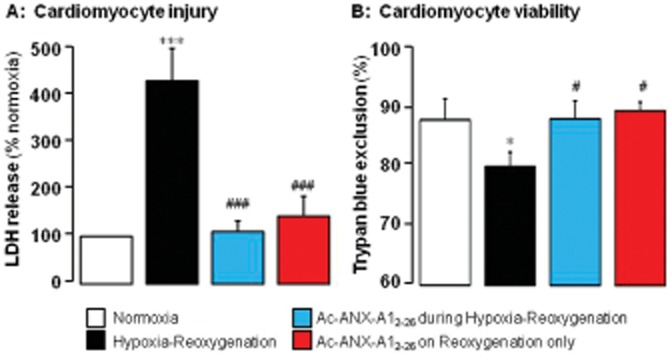
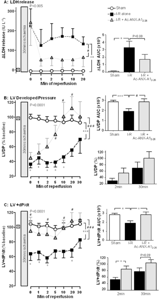
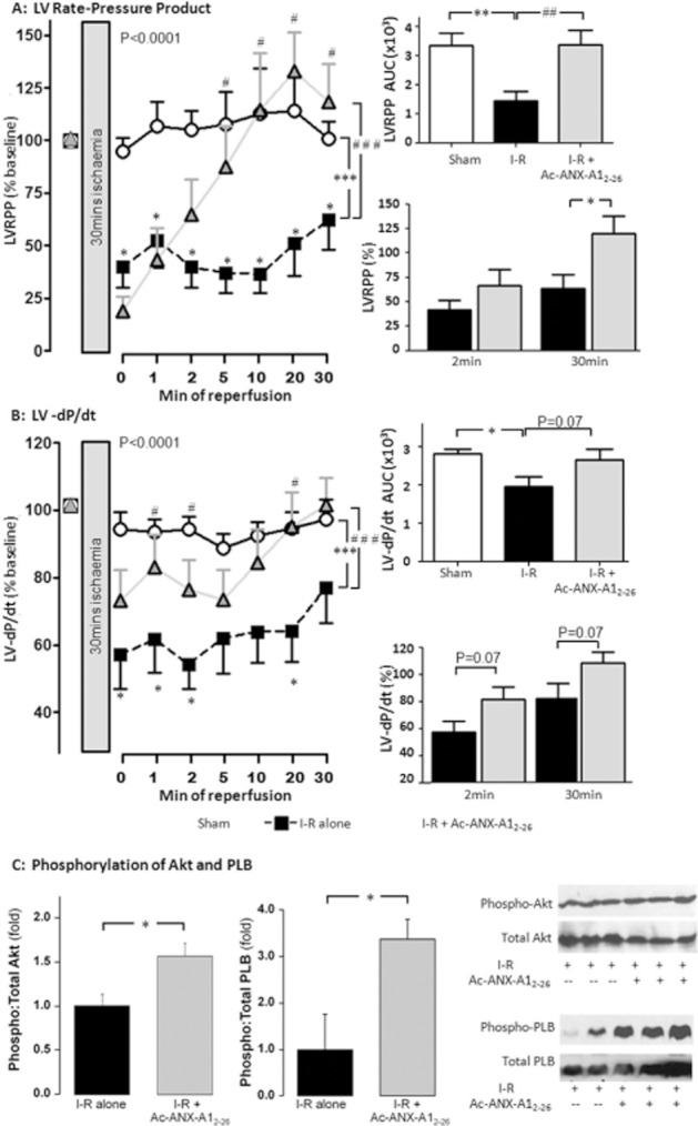
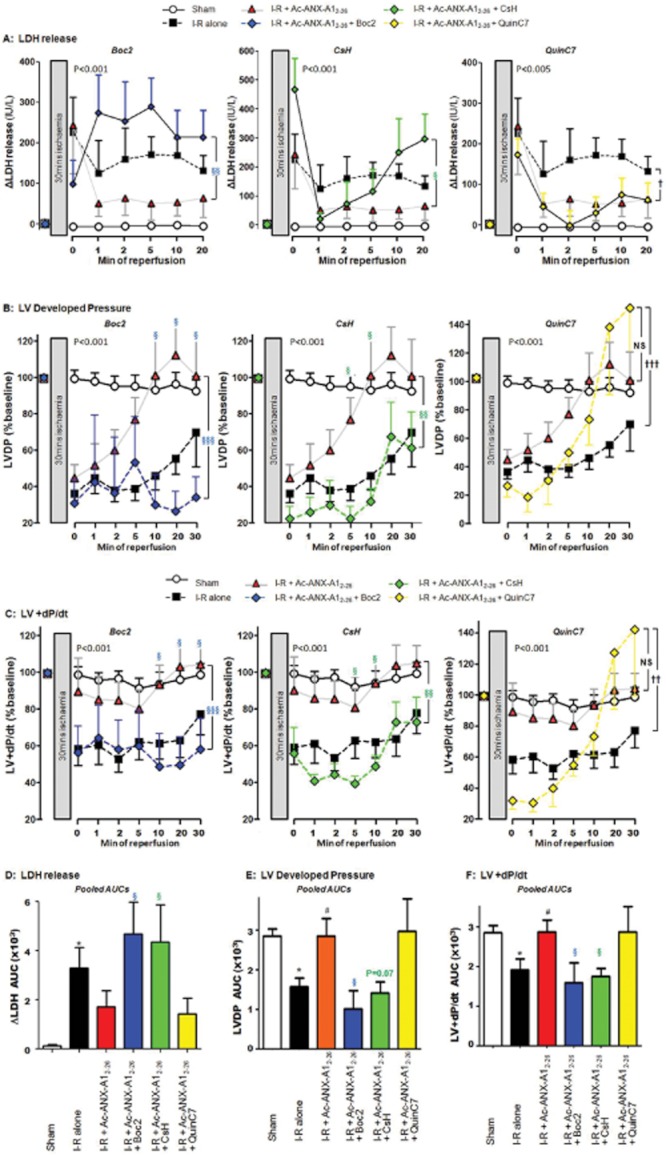
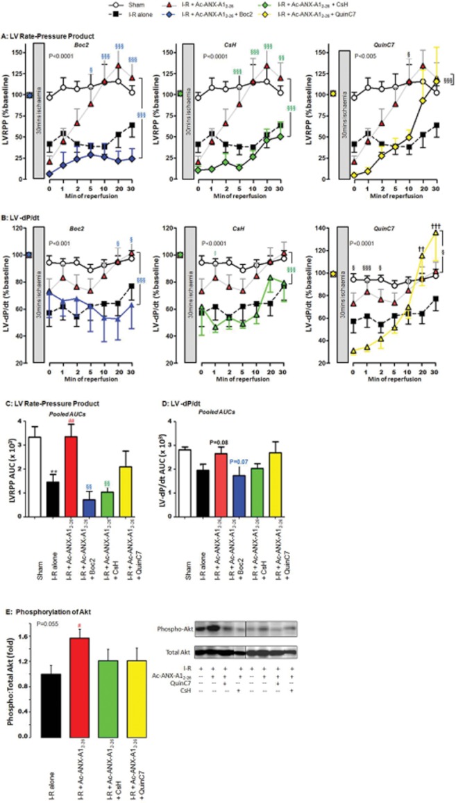
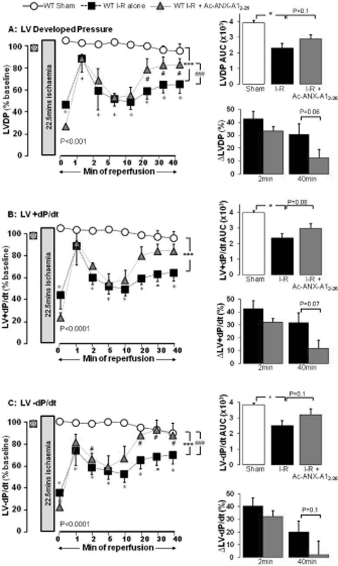
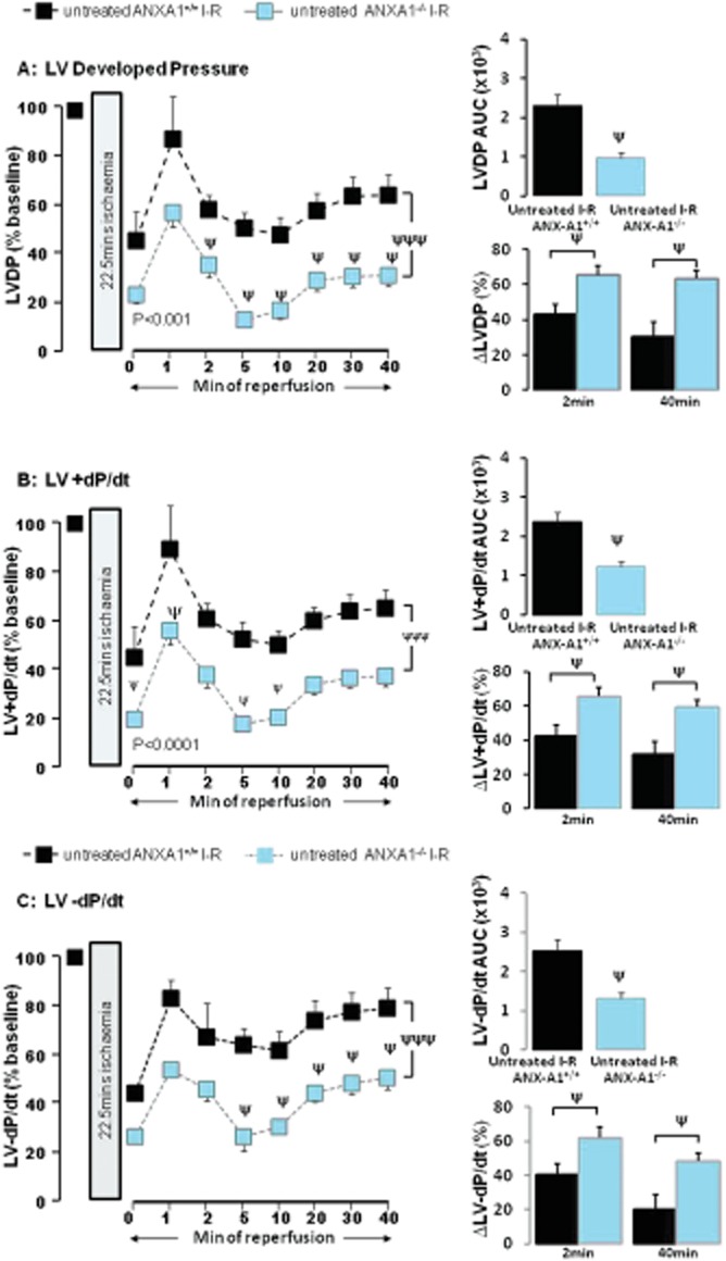
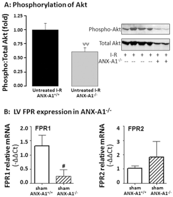
Key results: Ischaemia-reperfusion markedly impaired both cardiomyocyte viability and recovery of LV function by 60%. Treatment with exogenous Ac-ANX-A1(2-26) at the onset of reperfusion prevented cardiomyocyte injury and significantly improved recovery of LV function, in both intact rat and wild-type mouse hearts. Ac-ANX-A1(2-26) cardioprotection was abolished by either formyl peptide receptor (FPR)-nonselective or FPR1-selective antagonists, Boc2 and cyclosporin H, but was relatively insensitive to the FPR2-selective antagonist QuinC7. ANX-A1-induced cardioprotection was associated with increased phosphorylation of the cell survival kinase Akt. ANX-A1(-/-) exaggerated impairment of post-ischaemic recovery of LV function, in addition to selective LV FPR1 down-regulation.

Conclusions and implications: These data represent the first evidence that ANX-A1 affects myocardial function. Our findings suggest ANX-A1 is an endogenous regulator of post-ischaemic recovery of LV function. Furthermore, the ANX-A1-derived peptide Ac-ANX-A1(2-26) on reperfusion rescues LV function, probably via activation of FPR1. ANX-A1-based therapies may thus represent a novel clinical approach for the prevention and treatment of myocardial reperfusion injury.


Figures

Figure 1
Figure 1
(A) Ac-ANX-A12–26 (0.3 μM) prevents adult rat cardiomyocyte hypoxia–reoxygenation (H–R) injury in vitro (assessed by measuring LDH activity) whether present for the full duration of H–R (n = 11) or only post-H–R (n = 15 cardiomyocyte preparations). (B) H–R-induced loss of cardiomyocyte viability (assessed by measuring trypan blue exclusion) was also prevented by Ac-ANX-A12–26 at both time points (n = 8 cardiomyocyte preparations). *P < 0.05 and ***P < 0.001 versus paired untreated control, and #P < 0.05 and ###P < 0.001 Ac-ANX-A12–26-treated versus untreated H–R cardiomyocytes from the same cardiomyocyte preparation on SNK.
Figure 2
Figure 2
Ac-ANX-A12–26 (0.3 μM) addition at the onset of reperfusion attenuates I–R-induced impairment of myocardial viability and LV systolic function in rat isolated heart ex vivo. (A) Ac-ANX-A12–26 decreases the I–R-stimulated release of LDH from the myocardium (P < 0.005, untreated I–R, n = 16 vs. sham n = 4, and P < 0.05 untreated I–R vs. Ac-ANX-A12–26–treated I–R, n = 15, on two-way anova). The untreated I–R group also had a significantly increased myocardial release of LDH (as assessed by AUC analysis; P < 0.05), a trend that was absent in the presence of Ac-ANX-A12–26 (P = 0.08, right panel). (B) Ac-ANX-A12–26 accelerates recovery of LVDP up to 30 min post reperfusion (P < 0.001 untreated I–R, n = 15 vs. sham n = 9, and P < 0.001 vs. Ac-ANX-A12–26–treated I–R, n = 17, on two-way anova). The untreated I–R group also had a significantly impaired recovery of LVDP on AUC analysis (P < 0.001), which was prevented by Ac-ANX-A12–26 (P < 0.05, upper inset). Ac-ANX-A12–26–induced recovery of LVDP after 2 or 30 min reperfusion is shown in the lower inset (both P = NS). (C) Ac-ANX-A12–26 accelerates recovery of LV + dP/dt up to 30 min post reperfusion (P < 0.001 untreated I–R vs. sham, and P < 0.001 vs. Ac-ANX-A12–26–treated I–R, by two-way anova). The untreated I–R group had a significantly impaired recovery of LV + dP/dt on AUC analysis (P < 0.05), which was prevented by Ac-ANX-A12–26 (upper inset). Ac-ANX-A12–26 significantly improved recovery of LV + dP/dt after 2 min reperfusion (P < 0.05, lower inset). *P < 0.05 and ***P < 0.001 untreated I–R versus sham; #P < 0.05 and ##P < 0.001 Ac-ANX-A12–26–treated I–R versus untreated I–R rat hearts at each time point on SNK respectively.
Figure 3
Figure 3
Ac-ANX-A12–26 (0.3 μM) attenuates I–R-induced impairment of (A) LVRPP and (B) LV-dP/dt up to 30 min post reperfusion in the rat isolated heart ex vivo. Recovery of RPP and LV-dP/dt on AUC analysis is shown on the upper inset of each panel, and relative recovery of both parameters after 2 and 30 min reperfusion on the lower insets. (C) ANX-A12–26 also increases phosphorylation of the cell survival kinase, Akt (P < 0.05 ANX-A12–26 I–R, n = 14 vs. untreated I–R rat hearts n = 10, left-hand panel) and of the SERCA regulator, phospholamban (P < 0.05 ANX-A12–26 I–R, n = 4 vs. untreated I–R rat hearts, n = 3, right-hand panel), in rat hearts subjected to I–R. *P < 0.05, **P < 0.005 and ***P < 0.001 untreated I–R versus sham; #P < 0.05, ##P < 0.01 and ###P < 0.001 Ac-ANX-A12–26–treated I–R versus untreated I–R rat hearts at each time point on SNK respectively.
Figure 4
Figure 4
Role of FPR subtypes in Ac-ANX-A12–26 cardioprotection in the rat isolated heart ex vivo, on the time course of protection in the presence of Ac-ANX-A12–26 (0.3 μM), alone or in the presence of the non-selective FPR-antagonist Boc2 (10 μM), the FPR1-selective antagonist CsH (1 μM), or the FPR2-selective antagonist QuinC7 (10 μM) during reperfusion. (A) Both Boc2 and CsH (both P < 0.05, n = 6) significantly inhibited Ac-ANX-A12–26–induced protection of myocardial LDH release, but QuinC7 had no effect (P = NS, n = 6). Similar attenuation of the Ac-ANX-A12–26 effect by Boc2 and CsH, but not QuinC7, was also evident on recovery of both (B) LVDP and (C) LV + dP/dt. The AUC results for Ac-ANX-A12–26 cardioprotection ± FPR antagonists are also shown, on (D) LDH release, (E) LVDP and (F) LV + dP/dt. Sham n = 9, untreated I–R, n = 15 and Ac-ANX-A12–26–treated I–R, n = 17. *P < 0.05 untreated I–R versus sham; #P < 0.05 Ac-ANX-A12–26–treated I–R versus untreated I–R rat hearts; §P < 0.05, §§P < 0.01 and §§§P < 0.001 antagonist + Ac-ANX-A12–26–treated I–R versus Ac-ANX-A12–26–treated I–R and P < 0.05, ††P < 0.01 and †††P < 0.001 antagonist + Ac-ANX-A12–26–treated I–R versus untreated I–R rat hearts respectively.
Figure 5
Figure 5
FPR antagonism attenuates the cardioprotective effects of Ac-ANX-A12–26 on recovery of both (A) RPP and (B) LV-dP/dt (sham n = 9, untreated I–R n = 15, Ac-ANX-A12–26–treated I–R n = 17 and all antagonist groups n = 6). Recovery of (C) RPP and (D) LV-dP/dt on AUC analysis is also shown, as well as (E) Ac-ANX-A12–26-induced phosphorylation of Akt (all n = 5). *P < 0.05 and **P < 0.005 untreated I–R versus sham; ##P < 0.01 Ac-ANX-A12–26–treated I–R versus untreated I–R rat hearts, §P < 0.05, §§P < 0.005 and §§§P < 0.001 antagonist + Ac-ANX-A12–26–treated I–R versus Ac-ANX-A12–26–treated I–R and ††P < 0.01 and †††P < 0.001 antagonist + Ac-ANX-A12–26–treated I–R versus untreated I–R rat hearts respectively.
Figure 6
Figure 6
ANX-A12–26 (0.3 μM) addition at the onset of reperfusion attenuates I–R-induced impairment of LV systolic function in the isolated wild-type mouse heart ex vivo. Recovery of LVDP, LV + dP/dt and LV-dP/dt on AUC analysis is shown on the upper inset of each panel, and the relative impairment (i.e. the relative reduction in LV function as % of baseline) of all parameters after 2 and 30 min reperfusion are shown on the lower insets. (A) Ac-ANX-A12–26 accelerates recovery of LVDP up to 40 min post reperfusion (P < 0.001 untreated I–R, n = 11 versus sham n = 10 and P < 0.001 versus ANX-A12–26–treated I–R, n = 6, both on two-way anova). Untreated I–R also significantly impaired recovery of LVDP on AUC analysis (P < 0.05), which tended to be prevented by Ac-ANX-A12–26 (P = 0.1, upper inset). The impairment in recovery of LVDP in untreated versus Ac-ANX-A12–26–treated I–R was not different after 2 min reperfusion (P = NS), but a greater recovery of LVDP in Ac-ANX-A12–26–treated hearts was evident after 40 min reperfusion (P = 0.06, lower inset). (B) Ac-ANX-A12–26 accelerates recovery of LV + dP/dt up to 40 min post reperfusion (untreated I–R, n = 11 P < 0.001 versus sham, n = 10 and P = 0.089 versus ANX-A12–26–treated I–R, n = 6, on two-way anova). Untreated I–R significantly impaired recovery of LV + dP/dt on AUC analysis (P < 0.05), which tended to be prevented by Ac-ANX-A12–26 (P = 0.08, upper inset). Ac-ANX-A12–26 tended to improve recovery of LV + dP/dt after 40 min reperfusion (P = 0.08) but not after 2 min reperfusion (P = NS, lower inset). (C) Ac-ANX-A12–26 accelerates recovery of LV-dP/dt up to 40 min post reperfusion (untreated I–R n = 11 P < 0.001 vs. sham n = 10 and P < 0.001 vs. ANX-A12–26–treated I–R n = 6, both by two-way anova). Untreated I–R significantly impaired recovery of LV-dP/dt on AUC analysis (P < 0.05), which Ac-ANX-A12–26 tended to prevent (P = 0.1, upper inset). Ac-ANX-A12–26 tended to improve recovery of LV-dP/dt after 40 min reperfusion (P = 0.1) but not after 2 min reperfusion (P = NS, lower inset). *P < 0.05 and ***P < 0.001 untreated I–R versus sham and #P < 0.05 and ###P < 0.001 ANX-A12–26–treated I–R versus untreated I–R mouse hearts respectively.
Figure 7
Figure 7
Deficiency of endogenous ANX-A1 exaggerates I–R-induced impairment of LV systolic function in the intact mouse heart ex vivo. Recovery of LVDP, LV + dP/dt and LV-dP/dt on AUC analysis is shown on the upper inset of each panel, and the relative impairment (i.e. the relative reduction in LV function as % of baseline) of all parameters after 2 and 40 min reperfusion are shown on the lower insets. (A) The time course of recovery of LVDP is significantly impaired in ANX-A1−/− (n = 14) versus ANX-A1+/+ mouse hearts (n = 11, P < 0.001 on two-way anova). ANX-A1−/− also significantly exaggerated the impaired recovery of LVDP on AUC analysis (P < 0.05, upper inset), which was evident at both 2 and 40 min reperfusion (both P < 0.05, lower inset). (B) ANX-A1−/− also significantly delayed recovery of LV + dP/dt compared with ANX-A1+/+ mouse hearts (P < 0.0001 on two-way anova), which was also evident on AUC analysis (P < 0.05, upper inset), and at both 2 and 40 min reperfusion (both P < 0.05, lower inset). (C) ANX-A1−/− similarly delayed recovery of LV-dP/dt compared with ANX-A1+/+ mouse hearts (P < 0.001 on two-way anova), which was also evident on AUC analysis (P < 0.05, upper inset), and at both 2 and 40 min reperfusion (both P < 0.05, lower inset). ψP < 0.001 and ψψψP < 0.001 untreated I–R ANX-A1+/+ versus untreated I–R ANX-A1−/− mouse hearts.
Figure 8
Figure 8
Deficiency of ANX-A1 (A) suppresses phosphorylation of the cell survival kinase Akt following I–R in mouse hearts (both n = 6, ψψP < 0.01) and (B) down-regulates gene expression of FPR1; FPR2 expression is unaffected (n = 4–5 per group).

References

    1. Burli RW, Xu H, Zou XM, Muller K, Golden J, Frohn M, et al. Potent hFPRL1 (ALXR) agonists as potential anti-inflammatory agents. Bioorg Med Chem Lett. 2006;16:3713–3718. - PubMed
    1. D'Amico M, Di Filippo C, La M, Solito E, McLean PG, Flower RJ, et al. Lipocortin 1 reduces myocardial ischemia-reperfusion injury by affecting local leukocyte recruitment. FASEB J. 2000;14:1867–1869. - PubMed
    1. Dreier R, Schmid KW, Gerke V, Riehemann K. Differential expression of annexins I, II and IV in human tissues: an immunohistochemical study. Histochem Cell Biol. 1998;110:137–148. - PubMed
    1. Dufton N, Perretti M. Therapeutic anti-inflammatory potential of formyl-peptide receptor agonists. Pharmacol Ther. 2010;127:175–188. - PubMed
    1. Dufton N, Hannon R, Brancaleone V, Dalli J, Patel HB, Gray M, et al. Anti-inflammatory role of the murine formyl-peptide receptor 2: ligand-specific effects on leukocyte responses and experimental inflammation. J Immunol. 2010;184:2611–2619. - PMC - PubMed

Publication types

MeSH terms