. 2012 Sep 25;6(9):7703-11.
doi: 10.1021/nn3025699. Epub 2012 Aug 27.

Unzipping a functional microbial amyloid

Affiliations

Unzipping a functional microbial amyloid

David Alsteens et al. ACS Nano. .

Abstract

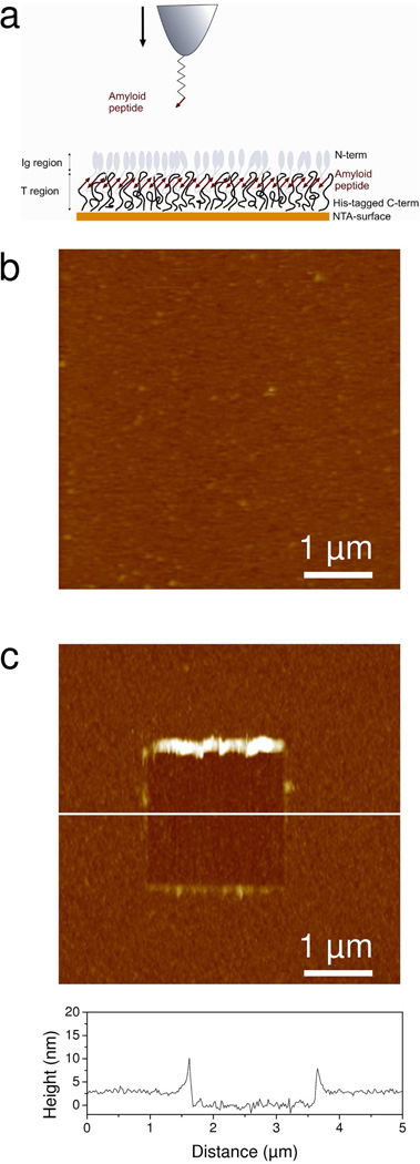
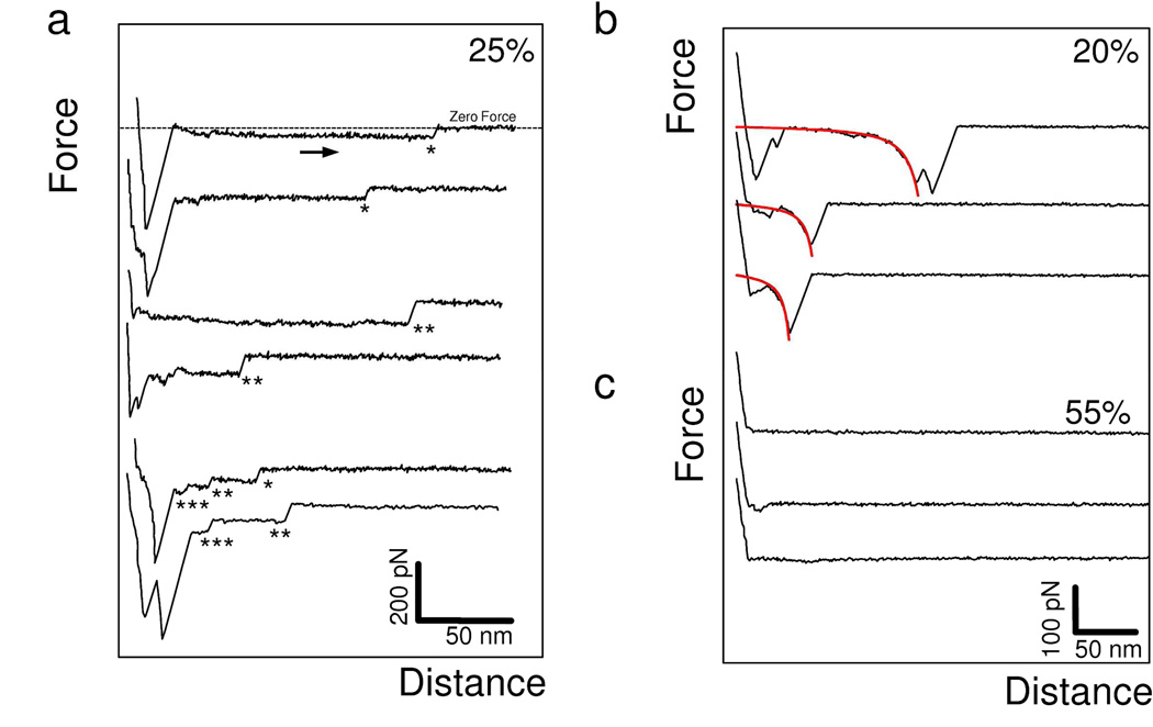
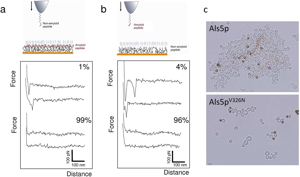
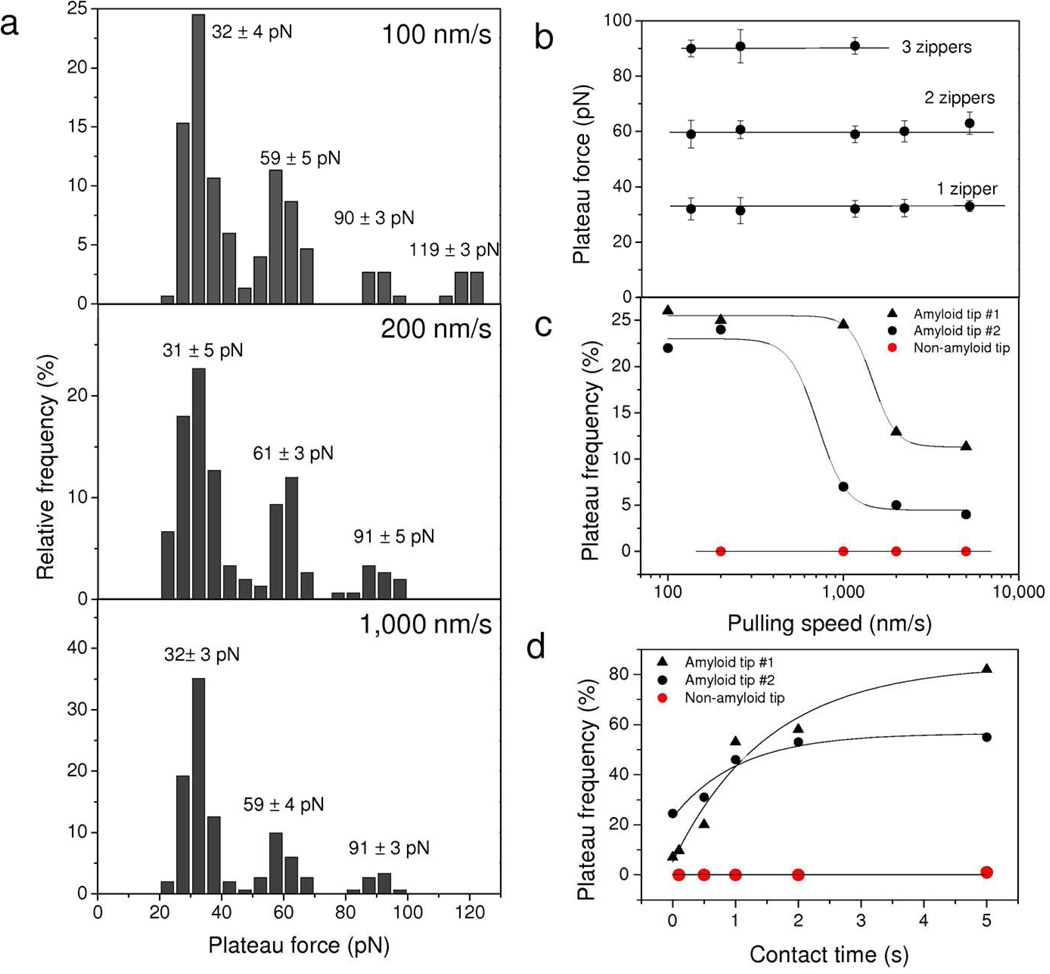
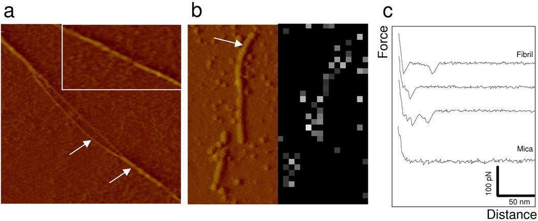
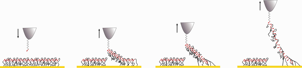
Bacterial and fungal species produce some of the best-characterized functional amyloids, that is, extracellular fibres that play key roles in mediating adhesion and biofilm formation. Yet, the molecular details underlying their mechanical strength remain poorly understood. Here, we use single-molecule atomic force microscopy to measure the mechanical properties of amyloids formed by Als cell adhesion proteins from the pathogen Candida albicans. We show that stretching Als proteins through their amyloid sequence yields characteristic force signatures corresponding to the mechanical unzipping of β-sheet interactions formed between surface-arrayed Als proteins. The unzipping probability increases with contact time, reflecting the time necessary for optimal inter β-strand associations. These results demonstrate that amyloid interactions provide cohesive strength to a major adhesion protein from a microbial pathogen, thereby strengthening cell adhesion. We suggest that such functional amyloids may represent a generic mechanism for providing mechanical strength to cell adhesion proteins. In nanotechnology, these single-molecule manipulation experiments provide new opportunities to understand the molecular mechanisms driving the cohesion of functional amyloid-based nanostructures.


Figures

Figure 1
Figure 1
Strategy for measuring the nanomechanical response of Als amyloids. (a) Principle of the single-molecule manipulation experiments. Each Als protein protruding from the yeast surface contains a ligand-binding Ig-like region (Ig) made of two β-sheet-rich domains followed by an amyloidogenic threonine-rich region (T). To mimic this orientation, Ig-T fragments terminated with a His-tag at the C terminal were assembled on a gold surface terminated with Ni2+-NTA (10 %) and EG3 (90 %) groups. Note that the cartoon is idealized and that the surface may be less structured than depicted. The surface-displayed Ig-T molecules were stretched via their amyloid region using an AFM tip functionalized with the amyloid-forming peptide SNGIVIVATTRTV. (b) AFM deflection image (5 µm × 5 µm) in PBS confirming the presence of a homogeneous, featureless Ig-T layer. (c) To determine the layer thickness, 2 µm × 2 µm square areas were first scanned at large forces (10 nN), followed by imaging 5 µm × 5 µm images of the same areas under smaller forces. A cross-section taken in the corresponding height image along the white line is shown under the image.
Figure 2
Figure 2
Unzipping individual β-sheets. (a-c) Representative retraction force curves recorded in buffer between an amyloid-tip and an Ig-T surface, using a pulling speed of 200 nm/s and an interaction time of 250 ms. (a) A substantial fraction (25 %) of the curves showed constant force plateaus, sometimes superimposed onto one another. The dashed line on the top curve represents the zero force line. The number of force plateaus superimposed is highlighted by a corresponding number of asterisks (*). (b) Another fraction of the curves (20 %) recorded in the same conditions showed elastic responses that were well-described by the worm-like-chain model using a persistence length of 0.4 nm (see fits in red). (c) The remaining curves (55 %) did not show any adhesion events. The data shown are from a total of 256 force curves recorded on different locations of a sample. Similar data were obtained using more than 3 different tips and 3 independent samples (> 3,072 force curves).
Figure 3
Figure 3
Unzipping interactions are abolished with a single-site mutation in the amyloid sequence. (a, b) Representatives force-distance curves recorded in buffer between a tip bearing a non-amyloid peptide with a V5N substitution (SNGINIVATTRTV) and Ig-T proteins (a), and between a tip bearing the amyloid peptide (SNGIVIVATTRTV) and non-amyloid Ig-T mutant proteins with a V326N substitution in the amyloid sequence (b). The pulling rate was 200 nm/s and the interaction time 250 ms. Both conditions lead to a dramatic reduction in the number of curves with adhesion events and to the disappearance of force plateau signatures, indicating that molecular zippers are formed through amyloid bonds. (c) Effect of the V326N substitution on adhesion and aggregation of S. cerevisiae yeast cells expressing Als5p. The brown spheres are BSA-coated ferromagnetic beads, 2 µm in diameter. This substitution in the 1419-residue protein greatly reduce cellular aggregation and amyloid-formation.,
Figure 4
Figure 4
Dynamics of the zipper interaction. (a) Histograms showing the distribution of plateau forces measured between an amyloid-tip and an Ig-T surface at different pulling speeds (100 nm/s, 200 nm/s and 1,000 nm/s; surface delay = 0 ms). ). For each loading rate, the data correspond to 150 plateau curves from a total of 3,072 force curves recorded using three different tips and samples. (b, c) Dependence of the mean plateau forces (b) and plateau force frequency (c) on the pulling speed. Each data point in (b) represents the mean ± σ calculated from 150 plateau curves from a total of 3,072 force curves obtained using three different tips and samples. (d) Dependence of the plateau force frequency on contact time measured at a constant pulling speed of 1,000 nm/s. The plateau force probability showed a strong dependence both on the pulling speed and the interaction time. Each value in (c) and (d) was obtained from a total of 1,024 forces curves. The two sets of black symbols represent two independent experiments with amyloid-tips, while the red symbols correspond to control experiments with tips bearing non-amyloid peptides.
Figure 5
Figure 5
Morphology and mechanical response of mature amyloid fibrils. (a) AFM deflection image (1 µm × 1 µm) recorded with a silicon nitride tip in buffer for a preparation of Ig-T molecules (0.2 mg/ml) incubated during 4 days at 4°C in PBS buffer and adsorbed on mica. Arrows show an amyloid fiber separated into two strands. The inset is an enlarged view of a fiber showing helical markings. (b) Deflection image and adhesion force map (16 × 32 force curves on a 0.5 µm × 1 µm area) recorded in buffer with an amyloid-tip. The arrow emphasizes a fibril. Bright pixels in the map correspond to binding events. (c) Representative force curves recorded on mica and on a fibril, documenting single unbinding forces (32 ± 5 pN) without any evidence for unzipping interactions.
Figure 6
Figure 6
Nanomechanics of Als amyloid zippers. Model showing homotypic binding of lateral Als adhesins through antiparallel β-strands (red arrows) leading to the formation of a surface amyloid zipper (for sake of clarity, Ig regions are not shown). Note that the surface may be less structured than depicted, and we do not know whether the amyloid-like interactions result from association of parallel or anti-parallel β-strands. From left to right: approaching an amyloid peptide towards the Ig-T β-strands leads to the formation of an amyloid bond; retracting the probing peptide stretches and lifts amyloid regions from the surface, breaks H bonds sequentially between two strands (30 pN) or more (60 pN or 90 pN) at a time. In nature, this molecular zipper provides a powerful mechanism to strengthen cell-cell adhesion.

References

    1. Dobson CM. Protein Misfolding, Evolution and Disease. Trends Biochem. Sci. 1999;24:329–332. - PubMed
    1. Dobson CM. Structural Biology: Prying into Prions. Nature. 2005;435:747–749. - PubMed
    1. Shewmaker F, McGlinchey RP, Wickner RB. Structural Insights into Functional and Pathological Amyloid. J. Biol. Chem. 2011;286:16533–16540. - PMC - PubMed
    1. Nelson R, Sawaya MR, Balbirnie M, Madsen AØ, Riekel C, Grothe R, Eisenberg D. Structure of the Cross-β Spine of Amyloid-Like Fibrils. Nature. 2005;435:773–778. - PMC - PubMed
    1. Sawaya MR, Sambashivan S, Nelson R, Ivanova MI, Sievers SA, Apostol MI, Thompson MJ, Balbirnie M, Wiltzius JJW, McFarlane HT, et al. Atomic Structures of Amyloid Cross-β Spines Reveal Varied Steric Zippers. Nature. 2007;447:453–457. - PubMed

Publication types

MeSH terms

Substances