. 2012 Sep 26;134(38):15765-72.
doi: 10.1021/ja303612d. Epub 2012 Sep 17.

Engineered synthetic polymer nanoparticles as IgG affinity ligands

Affiliations

Engineered synthetic polymer nanoparticles as IgG affinity ligands

Shih-Hui Lee et al. J Am Chem Soc. .

Abstract

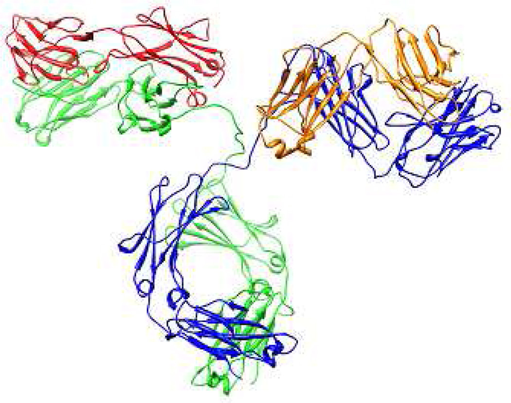
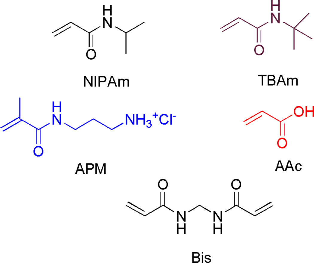
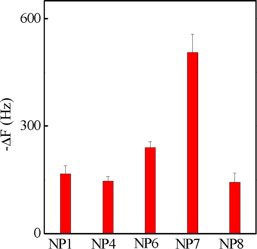
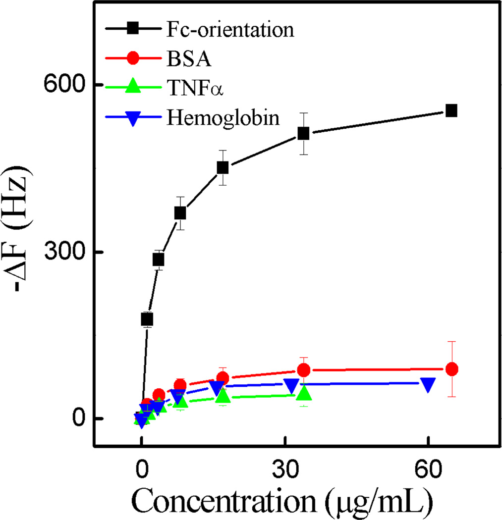
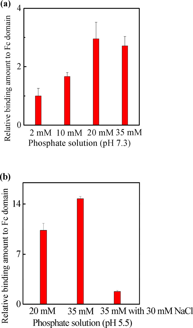
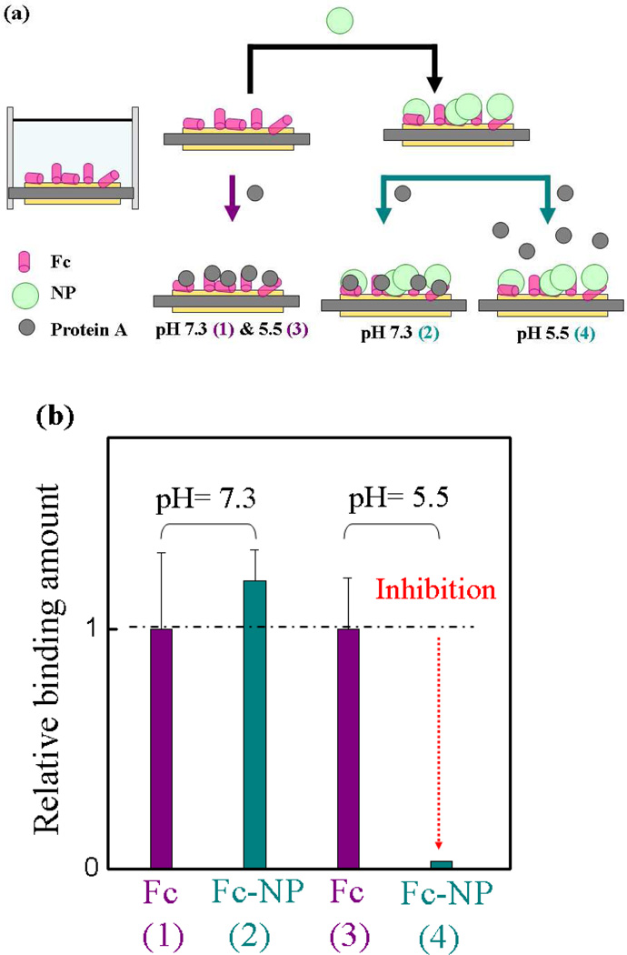
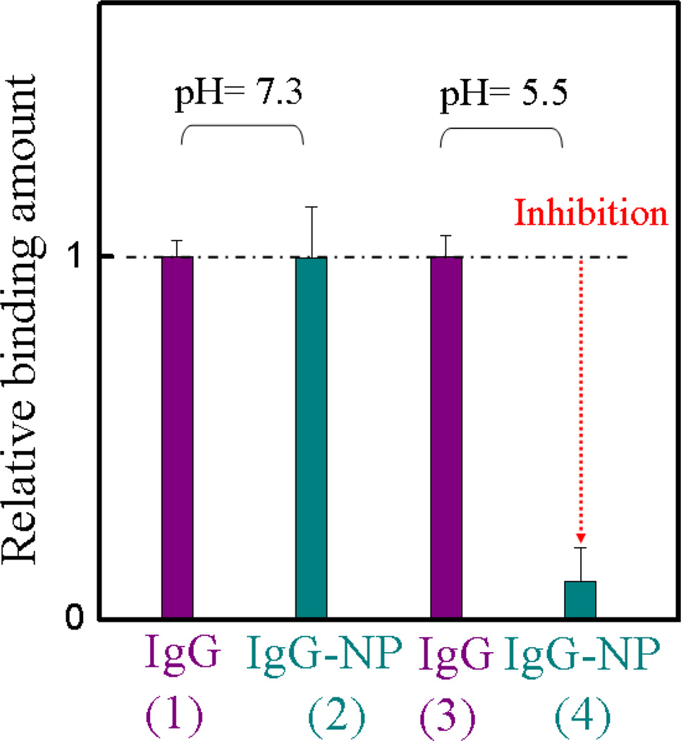
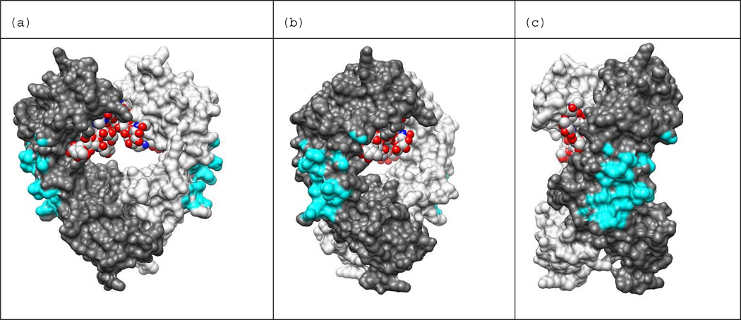
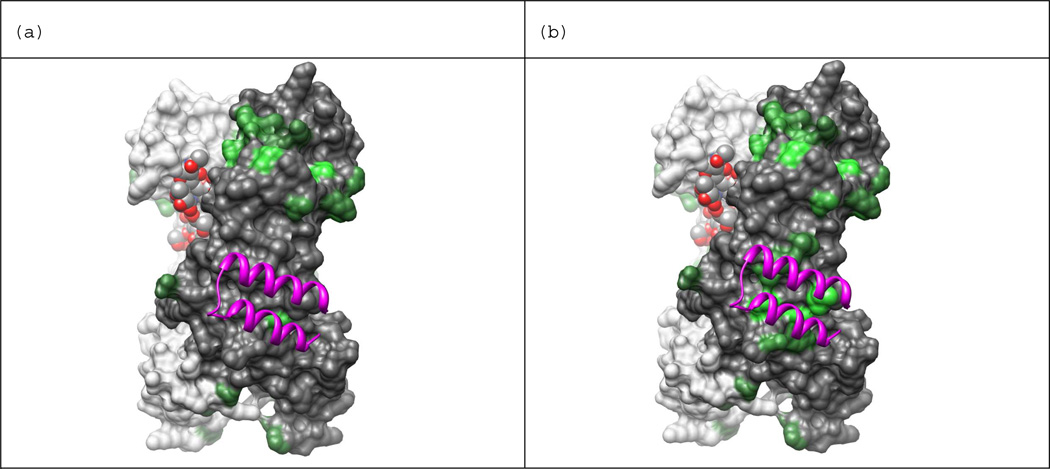
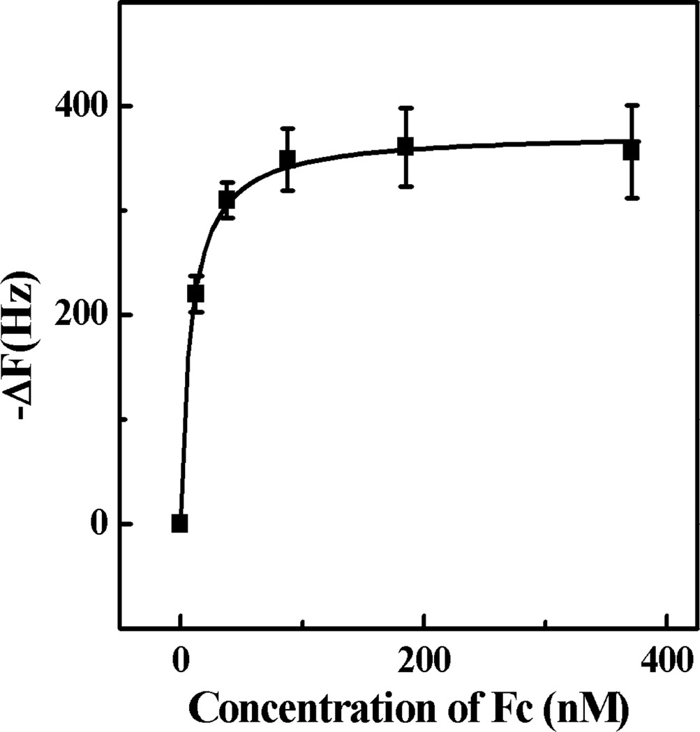
A process for the preparation of an abiotic protein affinity ligand is described. The affinity ligand, a synthetic polymer hydrogel nanoparticle (NP), is formulated with functional groups complementary to the surface presentation of the target protein. An iterative process is used to improve affinity by optimizing the composition and proportion of functional monomers. Since the polymer NPs are formed by a kinetically driven process, the sequence of functional monomers in the polymer chain is not controlled; only the average composition can be adjusted by the stoichiometry of the monomers in the feed. To compensate for this the hydrogel NP is lightly cross-linked resulting in chain flexibility that takes place on a submillisecond time scale allowing the polymer to "map" onto a protein surface with complementary functionality. In this study, we report a lightly cross-linked (2%) N-isopropyl acrylamide (NIPAm) synthetic polymer NP (50-65 nm) incorporating hydrophobic and carboxylate groups that binds with high affinity to the Fc fragment of IgG. The affinity and amount of NP bound to IgG is pH dependent. The hydrogel NP inhibits protein A binding to the Fc domain at pH 5.5, but not at pH 7.3. A computational analysis was used to identify potential NP-protein interaction sites. Candidates include a NP binding domain that overlaps with the protein A-Fc binding domain at pH 5.5. The computational analysis supports the inhibition experimental results and is attributed to the difference in the charged state of histidine residues. Affinity of the NP (3.5-8.5 nM) to the Fc domain at pH 5.5 is comparable to protein A at pH 7. These results establish that engineered synthetic polymer NPs can be formulated with an intrinsic affinity to a specific domain of a large biomacromolecule.


Figures

Figure 1
Figure 1
The structure of IgG with heavy chains in blue and green and light chains in orange and red. The Fc-Fc dimer is shown at the bottom of the image. The PDB ID is 1HZH.
Figure 2
Figure 2
Monomers used for NP synthesis
Figure 3
Figure 3
QCM screening of the library of polymer NPs containing 40% TBAm and varying amounts of AAc with Fc-oriented IgG in PBS (35 mM, pH 7.3 with 150 mM NaCl). Standard deviations were calculated from three independent measurements.
Figure 4
Figure 4
The interactions between NP7 and IgG, BSA, TNFα and hemoglobin in each of which were immobilized on a QCM surface in PBS (35 mM, pH 7.3 with 150 mM NaCl). Standard deviations were calculated from three independent measurements.
Figure 5
Figure 5
The interaction between the Fc domain and NP7 by QCM at various phosphate concentrations at (a) pH 7.3; and (b) pH 5.5. The relative amount is related to the amount of NP7 bound to the Fc domain at various salt concentrations compared to the amount bound in 2 mM phosphate buffer at pH 7.3. Standard deviations were calculated from three independent measurements.
Figure 6
Figure 6
(a) Schematic of the competitive binding of protein A to Fc in the presence and absence of NP7. (b) The interaction of the Fc fragment and protein A at pH 7.3 and 5.5 in PBS (35 mM, without salt), respectively, following treatment with NP7. The arrow color in (a) corresponds to the color of the column in (b). Cyan column: the frequency change resulting from protein A binding to the Fc domain in the presence of NP7 at pH 7.3 (2) and 5.5 (4), respectively. Purple column: control experiment under the same conditions omitting NPs at pH 7.3 (1) and 5.5 (3), respectively. Relative binding is defined by comparing the binding amount of the NP to that of protein A for the Fc domain. Standard deviations were calculated from three independent measurements.
Figure 7
Figure 7
Interaction between the Fc fragment of IgG and protein A at pH 7.3 and 5.5 in 35 mM phosphate buffer, before and after addition of NP7. The experimental design is similar to Figure 6a. Whole IgG was used to functionalize the QCM cell. The color of the column in the graph corresponds to the color of the arrowhead in Figure 6a. Cyan column: the frequency change was caused by protein A binding to the Fc domain of IgG in the presence of NP7 at pH 7.3 (2) and 5.5 (4), respectively. Purple column: control experiment under the same conditions except omitting the introduction of NPs at pH 7.3 (1) and 5.5 (3). Relative binding is defined as comparing the binding amount of the NP to that of protein A for whole IgG. Standard deviations were calculated from three independent measurements.
Figure 8
Figure 8
Protein A binding site shown on IgG1-Fc dimer. Binding site atoms are colored cyan. An Fc atom is considered in contact with protein A if it is within 5 Å of any heavy atom of protein A. The holo structure PDB ID: 1FC2 was used to calculate distances between Fc and protein A. The N-terminus of Fc is at the top of the view and C-terminus is at the bottom. Chain H is colored dark gray, chain K is colored light gray, and the carbohydrates bound in the Fc dimer core (red) are shown in the space filled representation. Images generated from PDB ID: 1HZH with UCSF Chimera. The view in (b) is rotated 45 degrees about the vertical axis with respect to the view in (a), and the view in (c) is rotate 90 degrees with respect to (a).
Figure 9
Figure 9
Computationally selected NP binding sites at (a) neutral and (b) low pH. Computational results indicate NP7 is more likely to bind the protein A binding site at lower pH in agreement with experimental observations. Residue side-chains are colored green if the combined score of its surface region is greater than 0.0, and brighter shades of green indicate higher scores. (a) presents results calculated assuming neutral pH and (b) at low pH where His residues can carry a positive charge. Chain H is colored dark gray, chain K is colored light gray, and the carbohydrates bound in the Fc dimer core are shown in space fill representation. Protein A appears as purple ribbons. Images generated from PDB ID: 1HZH with UCSF Chimera.
Figure 10
Figure 10
Binding isotherm of Fc domain to NP7 immobilized on QCM surface in water (pH 5.5) Standard deviations were calculated from three independent measurements.

References

    1. Meenach SA, Hilt JZ, Anderson KW. Acta Biomater. 2010;6:1039–1046. - PubMed
    1. Bhojani MS, Van Dort M, Rehemtulla A, Ross BD. Mol. Pharmaceut. 2010;7:1921–1929. - PMC - PubMed
    1. Tong R, Christian DA, Tang L, Cabral H, Baker JR, Kataoka K, Discher DE, Cheng JJ. Mrs Bull. 2009;34:422–431.
    1. Messerschmidt SKE, Musyanovych A, Altvater M, Scheurich P, Pfizenmaier K, Landfester K, Kontermann RE. J. Control. Release. 2009;137:69–77. - PubMed
    1. Davis ME, Chen Z, Shin DM. Nat. Rev. Drug Discov. 2008;7:771–782. - PubMed

Publication types