

A retrospective: use of Escherichia coli as a vehicle to study phospholipid synthesis and function
- PMID: 22925633
- PMCID: PMC3513495
- DOI: 10.1016/j.bbalip.2012.08.007
A retrospective: use of Escherichia coli as a vehicle to study phospholipid synthesis and function
Abstract
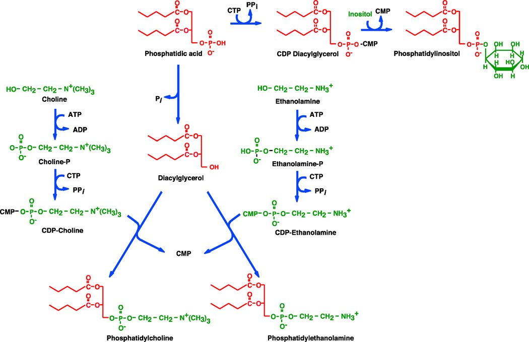
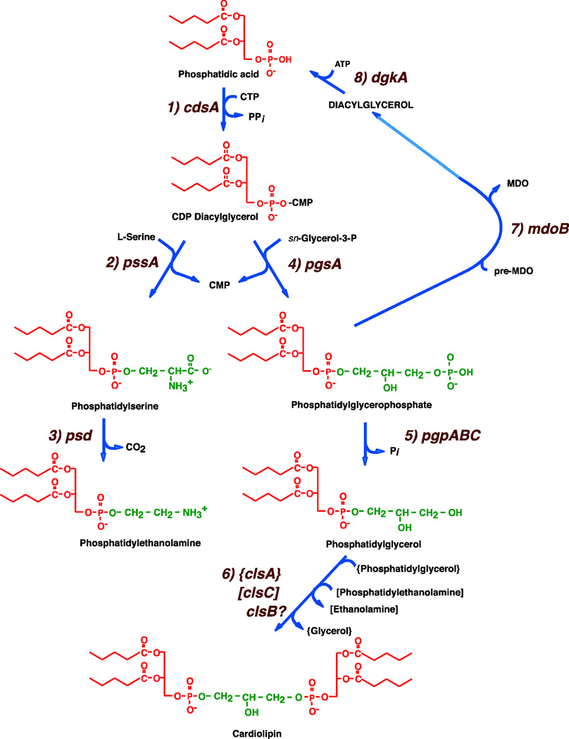
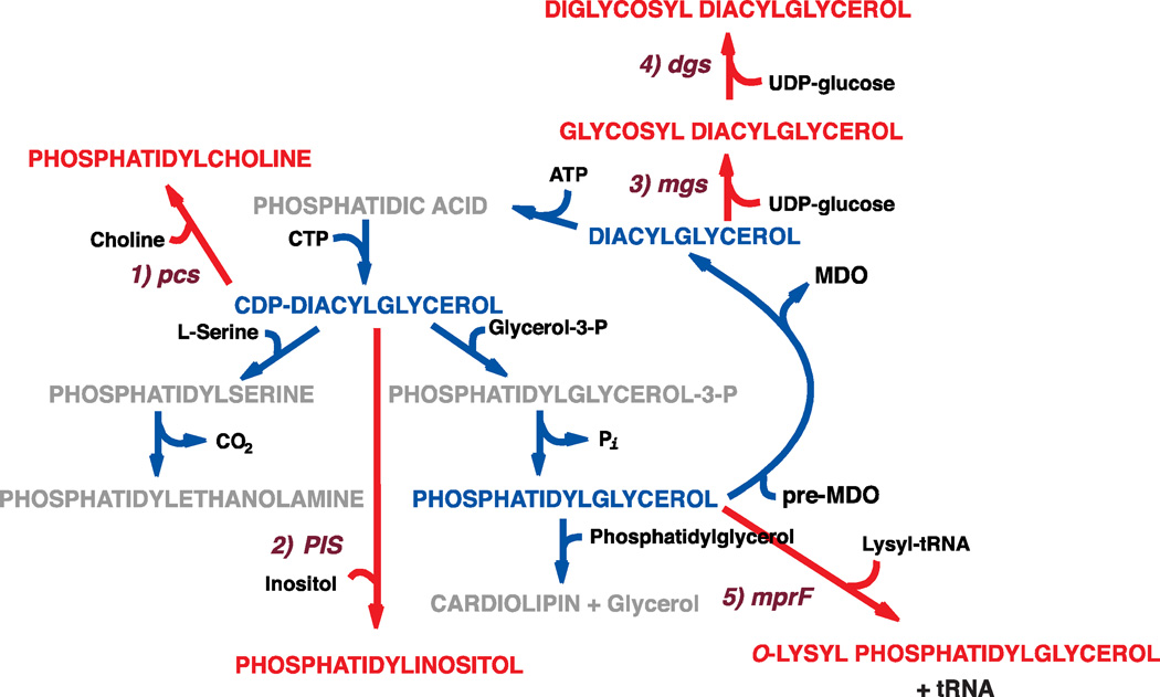
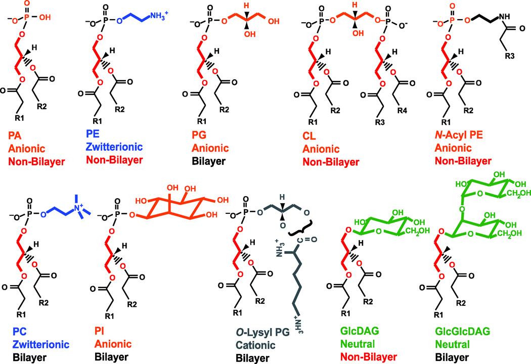
Although the study of individual phospholipids and their synthesis began in the 1920s first in plants and then mammals, it was not until the early 1960s that Eugene Kennedy using Escherichia coli initiated studies of bacterial phospholipid metabolism. With the base of information already available from studies of mammalian tissue, the basic blueprint of phospholipid biosynthesis in E. coli was worked out by the late 1960s. In 1970s and 1980s most of the enzymes responsible for phospholipid biosynthesis were purified and many of the genes encoding these enzymes were identified. By the late 1990s conditional and null mutants were available along with clones of the genes for every step of phospholipid biosynthesis. Most of these genes had been sequenced before the complete E. coli genome sequence was available. Strains of E. coli were developed in which phospholipid composition could be changed in a systematic manner while maintaining cell viability. Null mutants, strains in which phospholipid metabolism was artificially regulated, and strains synthesizing foreign lipids not found in E. coli have been used to this day to define specific roles for individual phospholipid. This review will trace the findings that have led to the development of E. coli as an excellent model system to study mechanisms underlying the synthesis and function of phospholipids that are widely applicable to other prokaryotic and eukaryotic systems. This article is part of a Special Issue entitled Phospholipids and Phospholipid Metabolism.
Copyright © 2012 Elsevier B.V. All rights reserved.
Figures
Similar articles
-
Molecular genetics of membrane phospholipid synthesis.Annu Rev Genet. 1986;20:253-95. doi: 10.1146/annurev.ge.20.120186.001345. Annu Rev Genet. 1986. PMID: 3545060 Review.
-
Overproduction of a foreign membrane protein in Escherichia coli stimulates and depends on phospholipid synthesis.Eur J Biochem. 1996 Oct 15;241(2):691-6. doi: 10.1111/j.1432-1033.1996.00691.x. Eur J Biochem. 1996. PMID: 8917473
-
Involvement of the YneS/YgiH and PlsX proteins in phospholipid biosynthesis in both Bacillus subtilis and Escherichia coli.BMC Microbiol. 2007 Jul 24;7:69. doi: 10.1186/1471-2180-7-69. BMC Microbiol. 2007. PMID: 17645809 Free PMC article.
-
Understanding phospholipid function: Why are there so many lipids?J Biol Chem. 2017 Jun 30;292(26):10755-10766. doi: 10.1074/jbc.X117.794891. Epub 2017 May 10. J Biol Chem. 2017. PMID: 28490630 Free PMC article. Review.
-
Mutants of Escherichia coli defective in membrane phospholipid synthesis. Effects of cessation and reinitiation of phospholipid synthesis on macromolecular synthesis and phospholipid turnover.J Biol Chem. 1977 Jul 10;252(13):4487-93. J Biol Chem. 1977. PMID: 326776 No abstract available.
Cited by
-
Making a membrane on the other side of the wall.Biochim Biophys Acta Mol Cell Biol Lipids. 2017 Nov;1862(11):1386-1393. doi: 10.1016/j.bbalip.2016.10.004. Epub 2016 Oct 11. Biochim Biophys Acta Mol Cell Biol Lipids. 2017. PMID: 27742351 Free PMC article. Review.
-
Lipid-Assisted Membrane Protein Folding and Topogenesis.Protein J. 2019 Jun;38(3):274-288. doi: 10.1007/s10930-019-09826-7. Protein J. 2019. PMID: 30937648 Free PMC article. Review.
-
Lipids and topological rules governing membrane protein assembly.Biochim Biophys Acta. 2014 Aug;1843(8):1475-88. doi: 10.1016/j.bbamcr.2013.12.007. Epub 2013 Dec 14. Biochim Biophys Acta. 2014. PMID: 24341994 Free PMC article. Review.
-
Defects in The First Step of Lipoprotein Maturation Underlie The Synthetic Lethality of Escherichia coli Lacking The Inner Membrane Proteins YciB And DcrB.J Bacteriol. 2021 Mar 15;203(6):e00640-20. doi: 10.1128/JB.00640-20. Epub 2021 Jan 11. J Bacteriol. 2021. PMID: 33431434 Free PMC article.
-
Marginally hydrophobic transmembrane α-helices shaping membrane protein folding.Protein Sci. 2015 Jul;24(7):1057-74. doi: 10.1002/pro.2698. Epub 2015 May 30. Protein Sci. 2015. PMID: 25970811 Free PMC article. Review.
References
-
- Levene PA, West CJ. Lecithin. I: "Hydrolecithin" and its bearing on the constitution of cephalin. J Biol Chem. 1918;33:111–117.
-
- Levene PA, West CJ. Cephalin V. Hydrocephalin of the egg yolk. J Biol Chem. 1918;35:285–290.
Publication types
MeSH terms
Substances
Grants and funding
LinkOut - more resources
Full Text Sources
Other Literature Sources
Molecular Biology Databases
