. 2012 Oct;145(1):44-54.
doi: 10.1016/j.clim.2012.07.010. Epub 2012 Aug 3.

Trauma patients' elevated tumor necrosis related apoptosis inducing ligand (TRAIL) contributes to increased T cell apoptosis

Affiliations

Trauma patients' elevated tumor necrosis related apoptosis inducing ligand (TRAIL) contributes to increased T cell apoptosis

Gautam Bandyopadhyay et al. Clin Immunol. 2012 Oct.

Abstract

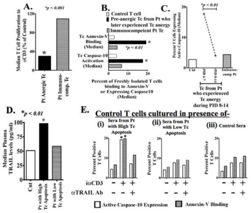
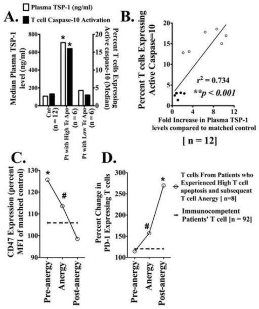
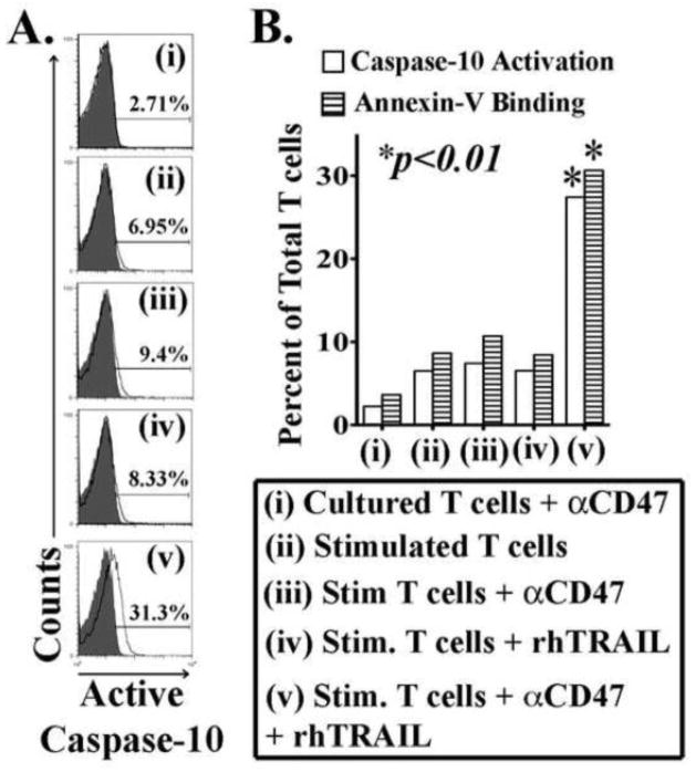
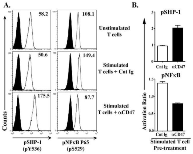
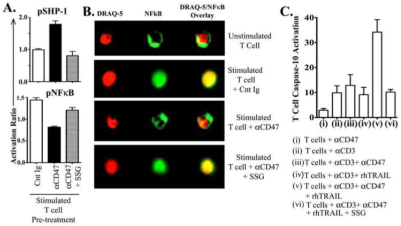
Immunosuppression resulting from excessive post-trauma apoptosis of hyperactivated T cells is controversial. TRAIL mediated T cell apoptosis decreases highly activated T cells' responses. Caspase-10, a particular TRAIL target, was increased in trauma patients' T cells with concomitantly elevated plasma TRAIL levels. These patients' T cells developed anergy, implicating increased TRAIL-mediated T cell apoptosis in post-trauma T cell anergy. Control T cells cultured with patients' sera containing high TRAIL levels increased their caspase-10 activity and apoptosis. Stimulated primary T cells are TRAIL apoptosis resistant. Increased plasma thrombospondin-1 and T cell expression of CD47, a thrombospondin-1 receptor, preceded patients' T cell anergy. CD47 triggering of T cells increased their sensitivity to TRAIL-induced apoptosis. Augmentation of T cell TRAIL-induced apoptosis was secondary to CD47 triggered activation of the Src homology-containing phosphatase-1 (SHP-1) and was partially blocked by a SHP-1 inhibitor. We suggest that combined post-trauma CD47 triggering, SHP-1 mediated NFκB suppression, and elevated TRAIL levels increase patients' CD47 expressing T cell apoptosis, thus contributing to subsequent T cell anergy.


Figures

Figure 1
Figure 1. Excessive post-injury T-cell apoptosis that presage the development of T-cell anergy is TRAIL-induced
(A) Freshly isolated T-cells from trauma patients (Pt) and age, sex and ethnicity matched controls (Cnt) were stimulated with plate-bound αCD3 to assess T-cell (Tc) proliferation. Pt T-cells were considered anergic when proliferation to αCD3 was less than 50% of Cnt T-cell response. Data show the median value from 17 Pt with T-cell anergy and 96 immunocompetent Pt; p<0.001 by t test and ANOVA. (B) Trauma patients who experienced T-cell hyporesponsiveness showed earlier pre-anergy elevated T-cell apoptosis. Freshly isolated Patients’ (Pt) and matched Controls’ (Cnt) T-cells were incubated with annexin-V + 7AAD or FAM-AEVD-FMK peptides (binds specifically to human active Caspase-10) and assayed by flowcytometry. Data shows the median value. *p<0.01 by t test and ANOVA compared to controls’ and immunocompetent patients’ T-cells. (C) Only Pt with early (PID- 4–7) excessive T-cell apoptosis later (PID- 8–14) developed T-cell anergy. Pt actual anergic T-cell (PID- 8–14) did not show increased apoptosis. *p<0.01 by t test and ANOVA. (D) Patients experiencing increased T-cell apoptosis also had increased plasma TRAIL levels as determined by ELISA *p<0.01 by Mann-Whitney U test compared to matched controls. (E) Freshly isolated control T-cells were stimulated with iαCD3 in culture with IMDM medium plus (i) sera from Pt who experienced elevated T-cell apoptosis, (ii) sera from Pt with low T-cell apoptosis, or (iii) sera from matched healthy control volunteers. In the indicated experimental conditions, sera were pre-treated with αTRAIL blocking antibody to block existing TRAIL in the sera. After 5 days of culture, T-cell Caspase-10 activation and annexin-V binding were assessed n=3 for each Pt group; n=6 for Cnt *p < 0.05.
Figure 2
Figure 2. Post trauma increased T cell apoptosis positively correlated with increased plasma Thrombospondin-1 levels
(A) Patients experiencing elevated T cell apoptosis also had increased plasma Thrombospondin-1 levels. Elevated plasma TSP-1 levels in patients with increased T-cell apoptosis as determined by ELISA. T cell apoptosis was determined by assessing active Caspase-10 binding to FAM-AEVD-FMK peptide. *p<0.01 vs. control and immunocompetent Patients (Pt) T cells by Mann-Whitney U test compared to matched controls; n = 6 for each patient group. (B) Linear regression plot showing significant correlation between elevated plasma TSP-1 levels and T cell Caspase-10 activation. r2 = 0.734, **p < 0.001, n = 12. (C–D) Elevated T cell CD47 and PD-1 expression levels in patients who experienced T cell anergy. Freshly isolated T-cells from patients and matched controls were stained for CD47 (C) and PD-1 (D) by flowcytometry. Percent changes compared to parallely assessed T cells form matched controls. Dashed lines represent similar percent change values from immunocompetent patients’ T cells. All datapoints represent median value. *p<0.01 & #p < 0.05 by Mann Whitney U test, n = 8 for patients experiencing T cell anergy and 92 for patients’ immunocompetent T cells.
Figure 3
Figure 3. Stimulated control T-cells expressed increased active Caspase-10 following combined treatment with rhTRAIL & αCD47 antibody
Freshly isolated T-cells were cultured with (i) 10μg/ml αCD Ab only, or (ii) immobilized αCD3 antibody alone, or (iii) αCD3+ αCD Ab, or (iv) αCD3 + rhTRAIL [1 μg/ml], or (v) αCD3 + rhTRAIL + αCD Ab for five days. Cleaved Caspase-10 levels were determined as described previously. Annexin-V binding levels were determined by flowcytometry using an AnnexinV + 7AAD kit. (A) Histograms showing active Caspase-10 levels in T-cells. (B) Graphical representation & statistics of above experiments *p < 0.01 by t test and ANOVA (n=8).
Figure 4
Figure 4. CD47 engagement in control T-cells inhibits TCR induced NFκB activation
Freshly isolated T-cells from healthy control donors were pre-incubated with αCD47 antibody or an isotype matched nonspecific control immunoglobulin (Cnt Ig) for 30 minutes, then stimulated with plate-bound αCD3 Ab for 3 hours. Unstimulated T-cells served as controls for background phosphorylation levels. Cells were harvested and stained for intracellular pSHP-1 (pY536) and pNFκB p65 (pS529) and assessed by flowcytometry. (A) Histograms showing SHP-1 and NFκB phosphorylation levels in T-cells. (B) Graphs showing the activation ratio of SHP-1 and NFκB in αCD47 or Cnt Ig treated TCR-stimulated T-cells. Activation ratio has been calculated as- [Mean fluorescence intensity (MFI) in treated T-cells/MFI in unstimulated T-cells]. Data represented as Mean ± SEM, n=3.
Figure 5
Figure 5. Inhibition of SHP-1 by the pharmacological inhibitor, Sodium Stibogluconate (SSG), prevents CD47-induced NFκB deactivation and rescues T-cells from TRAIL-induced apoptosis
Freshly isolated T-cells from healthy control donors were pre-incubated either with a control immunoglobulin (Cnt Ig), or αCD47 antibody (Ab) or αCD47 AB + 15 μg/ml SSG for 30 minutes, then stimulated with plate-bound αCD3 Ab for 3 hours. (A) Activation ratio of intracellular pSHP-1 and pNFκB p65 were assessed as described above. Activation ratio has been calculated as [Mean fluorescence intensity (MFI) in treated T-cells/MFI in unstimulated T-cells]. Data represented as Mean±SEM, n=3. (B) Cells were stained for intracellular NFκB p65 (total protein) and nuclear dye DRAQ-5 to assess nuclear/cytoplasmic localization of NFκB by Amnis Image-cytometry. Data are representative of three experiments with similar results. (C) Freshly isolated T-cells were cultured with (i) 10μg/ml αCD47 Ab only, or (ii) immobilized αCD3 antibody alone, or (iii) αCD3 + αCD47 Ab, or (iv) αCD3 + rhTRAIL [1 μg/ml], or (v) αCD3 + rhTRAIL + αCD47 Ab, or (vi) αCD3 + rhTRAIL + αCD47 Ab + 15 μg/ml SSG for five days. Active Caspase-10 levels were determined as previously described. Data shown as Mean±SEM, n=3.

References

    1. Hsieh YC, Athar M, Chaudry IH. When apoptosis meets autophagy: deciding cell fate after trauma and sepsis. Trends Mol Med. 2009;15:129–138. - PMC - PubMed
    1. Unsinger J, Kazama H, McDonough JS, Griffith TS, Hotchkiss RS, Ferguson TA. Sepsis-induced apoptosis leads to active suppression of delayed-type hypersensitivity by CD8+ regulatory T cells through a TRAIL-dependent mechanism. J Immunol. 2010;184:6766–6772. - PMC - PubMed
    1. Hostmann A, Jasse K, Schulze-Tanzil G, Robinson Y, Oberholzer A, Ertel W, et al. Biphasic onset of splenic apoptosis following hemorrhagic shock: critical implications for Bax, Bcl-2, and Mcl-1 proteins. Crit Care. 2008;12:R8. - PMC - PubMed
    1. Wesche DE, Lomas-Neira JL, Perl M, Chung C-S, Ayala A. Leukocyte apoptosis and its significance in sepsis and shock. J Leukoc Biol. 2005;78:325–337. - PubMed
    1. Zhang Y, Liang L, Wu W, Gao Y, Chen ZB, Liang ZY, et al. Resuscitation with hydroxyethyl starch solution prevents CD4+ T-lymphocyte apoptosis and modulates the balance of T helper type 1 and T helper type 2 responses in the rat with traumatic virgule/shill hemorrhagic shock. Shock. 2008;30:692–698. - PubMed

Publication types

MeSH terms