. 2012 Nov 15;371(2):180-90.
doi: 10.1016/j.ydbio.2012.08.007. Epub 2012 Aug 25.

Paladin is an antiphosphatase that regulates neural crest cell formation and migration

Affiliations

Paladin is an antiphosphatase that regulates neural crest cell formation and migration

Julaine Roffers-Agarwal et al. Dev Biol. .

Abstract

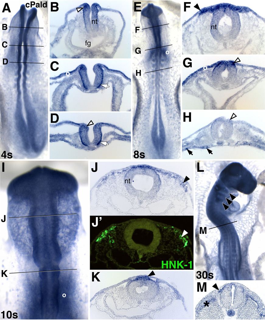
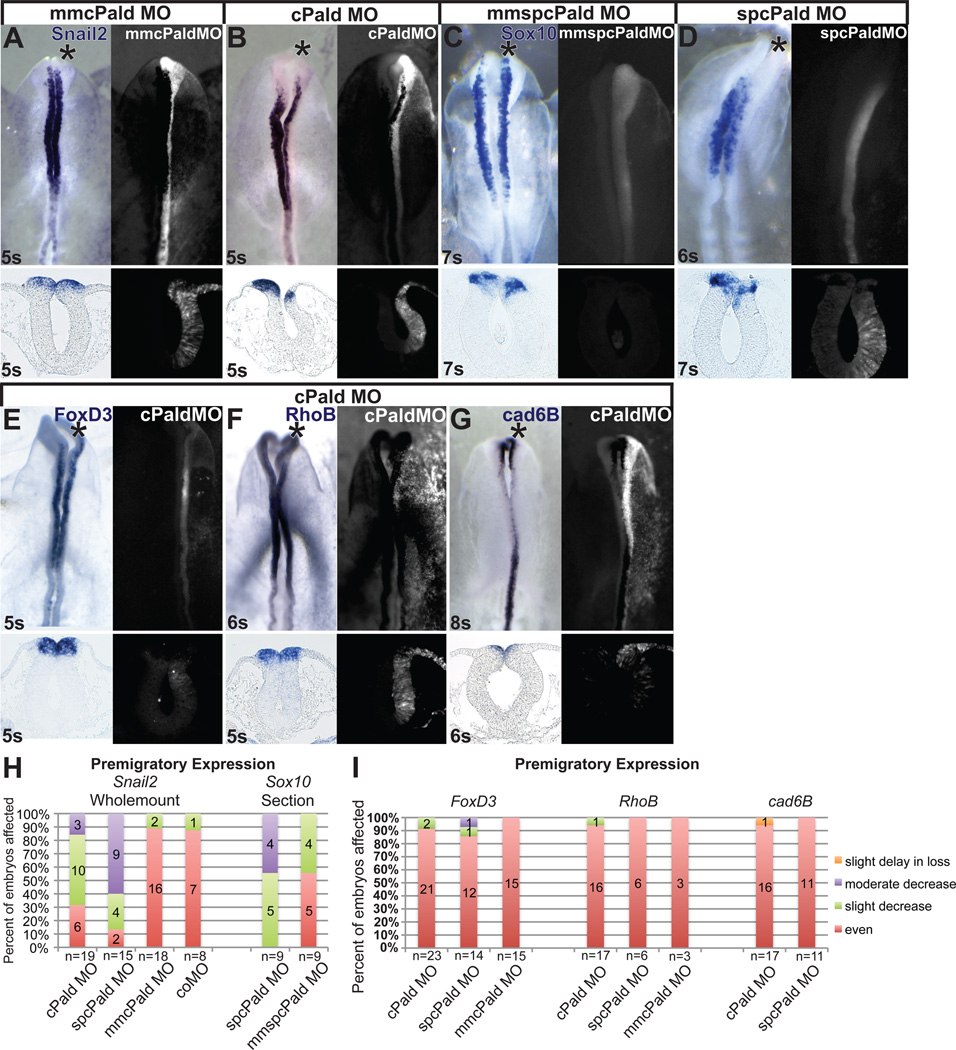
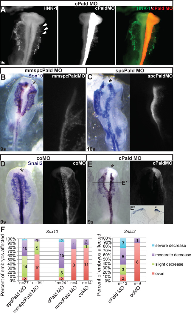
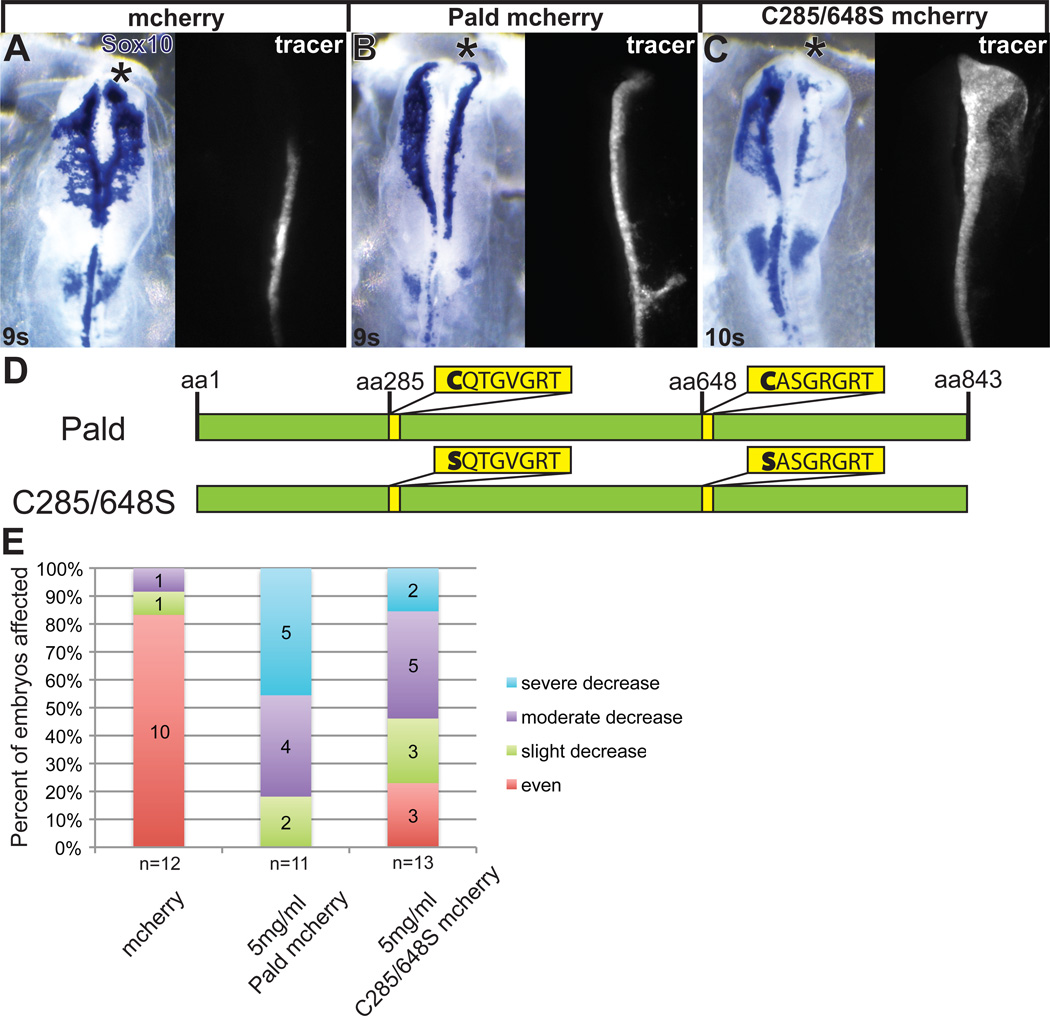
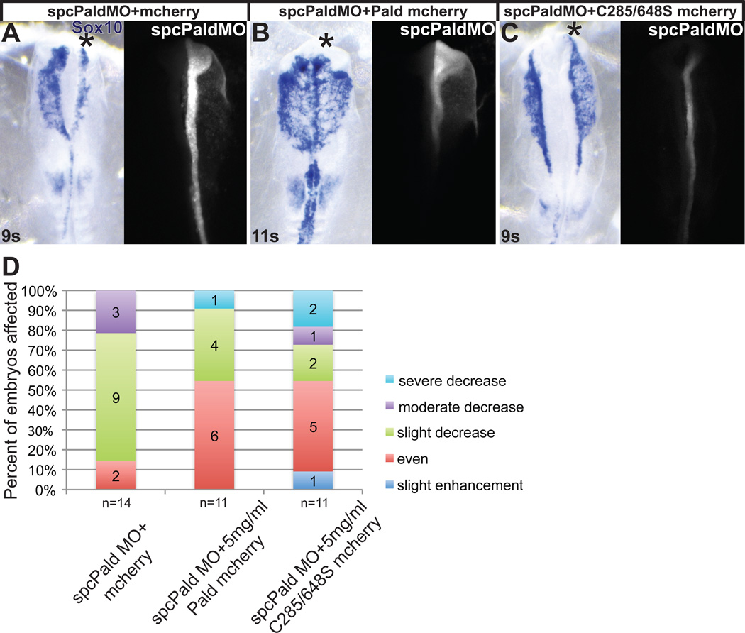
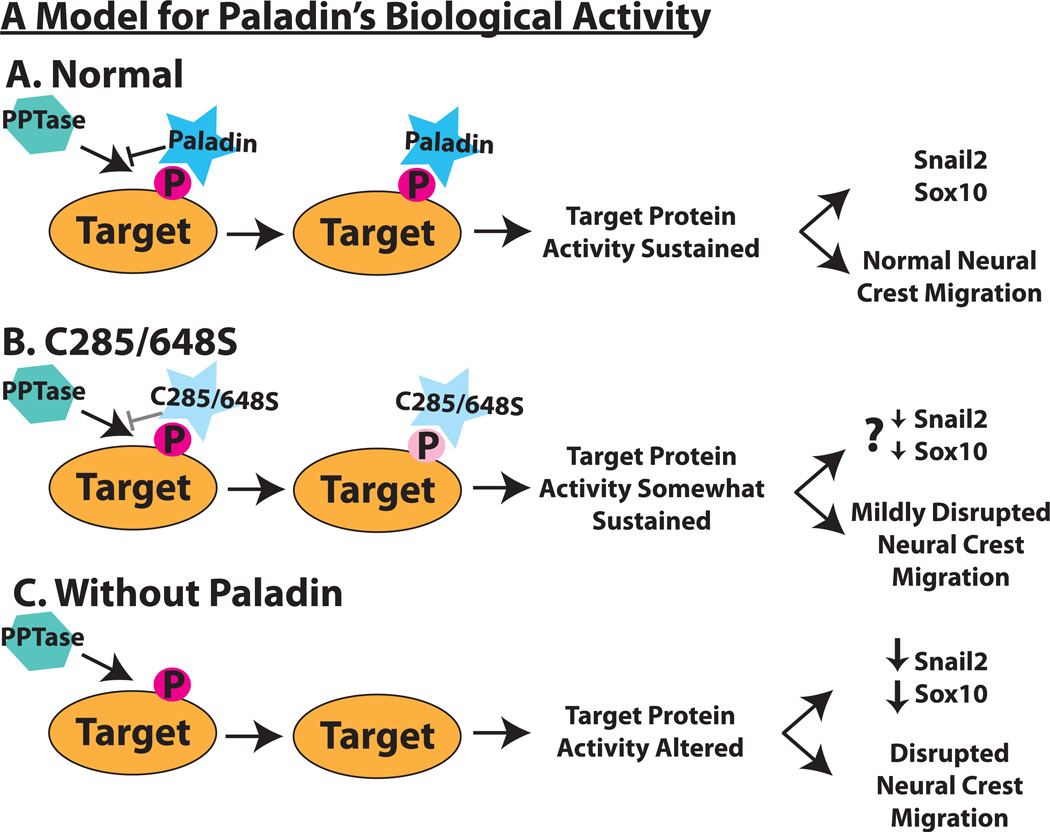
Although a network of transcription factors that specifies neural crest identity in the ectoderm has been defined, expression of neural crest transcription factors does not guarantee eventual migration as a neural crest cell. While much work has gone into determining regulatory relationships within the transcription factor network, the ability of protein modifications like phosphorylation to modulate the function of neural crest regulatory factors and determine when and where they are active also has crucial implications. Paladin, which was previously classified as a phosphatase based on sequence similarity, is expressed in chick neural crest precursors and is maintained throughout their epithelial to mesenchymal transition and migration. Loss of Paladin delays the expression of transcription factors Snail2 and Sox10 in premigratory neural crest cells, but does not affect accumulation of FoxD3, Cad6B or RhoB, indicating that Paladin differentially modulates the expression of genes previously thought to be coregulated within the neural crest gene regulatory network. Both gain and loss of Paladin function result in disrupted neural crest migration, reinforcing the importance of precisely regulated phosphorylation for neural crest migration. Mutation of critical, catalytic cysteine residues within Paladin's predicted phosphatase active site motifs did not abolish the function of Paladin in the neural crest. Collectively, these data indicate that Paladin is an antiphosphatase that modulates the activity of specific neural crest regulatory factors during neural crest development. Our work identifies a novel regulator of phosphorylation status that provides an additional layer of regulation in the neural crest.


Figures

Figure 1
Figure 1. Paladin is expressed in premigratory and migratory neural crest cells
(A–D) Cranial (B, C) and trunk (D) premigratory neural crest cells (open arrowhead) express Pald mRNA at 4 somites (4s). (E–M) At 8s (E–H), 10s (I–K), and 30s (L, M), Pald expression persists in hindbrain (G) and trunk premigratory neural crest cells (H, open arrowheads) and is maintained during delamination and migration of midbrain (F,I,J) hindbrain (K,L) and trunk neural crest cells (M; arrowheads). Pald expressing cells (J, arrowhead) are positive by immunofluorescence for the migratory neural crest marker HNK-1 (J’, arrowhead), confirming their identification as neural crest cells. Pald is also expressed in the basal surface of the neural tube (white arrows), blood vessels (black arrows), and dermomyotome (asterisk). A, E, I, dorsal view; L, dorsolateral view; B–D, F–H, J, K, M, transverse section at the level indicated in the accompanying whole mount. I, higher magnification view. Fg, foregut; nt, neural tube; o, otic placode.
Figure 2
Figure 2. Pald is required for premigratory expression of Snail2 and Sox10
Embryos were unilaterally electroporated at HH stage 4–5 with mmcPald MO (A), cPald MO (B, E, F, G), mmspcPald MO (C) or spcPald MO (D). After reincubation to 4–8 somites (s), in situ hybridization was performed to assess expression of Snail2 (A, B), Sox10 (C, D), FoxD3 (E), RhoB (F), or Cad6B (G). (A–D) Representative examples of Snail2 and Sox10 expression inhibition following Pald knock down. Snail2 expression is unaffected in embryos electroporated with mmcPald MO (A) but reduced on the side of the neural tube targeted with cPald MO (B). Likewise, Sox10 expression is unaffected in embryos electroporated with mmspcPaldMO (C) but reduced on the side of the neural tube targeted with spcPald MO (D). (E–G) Representative examples of neural crest regulators unaffected by Pald knock down. Expression of the transcription factor FoxD3 (E) and neural crest effector genes RhoB (F) and Cad6B (G) is equivalent on cPaldMO targeted and untargeted sides of the neural tube. (H) Stacked bar graphs depicting the frequency and severity of Snail2 and Sox10 expression defects in embryos electroporated with cPald MO, spcPald MO, mmcPald MO, mmspcPald MO, or control (co) MO when assayed in whole mount (Snail2) or sections (Sox10). (I) Stacked bar graphs reveal the absence of FoxD3, Cad6B, and RhoB expression defects in nearly all embryos assayed. In A-G, top row: dorsal views of in situ hybridization in left panel, fluorescent MO targeting in right panel; bottom row: transverse sections at the level of the midbrain. Note that the section shown is not necessarily from the wholemount embryo pictured. Asterisk, targeted side of the embryo.
Figure 3
Figure 3. Loss of Pald impedes neural crest migration
Embryos were unilaterally electroporated at HH stage 4–5 with cPaldMO, mmspcPald MO, spcPald MO, or standard control MO (coMO) and reincubated to 8-11 somites (s). (A) Neural crest migration defects visualized by HNK-1 immunofluorescence following Pald knock down. cPald MO-targeted neural crest cells (red; arrowheads) do not migrate as far as HNK-1 positive neural crest cells (green) on the untargeted side. (B, C) A representative example of neural crest migration defects visualized by Sox10 in situ hybridization following Pald knockdown. While mmspcPald MO does not affect neural crest migration (A), spcPald MO-targeted neural crest cells’ migration distance is reduced compared to the untargeted side (B). (D, E) A representative example of neural crest migration defects visualized by Snail2 in situ hybridization following Pald knockdown. While coMO does not affect neural crest migration (D), cPald MO-targeted neural crest cells’ migration distance is reduced compared to the untargeted side (E), transverse section at the level of the line indicated in E’. (B–E) Dorsal views of in situ hybridization in left panel, fluorescent MO targeting in right panel. Asterisk, targeted side of the embryo. (F) Stacked bar graphs depicting the severity and frequency of migration impairment in embryos electroporated with cPald MO, spcPald MO, mmcPald MO, mmspcPald MO, or coMO and visualized by Sox10 (left) or Snail2 (right) in situ hybridization. Migration of neural crest cells is mildly disrupted by electroporation of mmspcPald MO, indicating that 1.0mM spcPald MO is the upper limit of its effective and specific dose.
Figure 4
Figure 4. Pald overexpression disrupts neural crest migration
Embryos were unilaterally electroporated at HH stage 4–5 with pMES-mcherry expression constructs mixed with 1.0 mM FITC-labeled standard control MO (coMO). The MO served as a lineage tracer and a carrier for the DNA. At 8-11 somites (s), embryos were harvested and neural crest cells visualized by in situ hybridization for Sox10. (A–C) Representative embryos showing inhibition of migration by Pald overexpression. Whereas neural crest migration is unaffected by pMES-mcherry electroporation (A), electroporation of Paldmcherry (B) or C285/648S phosphatase domain mutant Pald (C) disrupt neural crest migration on the targeted side. Dorsal views of in situ hybridization in left panel, fluorescent MO in right panel. Asterisk, targeted side of the embryo. (D) Schematic of Pald sequence. Phosphotyrosine phosphatase sites in yellow, active site cysteine is mutated in both domains in C285/648S. (E) Stacked bar graphs depicting the severity and frequency of migration defects in embryos overexpressing either wildtype or phosphatase mutant Pald.
Figure 5
Figure 5. Phosphatase domain catalytic cysteine residues are not essential for Paladin activity in neural crest cells
Embryos were unilaterally electroporated at HH stage 4–5 with spcPald MO mixed with pMES-mcherry vector DNA, pMES-Pald-mcherry or pMES-C285/648S-mcherry and reincubated. At 8–11 somites (s) embryos were harvested and neural crest cells visualized by in situ hybridization for Sox10. (A–C) Representative examples of spcPald MO rescue. While neural crest migration is disrupted upon unilateral coelectroporation of empty pMES-mcherry (A), neural crest cells migrate normally following coelectroporation of 5 mg/ml pMES-Pald-mcherry (B) or 5mg/ml C285/648S-mcherry (C). Dorsal views of in situ hybridization in left panel, fluorescent MO in right panel. Asterisk, targeted side of the embryo. (D) Stacked bar graphs depicting the severity and frequency of migration defects in embryos coelectroporated with spcPald MO and wildtype or phosphatase mutant Pald.
Figure 6
Figure 6. A model for Paladin’s role in neural crest development
(A) Predicted mechanism of Paladin’s biological activity. A Paladin target protein (orange oval) is tyrosine-phosphorylated (dark pink P) during neural crest development. Paladin (blue star) binds to the phosphotyrosine, protecting it from removal by phosphatases (turquoise hexagon) and sustaining target protein activity. (B) In a C285/648S Paladin mutant, we predict that the loss of the critical cysteine reduces the affinity or specificity of mutant Paladin (light blue star) for the target phosphotyrosine, making protection from dephosphorylation less efficient (light pink P). We predict that expression of C285/648S mutant Paladin results in partial dephosphorylation of target protein(s), which we expect would lead to a slight decrease in Snail2 and Sox10 as well as the observed mild disruption of neural crest migration and less effective rescue. (C) Paladin knockdown results in unprotected target phosphotyrosines, allowing for dephosphorylation by a phosphatase. This alters target protein activity which impedes Snail2 and Sox10 expression and disrupts early phases of neural crest migration.

References

    1. Adams MS, Gammill LS, Bronner-Fraser M. Discovery of transcription factors and other candidate regulators of neural crest development. Dev Dyn. 2008;237:1021–1033. - PMC - PubMed
    1. Alonso A, Sasin J, Bottini N, Friedberg I, Osterman A, Godzik A, Hunter T, Dixon J, Mustelin T. Protein tyrosine phosphatases in the human genome. Cell. 2004;117:699–711. - PubMed
    1. Banerjee S, Gordon L, Donn TM, Berti C, Moens CB, Burden SJ, Granato M. A novel role for MuSK and non-canonical Wnt signaling during segmental neural crest cell migration. Development (Cambridge, England) 2011;138:3287–3296. - PMC - PubMed
    1. Basch M, Bronner-Fraser M, Garcia-Castro M. Specification of the neural crest occurs during gastrulation and requires Pax7. Nature. 2006;441:218–222. - PubMed
    1. Betancur P, Bronner-Fraser M, Sauka-Spengler T. Assembling neural crest regulatory circuits into a gene regulatory network. Annu Rev Cell Dev Biol. 2010;26:581–603. - PMC - PubMed

Publication types

Substances