. 2012 Dec 1;21(23):5106-17.
doi: 10.1093/hmg/dds356. Epub 2012 Aug 27.

Mutations of EFHC1, linked to juvenile myoclonic epilepsy, disrupt radial and tangential migrations during brain development

Affiliations

Mutations of EFHC1, linked to juvenile myoclonic epilepsy, disrupt radial and tangential migrations during brain development

Laurence de Nijs et al. Hum Mol Genet. .

Abstract

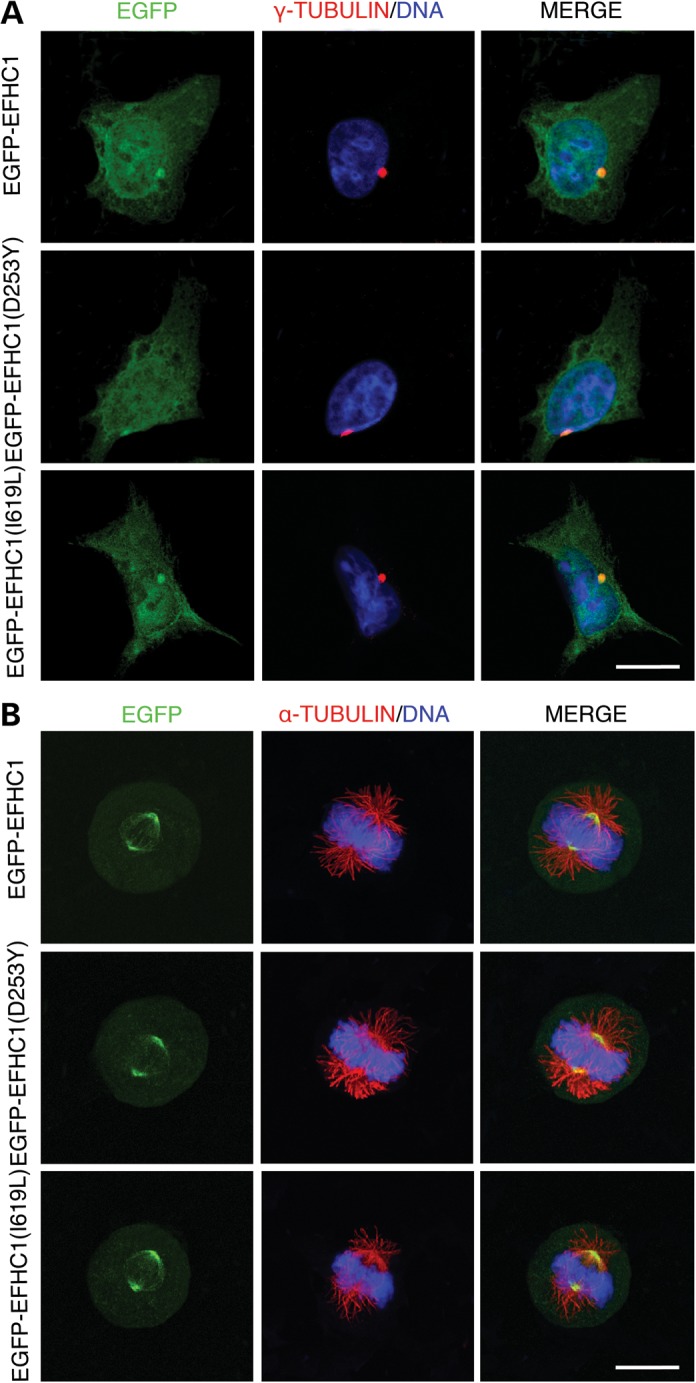
Heterozygous mutations in Myoclonin1/EFHC1 cause juvenile myoclonic epilepsy (JME), the most common form of genetic generalized epilepsies, while homozygous F229L mutation is associated with primary intractable epilepsy in infancy. Heterozygous mutations in adolescent JME patients produce subtle malformations of cortical and subcortical architecture, whereas homozygous F229L mutation in infancy induces severe brain pathology and death. However, the underlying pathological mechanisms for these observations remain unknown. We had previously demonstrated that EFHC1 is a microtubule-associated protein (MAP) involved in cell division and radial migration during cerebral corticogenesis. Here, we show that JME mutations, including F229L, do not alter the ability of EFHC1 to colocalize with the centrosome and the mitotic spindle, but act in a dominant-negative manner to impair mitotic spindle organization. We also found that mutants EFHC1 expression disrupted radial and tangential migration by affecting the morphology of radial glia and migrating neurons. These results show how Myoclonin1/EFHC1 mutations disrupt brain development and potentially produce structural brain abnormalities on which epileptogenesis is established.


Figures

Figure 1.
Figure 1.
Subcellular localization of mutant and polymorphic EFHC1 proteins. HEK293 cells expressing EGFP-tagged wild-type (EGFP-EFHC1), EGFP-EFHC1(D253Y) or EGFP-EFHC1(I619L) (green). EGFP-EFHC1(D253Y) and EGFP-EFHC1(I619L) images are representative of mutants and polymorphisms, respectively. (A) Interphase cells were stained for γ-tubulin (red) to visualize centrosomes. DNA displayed in blue. Merge panels show that all EGFP-tagged proteins colocalized with the centrosome. (B) Mitotic cells were stained for α-tubulin (red) to visualize microtubules. DNA displayed in blue. Merge panels show that all EGFP-tagged proteins associate with the mitotic spindle. Scale bars represent 20 μm.
Figure 2.
Figure 2.
EFHC1 mutations induce mitotic spindle defects. (A) Representative images of mitotic spindle defects observed in HEK293 cells expressing mutants EGFP-EFHC1 (green). Monopolar spindle and chromosome alignment failure on metaphase plate (congression abnormality, arrows) are shown. The cells were stained for α-tubulin (red) and DNA (blue). Scale bar represents 20 μm. (B) Quantification of abnormal spindles in cultures 2 days after transfections with EGFP, EGFP-tagged wild-type, mutants or polymorphics EFHC1. (C) Quantification of the mitotic index 2 days after transfections with EGFP, EGFP-EFHC1, mutant EGFP-EFHC1(D253Y) or polymorphic EGFP-EFHC1(I619L). Error bars show SEM. **P < 0.01.
Figure 3.
Figure 3.
Acute expression of EFHC1 mutations alters radial migration of projection neurons in the cerebral cortex. (A) Distribution of EGFP-positive cells in different cortical regions (VZ/SVZ, IZ and CP) 3 days after in utero electroporation at E14.5 of EGFP, EGFP-EFHC1, EGFP-EFHC1(D253Y) or EGFP-EFHC1(I619L) (green). EGFP-EFHC1(D253Y) and EGFP-EFHC1(I619L) images are representative of mutants and polymorphisms, respectively. EGFP-EFHC1(D253Y) panel shows radial migration defect in the CP. Scale bar represents 100 μm. (B) Quantification of EGFP-positive cells in different cortical regions (VZ/SVZ, IZ and CP) 3 days after in utero electroporation at E14.5 under different above conditions. Error bars show SEM. *P < 0.05, **P < 0.01.
Figure 4.
Figure 4.
Mutants EFHC1 do not influence proliferation and cell cycle exit of cortical progenitors. (A) Immunolabeling for PHH3 (red), a mitotic marker, of VZ/SVZ cells in brains 2 days after in utero electroporation at E14.5 with EGFP, EGFP-EFHC1, EGFP-EFHC1(D253Y) or EGFP-EFHC1(I619L) (green). (B) Quantification of the mitotic index under different above conditions. (C) Immunolabeling for BrdU (red) and Ki67 (blue), of VZ/SVZ cells in cortices 2 days after in utero electroporation at E14.5 with EGFP, EGFP-EFHC1, EGFP-EFHC1(D253Y) or EGFP-EFHC1(I619L) (green). (D) Quantification of the cell cycle exit index in the different above conditions. Scale bars represent 50 μm. Error bars show SEM.
Figure 5.
Figure 5.
Mutants EFHC1 affect the morphology of radial glia and radially migrating cells. (A) Immunolabeling for BLBP (red), a marker for radial glia, in cortices 3 days after in utero electroporation at E14.5 with EGFP, EGFP-EFHC1, EGFP-EFHC1(D253Y) or EGFP-EFHC1(I619L). EGFP-EFHC1(D253Y) panel shows a disruption of radial glia scaffold. (B) Morphology of cells in the IZ of brains 3 days after in utero electroporation at E14.5 with EGFP, EGFP-EFHC1, EGFP-EFHC1(D253Y) or EGFP-EFHC1(I619L) (green). Many cells expressing EGFP, EGFP-EFHC1 or EGFP-EFHC1(I619L) are bipolar (arrows), whereas most EGFP-EFHC1(D253Y) transfected cells remained with a multipolar morphology (arrow heads). (C) Quantification of bipolar and multipolar cells within the IZ under different above conditions. (D) Morphology of cells in the CP of brains 3 days after in utero electroporation at E14.5 with EGFP, EGFP-EFHC1, EGFP-EFHC1(D253Y) or EGFP-EFHC1(I619L) (green). Cells electroporated with EGFP, EGFP-EFHC1 or EGFP-EFHC1(I619L) present long leading processes, though EGFP-EFHC1(D253Y) transfected cells display abnormal leading processes: short and twisted (white arrows), harboring several bulges (white arrow heads), round-shaped cell lacking a leading process (red arrow heads). (E) Quantification of leading processes length of migrating cells within the CP under different above conditions. Scale bars represent 100 μm. Error bars show SEM. *P < 0.05, **P < 0.01.
Figure 6.
Figure 6.
Expression of mutants EFHC1 interferes with tangential migration of interneurons. (A) Distribution of EGFP-positive cells in coronal brain slices 3 days after focal electroporation at E17 of EGFP, EGFP-EFHC1, EGFP-EFHC1(D253Y) or EGFP-EFHC1(I619L) (green). EGFP-EFHC1(D253Y) and EGFP-EFHC1(I619L) images are representative of mutants and polymorphisms, respectively. EGFP-EFHC1(D253Y) panel shows tangential migration defect in the cortex. Scale bar represents 200 μm. (B) Quantification of EGFP-positive cells in the cortex 3 days after focal electroporation at E17 under different above conditions. (C) Morphology of migrating interneurons in brain slices 3 days after focal electroporation at E17 of EGFP, EGFP-EFHC1, EGFP-EFHC1(D253Y) or EGFP-EFHC1(I619L) (green). Most EGFP-EFHC1(D253Y) expressing cells appear with a short and twisted leading process. Scale bar represents 20 μm. (D) Quantification of leading processes length of migrating cells within the cortex under different above conditions. Error bars show SEM. *P < 0.05, **P < 0.01.

References

    1. Delgado-Escueta A.V., Enrile-Bacsal F. Juvenile myoclonic epilepsy of Janz. Neurology. 1984;34:285–294. - PubMed
    1. Delgado-Escueta A.V. Advances in genetics of juvenile myoclonic epilepsies. Epilepsy Curr. 2007;7:61–67. - PMC - PubMed
    1. Noebels J.L., Avoli M., Rogawski M.A., Olsen R.W., Delgado-Escueta A.V. The next decade of research in the basic mechanisms of the epilepsies. In: Noebels J.L., Avoli M., Rogawski M.A., Olsen R.W., Delgado-Escueta A.V., editors. Jasper's Basic Mechanisms of the Epilepsies. Oxford University Press; 2012. pp. 3–11.
    1. Suzuki T., Delgado-Escueta A.V., Aguan K., Alonso M.E., Shi J., Hara Y., Nishida M., Numata T., Medina M.T., Takeuchi T., et al. Mutations in EFHC1 cause juvenile myoclonic epilepsy. Nat. Genet. 2004;36:842–849. - PubMed
    1. Ma S., Blair M.A., Abou-Khalil B., Lagrange A.H., Gurnett C.A., Hedera P. Mutations in the GABRA1 and EFHC1 genes are rare in familial juvenile myoclonic epilepsy. Epilepsy Res. 2006;71:129–134. - PubMed

Publication types

MeSH terms

Substances