

Astrocytic expression of HIV-1 Nef impairs spatial and recognition memory
- PMID: 22926191
- PMCID: PMC3530662
- DOI: 10.1016/j.nbd.2012.08.007
Astrocytic expression of HIV-1 Nef impairs spatial and recognition memory
Abstract
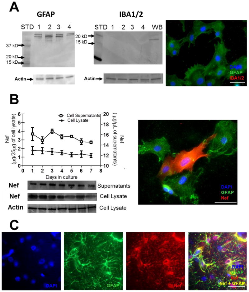
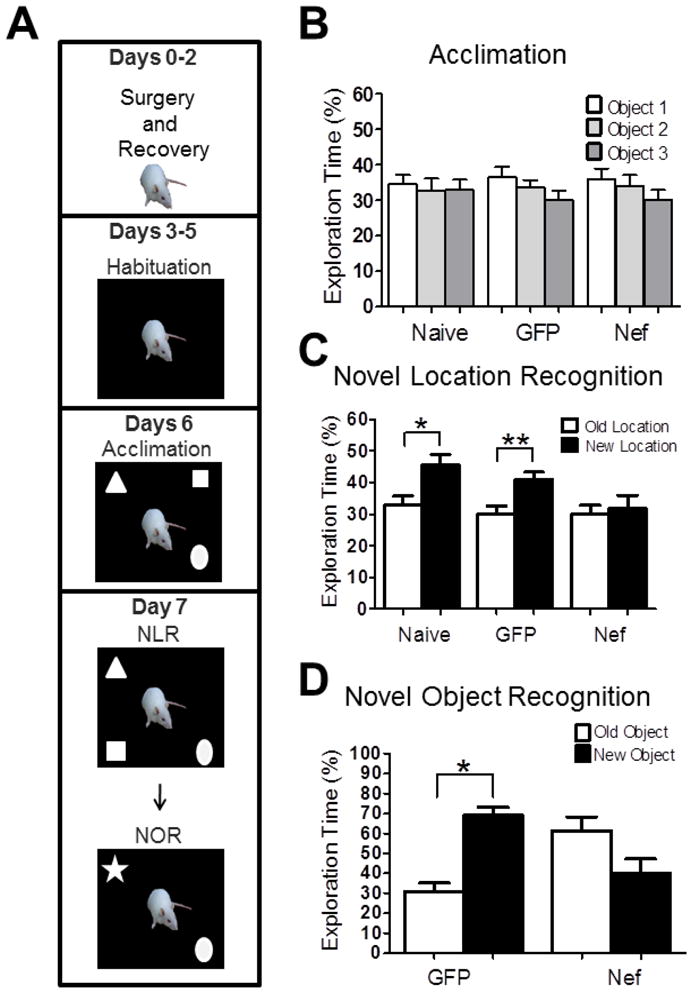
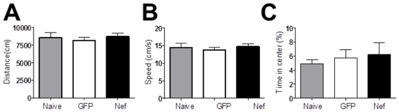
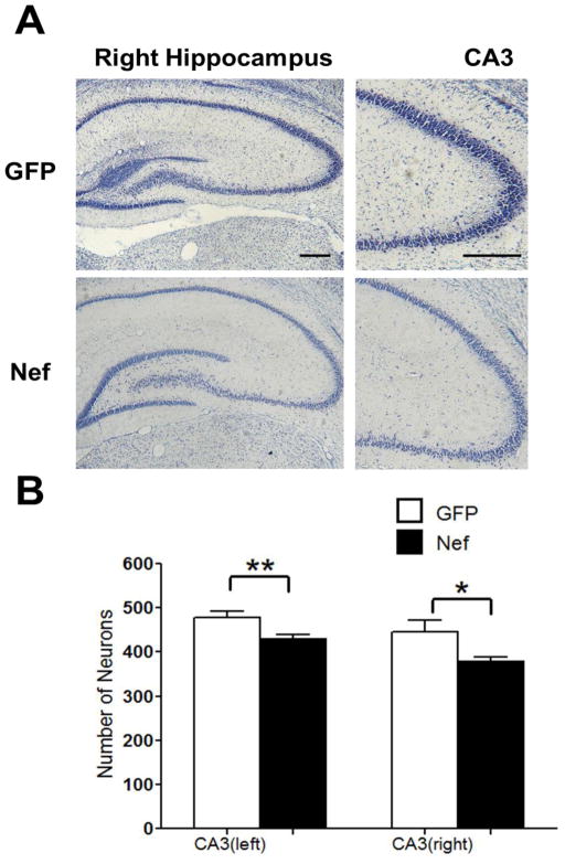
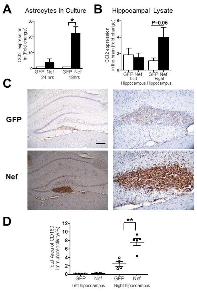
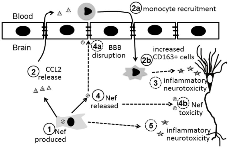
Despite the widespread use of antiretroviral therapy that effectively limits viral replication, memory impairment remains a dilemma for HIV infected people. In the CNS, HIV infection of astrocytes leads to the production of the HIV-1 Nef protein without viral replication. Post mortem studies have found Nef expression in hippocampal astrocytes of people with HIV associated dementia suggesting that astrocytic Nef may contribute to HIV associated cognitive impairment even when viral replication is suppressed. To test whether astrocytic expression of Nef is sufficient to induce cognitive deficits, we examined the effect of implanting primary rat astrocytes expressing Nef into the hippocampus on spatial and recognition memory. Rats implanted unilaterally with astrocytes expressing Nef showed impaired novel location and novel object recognition in comparison with controls implanted with astrocytes expressing green fluorescent protein (GFP). This impairment was correlated with an increase in chemokine ligand 2 (CCL2) expression and the infiltration of peripheral macrophages into the hippocampus at the site of injection. Furthermore, the Nef exposed rats exhibited a bilateral loss of CA3 neurons. These results suggest that Nef protein expressed by the implanted astrocytes activates the immune system leading to neuronal damage and spatial and recognition memory deficits. Therefore, the continued expression of Nef by astrocytes in the absence of viral replication has the potential to contribute to HIV associated cognitive impairment.
Keywords: Astrocyte; CCL2; CD163; Cognition; HAND.
Copyright © 2012 Elsevier Inc. All rights reserved.
Conflict of interest statement
Conflict of interest: None
Figures
Similar articles
-
TGFβRI antagonist inhibits HIV-1 Nef-induced CC chemokine family ligand 2 (CCL2) in the brain and prevents spatial learning impairment.J Neuroinflammation. 2019 Dec 11;16(1):262. doi: 10.1186/s12974-019-1664-4. J Neuroinflammation. 2019. PMID: 31829243 Free PMC article.
-
Astrocytic expression of HIV-1 viral protein R in the hippocampus causes chromatolysis, synaptic loss and memory impairment.J Neuroinflammation. 2014 Mar 22;11:53. doi: 10.1186/1742-2094-11-53. J Neuroinflammation. 2014. PMID: 24655810 Free PMC article.
-
Infusion of HIV-1 Nef-expressing astrocytes into the rat hippocampus induces enteropathy and interstitial pneumonitis and increases blood-brain-barrier permeability.PLoS One. 2019 Nov 27;14(11):e0225760. doi: 10.1371/journal.pone.0225760. eCollection 2019. PLoS One. 2019. PMID: 31774879 Free PMC article.
-
Nef-induced CCL2 Expression Contributes to HIV/SIV Brain Invasion and Neuronal Dysfunction.Front Immunol. 2019 Oct 15;10:2447. doi: 10.3389/fimmu.2019.02447. eCollection 2019. Front Immunol. 2019. PMID: 31681324 Free PMC article. Review.
-
Emerging Role of Nef in the Development of HIV Associated Neurological Disorders.J Neuroimmune Pharmacol. 2021 Jun;16(2):238-250. doi: 10.1007/s11481-020-09964-1. Epub 2020 Oct 29. J Neuroimmune Pharmacol. 2021. PMID: 33123948 Free PMC article. Review.
Cited by
-
The HIV-1 transgenic rat model of neuroHIV.Brain Behav Immun. 2015 Aug;48:336-49. doi: 10.1016/j.bbi.2015.02.020. Epub 2015 Feb 27. Brain Behav Immun. 2015. PMID: 25733103 Free PMC article. Review.
-
Enhanced antagonism of BST-2 by a neurovirulent SIV envelope.J Clin Invest. 2016 Jun 1;126(6):2295-307. doi: 10.1172/JCI83725. Epub 2016 May 9. J Clin Invest. 2016. PMID: 27159392 Free PMC article.
-
Neuro-HIV-New insights into pathogenesis and emerging therapeutic targets.FASEB J. 2023 Dec;37(12):e23301. doi: 10.1096/fj.202301239RR. FASEB J. 2023. PMID: 37942865 Free PMC article.
-
Rifaximin Improves Spatial Learning and Memory Impairment in Rats with Liver Damage-Associated Neuroinflammation.Biomedicines. 2022 May 28;10(6):1263. doi: 10.3390/biomedicines10061263. Biomedicines. 2022. PMID: 35740285 Free PMC article.
-
Impact of viral factors on subcellular distribution and RNA export activity of HIV-1 rev in astrocytes 1321N1.PLoS One. 2013 Sep 4;8(9):e72905. doi: 10.1371/journal.pone.0072905. eCollection 2013. PLoS One. 2013. PMID: 24023789 Free PMC article.
References
-
- Acevedo SF, Ohtsu H, Benice TS, Rizk-Jackson A, Raber J. Age-dependent measures of anxiety and cognition in male histidine decarboxylase knockout (Hdc−/−) mice. Brain Res. 2006;1071:113–23. - PubMed
-
- Anderson E, Zink W, Xiong H, Gendelman HE. HIV-1-associated dementia: a metabolic encephalopathy perpetrated by virus-infected and immune-competent mononuclear phagocytes. J Acquir Immune Defic Syndr. 2002;31(Suppl 2):S43–54. - PubMed
-
- Benice TS, Raber J. Object recognition analysis in mice using nose-point digital video tracking. J Neurosci Methods. 2008;168:422–30. - PubMed
-
- Benice TS, Rizk A, Kohama S, Pfankuch T, Raber J. Sex-differences in age-related cognitive decline in C57BL/6J mice associated with increased brain microtubule-associated protein 2 and synaptophysin immunoreactivity. Neuroscience. 2006;137:413–23. - PubMed
-
- Chauhan A, Turchan J, Pocernich C, Bruce-Keller A, Roth S, Butterfield DA, Major EO, Nath A. Intracellular human immunodeficiency virus Tat expression in astrocytes promotes astrocyte survival but induces potent neurotoxicity at distant sites via axonal transport. J Biol Chem. 2003;278:13512–13519. - PubMed
Publication types
MeSH terms
Substances
Grants and funding
LinkOut - more resources
Full Text Sources
Other Literature Sources
Medical
Research Materials
Miscellaneous
