

Uncoupling stimulus specificity and glomerular position in the mouse olfactory system
- PMID: 22926192
- PMCID: PMC3494770
- DOI: 10.1016/j.mcn.2012.08.006
Uncoupling stimulus specificity and glomerular position in the mouse olfactory system
Abstract
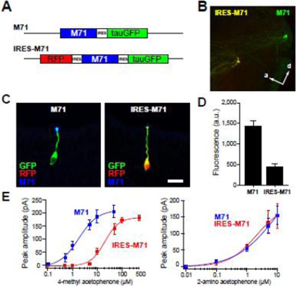
Sensory information is often mapped systematically in the brain with neighboring neurons responding to similar stimulus features. The olfactory system represents chemical information as spatial and temporal activity patterns across glomeruli in the olfactory bulb. However, the degree to which chemical features are mapped systematically in the glomerular array has remained controversial. Here, we test the hypothesis that the dual roles of odorant receptors, in axon guidance and odor detection, can serve as a mechanism to map olfactory inputs with respect to their function. We compared the relationship between response specificity and glomerular position in genetically-defined olfactory sensory neurons expressing variant odorant receptors. We find that sensory neurons with the same odor response profile can be mapped to different regions of the bulb, and that neurons with different response profiles can be mapped to the same glomeruli. Our data demonstrate that the two functions of odorant receptors can be uncoupled, indicating that the mechanisms that map olfactory sensory inputs to glomeruli do so without regard to stimulus specificity.
Copyright © 2012 Elsevier Inc. All rights reserved.
Figures
Similar articles
-
The olfactory bulb: coding and processing of odor molecule information.Science. 1999 Oct 22;286(5440):711-5. doi: 10.1126/science.286.5440.711. Science. 1999. PMID: 10531048 Review.
-
Precision and diversity in an odor map on the olfactory bulb.Nat Neurosci. 2009 Feb;12(2):210-20. doi: 10.1038/nn.2262. Epub 2009 Jan 18. Nat Neurosci. 2009. PMID: 19151709
-
Mapping odorant sensitivities reveals a sparse but structured representation of olfactory chemical space by sensory input to the mouse olfactory bulb.Elife. 2022 Jul 21;11:e80470. doi: 10.7554/eLife.80470. Elife. 2022. PMID: 35861321 Free PMC article.
-
Grouping and representation of odorant receptors in domains of the olfactory bulb sensory map.Microsc Res Tech. 2002 Aug 1;58(3):168-75. doi: 10.1002/jemt.10146. Microsc Res Tech. 2002. PMID: 12203695 Review.
-
Recovery of glomerular morphology in the olfactory bulb of young mice after disruption caused by continuous odorant exposure.Brain Res. 2017 Sep 1;1670:6-13. doi: 10.1016/j.brainres.2017.05.030. Epub 2017 Jun 2. Brain Res. 2017. PMID: 28583862
Cited by
-
Vapor detection and discrimination with a panel of odorant receptors.Nat Commun. 2018 Nov 1;9(1):4556. doi: 10.1038/s41467-018-06806-w. Nat Commun. 2018. PMID: 30385742 Free PMC article.
-
Multiple perceptible signals from a single olfactory glomerulus.Nat Neurosci. 2013 Nov;16(11):1687-91. doi: 10.1038/nn.3519. Epub 2013 Sep 22. Nat Neurosci. 2013. PMID: 24056698
-
The Odorant Receptor-Dependent Role of Olfactory Marker Protein in Olfactory Receptor Neurons.J Neurosci. 2016 Mar 9;36(10):2995-3006. doi: 10.1523/JNEUROSCI.4209-15.2016. J Neurosci. 2016. PMID: 26961953 Free PMC article.
-
The extremely broad odorant response profile of mouse olfactory sensory neurons expressing the odorant receptor MOR256-17 includes trace amine-associated receptor ligands.Eur J Neurosci. 2016 Mar;43(5):608-17. doi: 10.1111/ejn.13153. Epub 2016 Jan 28. Eur J Neurosci. 2016. PMID: 26666691 Free PMC article.
-
In Vitro Mutational Analysis of the β2 Adrenergic Receptor, an In Vivo Surrogate Odorant Receptor.PLoS One. 2015 Oct 29;10(10):e0141696. doi: 10.1371/journal.pone.0141696. eCollection 2015. PLoS One. 2015. PMID: 26513247 Free PMC article.
References
-
- Abaffy T, Malhotra A, Luetje CW. The molecular basis for ligand specificity in a mouse olfactory receptor: a network of functionally important residues. J Biol Chem. 2007;282:1216–1224. - PubMed
-
- Bozza T, McGann JP, Mombaerts P, Wachowiak M. In vivo imaging of neuronal activity by targeted expression of a genetically encoded probe in the mouse. Neuron. 2004;42:9–21. - PubMed
Publication types
MeSH terms
Substances
Grants and funding
LinkOut - more resources
Full Text Sources
Molecular Biology Databases
