

An essential role of metalloprotease-disintegrin ADAM12 in triple-negative breast cancer
- PMID: 22926263
- PMCID: PMC3470813
- DOI: 10.1007/s10549-012-2220-4
An essential role of metalloprotease-disintegrin ADAM12 in triple-negative breast cancer
Abstract
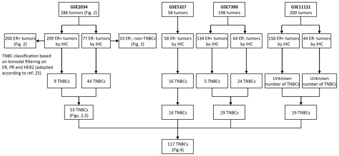
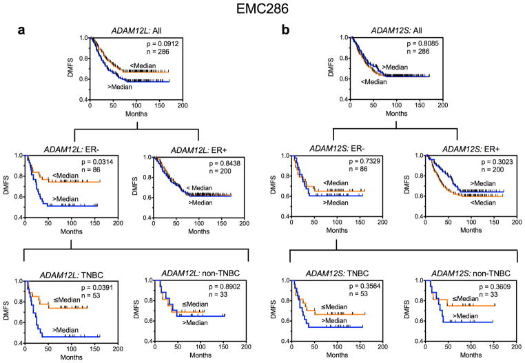
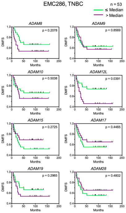
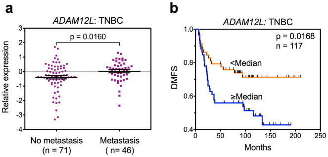
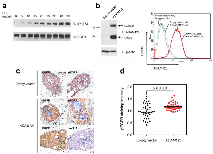
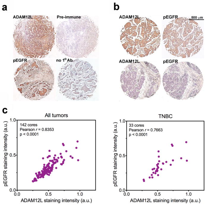
In the absence of HER2 overexpression, triple-negative breast cancers (TNBCs) rely on signaling by epidermal growth factor receptor (EGFR/ErbB1/HER1) to convey growth signals and stimulate cell proliferation. Soluble EGF-like ligands are derived from their transmembrane precursors by ADAM proteases, but the identity of the ADAM that is primarily responsible for ligand release and activation of EGFR in TNBCs is not clear. Using publicly available gene expression data for patients with lymph node-negative breast tumors who did not receive systemic treatment, we show that ADAM12L is the only ADAM with an expression level significantly associated with decreased distant metastasis-free survival times. Similar effect was not observed for patients with ER-negative non-TNBCs. There was a positive correlation between ADAM12L and HB-EGF and EGFR in TNBCs, but not in ER-negative non-TNBCs. We further demonstrate that ectopic expression of ADAM12L increased EGFR phosphorylation in a mouse intraductal xenograft model of early breast cancer. Finally, we detect strong correlation between the level of anti-ADAM12L and anti-phospho-EGFR immunostaining in human breast tumors using tissue microarrays. These studies suggest that ADAM12L is the primary protease responsible for the activation of EGFR in early stage, lymph node-negative TNBCs. Thus, our results may provide novel insight into the biology of TNBC.
Conflict of interest statement
The authors declare that they have no conflict of interest.
Figures
Similar articles
-
ADAM12 induces estrogen-independence in breast cancer cells.Breast Cancer Res Treat. 2012 Feb;131(3):731-41. doi: 10.1007/s10549-011-1431-4. Epub 2011 Mar 9. Breast Cancer Res Treat. 2012. PMID: 21387162 Free PMC article.
-
Metalloproteinase-disintegrin ADAM12 is associated with a breast tumor-initiating cell phenotype.Breast Cancer Res Treat. 2013 Jun;139(3):691-703. doi: 10.1007/s10549-013-2602-2. Epub 2013 Jun 16. Breast Cancer Res Treat. 2013. PMID: 23771733 Free PMC article.
-
Increased expression of ADAM12 and ADAM17 genes in laser-capture microdissected breast cancers and correlations with clinical and pathological characteristics.Acta Histochem. 2012 Feb;114(2):131-9. doi: 10.1016/j.acthis.2011.03.009. Epub 2011 Apr 17. Acta Histochem. 2012. PMID: 21501859
-
Prognostic and clinical significance of syndecan-1 expression in breast cancer: A systematic review and meta-analysis.Eur J Surg Oncol. 2019 Jul;45(7):1132-1137. doi: 10.1016/j.ejso.2018.12.019. Epub 2018 Dec 25. Eur J Surg Oncol. 2019. PMID: 30598194
-
Cellular roles of ADAM12 in health and disease.Int J Biochem Cell Biol. 2008;40(9):1685-702. doi: 10.1016/j.biocel.2008.01.025. Epub 2008 Feb 1. Int J Biochem Cell Biol. 2008. PMID: 18342566 Review.
Cited by
-
Alternative mRNA splicing generates two distinct ADAM12 prodomain variants.PLoS One. 2013 Oct 7;8(10):e75730. doi: 10.1371/journal.pone.0075730. eCollection 2013. PLoS One. 2013. PMID: 24116070 Free PMC article.
-
The Multiple Functions of HB-EGF in Female Reproduction and Related Cancer: Molecular Mechanisms and Targeting Strategies.Reprod Sci. 2024 Sep;31(9):2588-2603. doi: 10.1007/s43032-024-01454-6. Epub 2024 Feb 29. Reprod Sci. 2024. PMID: 38424408 Review.
-
Integrated bioinformatics analysis of key genes involved in progress of colon cancer.Mol Genet Genomic Med. 2019 Apr;7(4):e00588. doi: 10.1002/mgg3.588. Epub 2019 Feb 11. Mol Genet Genomic Med. 2019. PMID: 30746900 Free PMC article.
-
EGFR conjunct FSCN1 as a Novel Therapeutic Strategy in Triple-Negative Breast Cancer.Sci Rep. 2017 Nov 15;7(1):15654. doi: 10.1038/s41598-017-15939-9. Sci Rep. 2017. PMID: 29142206 Free PMC article.
-
Transcriptional Factor Repertoire of Breast Cancer in 3D Cell Culture Models.Cancers (Basel). 2022 Feb 17;14(4):1023. doi: 10.3390/cancers14041023. Cancers (Basel). 2022. PMID: 35205770 Free PMC article. Review.
References
Publication types
MeSH terms
Substances
Grants and funding
LinkOut - more resources
Full Text Sources
Other Literature Sources
Medical
Research Materials
Miscellaneous
