. 2012 Oct;160(2):1081-96.
doi: 10.1104/pp.112.201798. Epub 2012 Aug 27.

Nitric oxide-mediated maintenance of redox homeostasis contributes to NPR1-dependent plant innate immunity triggered by lipopolysaccharides

Affiliations

Nitric oxide-mediated maintenance of redox homeostasis contributes to NPR1-dependent plant innate immunity triggered by lipopolysaccharides

Aizhen Sun et al. Plant Physiol. 2012 Oct.

Abstract

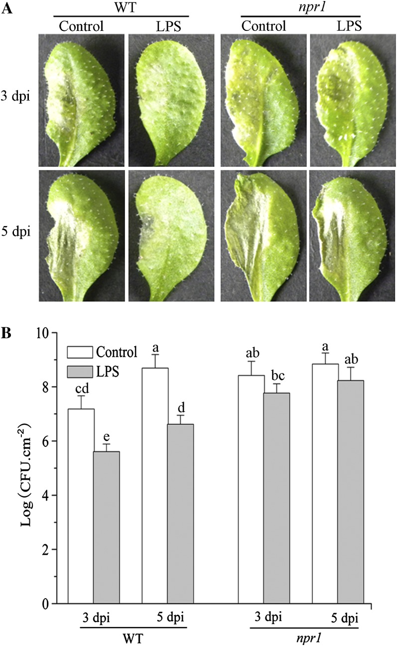
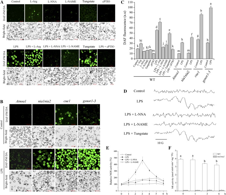
The perception of lipopolysaccharides (LPS) by plant cells can lead to nitric oxide (NO) production and defense gene induction. However, the signaling cascades underlying these cellular responses have not yet been resolved. This work investigated the biosynthetic origin of NO and the role of NONEXPRESSOR OF PATHOGENESIS-RELATED GENES1 (NPR1) to gain insight into the mechanism involved in LPS-induced resistance of Arabidopsis (Arabidopsis thaliana). Analysis of inhibitors and mutants showed that LPS-induced NO synthesis was mainly mediated by an arginine-utilizing source of NO generation. Furthermore, LPS-induced NO caused transcript accumulation of alternative oxidase genes and increased antioxidant enzyme activity, which enhanced antioxidant capacity and modulated redox state. We also analyzed the subcellular localization of NPR1 to identify the mechanism for protein-modulated plant innate immunity triggered by LPS. LPS-activated defense responses, including callose deposition and defense-related gene expression, were found to be regulated through an NPR1-dependent pathway. In summary, a significant NO synthesis induced by LPS contributes to the LPS-induced defense responses by up-regulation of defense genes and modulation of cellular redox state. Moreover, NPR1 plays an important role in LPS-triggered plant innate immunity.


Figures

Figure 1.
Figure 1.
Effect of LPS application on disease progression in leaves of wild-type (WT) and npr1 plants. A, After spraying with 250 μm MgCl2 and 100 μm CaCl2 (control solution) or LPS (100 μg mL−1 in 250 μm MgCl2 and 100 μm CaCl2) solution for 24 h, wild-type Arabidopsis and npr1 mutant plants were inoculated with pathogen Pma DG3 (OD600 = 0.01 in 10 mm MgCl2). Leaves were infected on their left halves, and samples were collected at 3 and 5 dpi. B, Bacterial growth quantification of Pma DG3-inoculated (OD600 = 0.0001) leaves after spraying with control solution or 100 μg mL−1 LPS. Samples were collected at 3 and 5 dpi for assay. Each value is the mean ± se of three replicates. Different letters indicate statistically significant differences between treatments (Duncan’s multiple range test: P < 0.05). CFU, Colony-forming units. [See online article for color version of this figure.]
Figure 2.
Figure 2.
LPS-elicited Arg-utilizing source of NO generation. A, Effects of mammalian NOS inhibitors and NO scavenger on NO level by LPS induction. Protoplasts prepared from wild-type plants were loaded with DAF-FM DA for 20 min prior to different treatments for 2 h. For each treatment, fluorescence and bright-field images are shown. B, Confocal images of DAF-FM fluorescence in protoplasts from Atnoa1, nia1nia2, cue1, and gsnor1-3 plants treated with control solution or 100 μg mL−1 LPS for 2 h. Bars = 50 µm. C, Quantitative analysis of NO-related DAF-FM fluorescence by a fluorescence spectrometer under various treatments for 2 h as shown in A and B. WT, Wild type. D, NO production was examined by EPR analysis. E, Effect of LPS and mammalian NOS inhibitors on NOS-like enzyme activity. F, Effect of LPS on NR activity of wild-type and nia1nia2 plants. Pr, Protein. Data are means ± se of three experiments. Different letters indicate statistically significant differences between treatments (Duncan’s multiple range test: P < 0.05). [See online article for color version of this figure.]
Figure 3.
Figure 3.
Induction of PR1 gene expression and callose deposition in Arabidopsis by LPS. A, Approximately 10-d-old transgenic PR1:GUS seedlings grown on MS medium were then transferred to 24-well plates containing 400 μL of liquid MS medium without 100 μg mL−1 LPS (control) for 12 h or with 100 μg mL−1 LPS for 6, 12, or 24 h and collected for histochemical GUS staining. Each experiment was performed with eight plants and repeated twice with similar results. B, Quantitative RT-PCR data showing the expression of the PR1 gene in wild-type (WT) and npr1 Arabidopsis. Total RNA was extracted from the leaves of Arabidopsis after spraying with control solution at 12 h post treatment or 100 μg mL−1 LPS at 0, 6, 12, and 24 h post treatment. Arabidopsis ACTIN2 was used as an internal control. Expression levels for each treatment were normalized to a LPS-treated (6 h) wild-type plant. Values represent means ± se of three independent experiments. C, Callose-staining imaging of leaves and roots from LPS-treated plants. D and E, Callose deposition in leaves (D) and roots (E) was quantified by determining the number of pixels (corresponding to LPS-induced callose) per million pixels in digital photographs. Data are means ± se of three experiments. F, Induction of CalS1 and CalS12 genes in wild-type and npr1 mutant Arabidopsis by LPS treatment. Total RNA was extracted from leaves treated with control solution (−) or 100 μg mL−1 LPS (+) for 12 h. ACTIN2 was used as an internal control. The experiment was performed three times with similar results. G, Quantitative analysis of CalS1 and CalS12 genes shown in F with ImageJ software. Three gel photographs were taken for quantitative analysis, and values represent means ± se. Different letters indicate statistically significant differences between treatments (Duncan’s multiple range test: P < 0.05). [See online article for color version of this figure.]
Figure 4.
Figure 4.
LPS induction results in nuclear localization of NPR1. Approximately 10-d-old transgenic 35S:NPR1-GFP seedlings grown on MS medium were then transferred to 24-well plates containing control solution, 300 μm l-NNA, 300 μm l-NAME, 1 mm cPTIO, 50 μm AIP, 100 μg mL−1 LPS, 100 μg mL−1 LPS + 300 μm l-NNA, 100 μg mL−1 LPS + 300 μm l-NAME, 100 μg mL−1 LPS + 1 mm cPTIO, or 100 μg mL−1 LPS + 50 μm AIP for 12 h. GFP fluorescence in guard cells was observed with a laser confocal scanning microscope. Results shown are representative. Bars = 20 µm. [See online article for color version of this figure.]
Figure 5.
Figure 5.
A to E, Transcript levels of AOX1a (A), AOX1b (B), AOX1c (C), AOX1d (D), and AOX2 (E) in wild-type Arabidopsis plants under LPS treatment. Total RNA was extracted from the leaves of full-grown Arabidopsis after spraying with control solution, 300 μm l-NNA, 300 μm l-NAME, 1 mm cPTIO, 100 μg mL−1 LPS, 100 μg mL−1 LPS + 300 μm l-NNA, 100 μg mL−1 LPS + 300 μm l-NAME, or 100 μg mL−1 LPS + 1 mm cPTIO at 3, 6, and 9 h post treatment. Arabidopsis ACTIN2 was used as an internal control. Asterisks indicate significant differences from the control (Duncan’s multiple range test: *P < 0.05, **P < 0.01). F, Transcript levels of AOX1a and AOX1b in wild-type and npr1 Arabidopsis. Asterisks indicate significant differences between the wild type and the mutant (Student’s paired t test: *P < 0.05, **P < 0.01). Expression levels for each gene were normalized to control treatment (0 h) of the wild-type plant. Values represent means ± se of three independent experiments.
Figure 6.
Figure 6.
A to C, Activities of antioxidant enzymes during LPS and NO scavenger treatment. Leaves were treated with control solution, 300 μm l-NNA, 300 μm l-NAME, 1 mm cPTIO, 100 μg mL−1 LPS, 100 μg mL−1 LPS + 300 μm l-NNA, 100 μg mL−1 LPS + 300 μm l-NAME, or 100 μg mL−1 LPS + 1 mm cPTIO for 3, 6, and 9 h and then collected for determination of SOD (A), POD (B), and CAT (C) activities. Asterisks indicate significant differences from the control (Duncan’s multiple range test: *P < 0.05, **P < 0.01). D, Activities of antioxidant enzymes in wild-type (WT) and npr1 Arabidopsis. Asterisks indicate significant differences between the wild-type and the mutant (Student’s paired t test: *P < 0.05, **P < 0.01). FW, Fresh weight; U, units. Data are means ± se of at least three independent experiments.
Figure 7.
Figure 7.
Changes in redox state and antioxidant capacity during various treatments. A, After spraying with control solution, 300 μm l-NNA, 300 μm l-NAME, 200 μm SHAM, 1 mm cPTIO, 100 μg mL−1 LPS, 100 μg mL−1 LPS + 300 μm l-NNA, 100 μg mL−1 LPS + 300 μm l-NAME, 100 μg mL−1 LPS + 200 μm SHAM, or 100 μg mL−1 LPS + 1 mm cPTIO for 12 h, leaves of transgenic mit-roGFP1 Arabidopsis were inoculated with Pma DG3 (OD600 = 0.01) and then collected at 12 h for measurement of mit-roGFP1 oxidation. B to F, Pma DG3-inoculated leaves of wild-type plants under various treatments were collected at 24 h for DAB staining indicating H2O2 generation (B) and quantitative analysis of DAB-stained areas (C) and were collected at 12 h for determination of glutathione levels (D), the ratios of GSH to GSSG (E), and total antioxidant capacity (F). Pr, Protein. Data are means ± se of at least three independent experiments. Different letters indicate statistically significant differences between treatments (Duncan’s multiple range test: P < 0.05). [See online article for color version of this figure.]
Figure 8.
Figure 8.
Effects of NO scavenger and mammalian NOS inhibitors on LPS-induced defense response. A, Effects of mammalian NOS inhibitors and NO scavenger on callose deposition. B, Callose deposition in leaves and roots was quantified by determining the number of pixels (corresponding to LPS-induced callose) per million pixels in digital photographs. Data are means ± se of three experiments. C, PR1 expression in leaves treated with control solution, 100 μg mL−1 LPS, 100 μg mL−1 LPS + 300 μm l-NNA, 100 μg mL−1 LPS + 300 μm l-NAME, or 100 μg mL−1 LPS + 1 mm cPTIO. Samples were harvested at 12 h post treatment for quantitative RT-PCR analysis. Arabidopsis ACTIN2 was used as an internal control. Expression levels for each treatment were normalized to plants treated with LPS + cPTIO. Each value is the mean ± se of three independent experiments. D, Bacterial growth quantification of Pma DG3-inoculated (OD600 = 0.0001) leaves after various treatments for 12 h. Samples were collected at 3 dpi for assay. Each value is the mean ± se of three replicates. Different letters indicate statistically significant differences between treatments (Duncan’s multiple range test: P < 0.05). CFU, Colony-forming units. [See online article for color version of this figure.]
Figure 9.
Figure 9.
Model showing the possible signaling pathway for LPS-induced plant innate immunity. The extracellular LPS are recognized by a receptor in the plant cell plasma membrane. LPS perceived by receptors result in an increase in cytosolic Ca2+ (Ali et al., 2007; Ma et al., 2009), which may activate NOS-like enzyme and then lead to an increase of NO level. NO functions to potentiate cyclic GMP/Ca2+-dependent PR1 gene expression (Durner et al., 1998) as well as up-regulation of antioxidant enzyme activity, and/or promote the nuclear translocation of NPR1 to induce PR1 expression (Lindermayr et al., 2010). LPS perception is mechanistically linked to NPR1-dependent defense responses. Induction by LPS results in the accumulation of SA and the activation of NPR1. Activated NPR1 then translocates into the nucleus, where it may interact with TGA transcription factors, thus leading to the induction of the PR1 gene, and it also activates the expression of CalS1 and CalS12 genes in the formation of callose by interacting with some unknown transcription factors. These directly induced PR1 expression and callose deposition by LPS constitute plant innate immunity, which protects plants against pathogen infection.

Similar articles

Cited by

References

    1. Ahn IP, Kim S, Lee YH, Suh SC. (2007) Vitamin B1-induced priming is dependent on hydrogen peroxide and the NPR1 gene in Arabidopsis. Plant Physiol 143: 838–848 - PMC - PubMed
    1. Ali R, Ma W, Lemtiri-Chlieh F, Tsaltas D, Leng Q, von Bodman S, Berkowitz GA. (2007) Death don’t have no mercy and neither does calcium: Arabidopsis CYCLIC NUCLEOTIDE GATED CHANNEL2 and innate immunity. Plant Cell 19: 1081–1095 - PMC - PubMed
    1. Apel K, Hirt H. (2004) Reactive oxygen species: metabolism, oxidative stress, and signal transduction. Annu Rev Plant Biol 55: 373–399 - PubMed
    1. Besson-Bard A, Pugin A, Wendehenne D. (2008) New insights into nitric oxide signaling in plants. Annu Rev Plant Biol 59: 21–39 - PubMed
    1. Bethke PC, Badger MR, Jones RL. (2004) Apoplastic synthesis of nitric oxide by plant tissues. Plant Cell 16: 332–341 - PMC - PubMed

Publication types

MeSH terms