Review
. 2013 Apr;70(8):1335-56.
doi: 10.1007/s00018-012-1115-1. Epub 2012 Aug 25.

Tumor cell migration in complex microenvironments

Affiliations
Review

Tumor cell migration in complex microenvironments

William J Polacheck et al. Cell Mol Life Sci. 2013 Apr.

Abstract

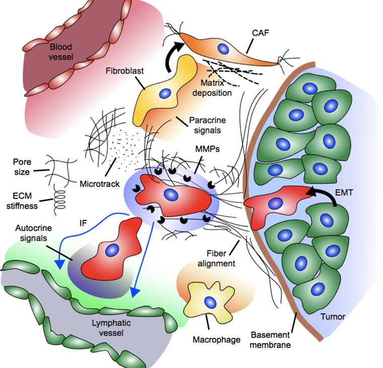
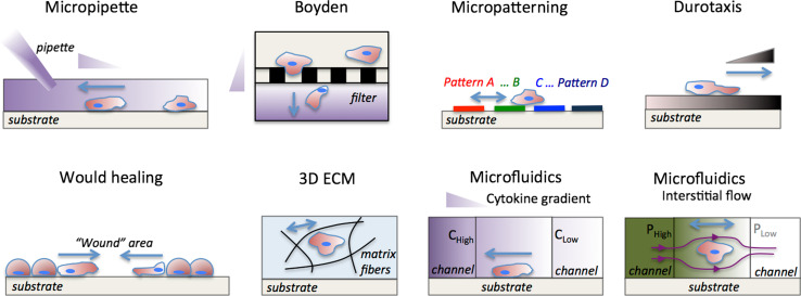
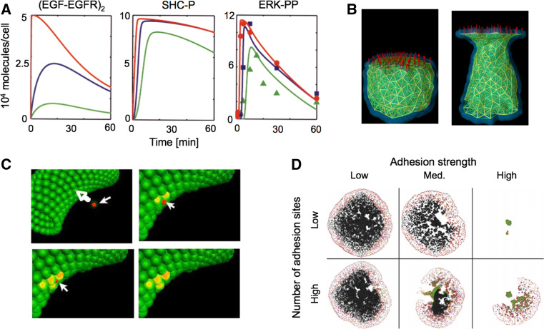
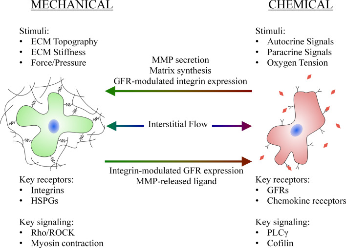
Tumor cell migration is essential for invasion and dissemination from primary solid tumors and for the establishment of lethal secondary metastases at distant organs. In vivo and in vitro models enabled identification of different factors in the tumor microenvironment that regulate tumor progression and metastasis. However, the mechanisms by which tumor cells integrate these chemical and mechanical signals from multiple sources to navigate the complex microenvironment remain poorly understood. In this review, we discuss the factors that influence tumor cell migration with a focus on the migration of transformed carcinoma cells. We provide an overview of the experimental and computational methods that allow the investigation of tumor cell migration, and we highlight the benefits and shortcomings of the various assays. We emphasize that the chemical and mechanical stimulus paradigms are not independent and that crosstalk between them motivates the development of new assays capable of applying multiple, simultaneous stimuli and imaging the cellular migratory response in real-time. These next-generation assays will more closely mimic the in vivo microenvironment to provide new insights into tumor progression, inform techniques to control tumor cell migration, and render cancer more treatable.


Figures

Fig. 1
Fig. 1
A host of biochemical and biophysical factors influence the migration of tumor cells. Mechanical signals include stiffness of the extracellular matrix (ECM), the pore size of the ECM, solid stress, fiber alignment, and fibroblast-generated matrix tension and microtracks. Fibroblasts are activated to assume the cancer-associated fibroblast (CAF) phenotype, and these cells secrete altered matrix components and generate tension. Chemical signals include autocrine gradients, MMPs, oxygen tension, and paracrine signals from the vasculature, lymphatics, and stromal cells (e.g., macrophages)
Fig. 2
Fig. 2
Experimental methods for investigating factors that influence tumor cell migration. In vitro tumor cell migration assays (reviewed in Table 1). Micropipette assay [21]: a pipette is placed in the vicinity of the cell and a chemoattractant solution is injected into the culture medium establishing a growth-factor gradient. Boyden [17] (or Transwell) chamber : cells are seeded in suspension in the top chamber and migrate through the porous filter (black rectangles) in response to a chemokine gradient, which is established by the different culture medium concentrations in the top and bottom chambers. Micropatterning [29]: cells are seeded on patterns of different geometry, size and surface coatings and their migration characteristics are monitored. Durotaxis [40]: cells are seeded on a substrate of variable stiffness and respond by changing traction forces, cell spread area, and migration direction. Wound healing [26]: a “wound” is formed on a confluent tumor monolayer, and the wound closure dynamics are monitored. 3D ECM [39]: cells are seeded inside the 3D ECM and migrate depending on the ECM architecture (stiffness, pore size, and ligand concentration); ECM fibers are outlined with black curved lines. Microfluidics [28, 32, 147, 206]: cytokine gradients can be established in a 3D matrix by flowing different chemokine concentration (C high − C low) solutions in the left and right microchannels; Interstitial flow can be established by adjusting the hydrostatic pressure (P high − P low) in the left and right microchannels; streamlines are indicated with dark magenta lines. Micropipette, Boyden chamber and microfluidics assays enable control of biochemical gradients. Durotaxis, 3D ECM and microfluidics assays enable control of biophysical forces (ECM stiffness and interstitial flow). Wound healing and micropatterning assay enable control of intercellular distances, whereas only micropatterning assays enable control of substrate topography. Growth-factor gradients are indicated by the purple triangles. ECM stiffness gradients are indicated by the dark brown triangle. Blue arrows indicate direction of tumor cell migration, and pressure gradients are indicated by the shades of green
Fig. 3
Fig. 3
Computational methods provide insight into tumor progression. a Cell signaling models capture the dynamics of protein activation and are useful in identifying key signaling molecules and events, such as the role of EGF concentration on ERK-1/2 phosphorylation. Colored lines represent different concentrations of EGF (red, blue, and green are 50, 0.5, and 0.125 ng/ml EGF, respectively). Symbols in panel 3 represent experimental data, demonstrating that the model well-captures the dynamics of EGF-induced ERK-1/2 phosphorylation (SHC-P is phosphorylated SHC, ERK-PP is phosphorylated ERK. Adapted with permission from [64]). b The subcellular element model recapitulates cell-level phenomena by discretizing the cell into a series of nodes and defining mechanical potentials that govern the interaction of the nodes. The panels demonstrate elongation of a cell under 1 nN of tensile force; the filaments indicate interactions between nodes, which lie at the intersection of the filaments (adapted with permission from [63]). c A multi-scale agent-based model captures the dynamics of tumor intravasation. The subset of models that contributed to the overall multi-scale model was comprised of a simplified set of differential equations for intracellular adhesion signaling, a modified Hertz model for intercellular adhesion mechanics, and the Langevin equation for multi-cellular interactions. The panels demonstrate a tumor cell (red) approaching the endothelium (green), forming nascent N-cadherin bonds (yellow), disrupting endothelial cell VE-cadherin bonds, and traversing the endothelium (adapted with permission from [66]). d An off-lattice hybrid discrete-continuum approach modeled the growth of a whole tumor and invasion at the tumor periphery. The model was comprised of a continuum model for nutrient transport, and a discrete model for single cell behavior such as motility, adhesion, and proliferation. The model demonstrated that single cell adhesion plays a role in determining the fate and morphology of the tumor mass (adapted with permission from [59])
Fig. 4
Fig. 4
Chemical factors influence the cellular response to mechanical factors and vice versa. MMP and matrix secretion modulates the stiffness and pore size of the surrounding matrix, while growth factor receptor (GFR) activation influences integrin expression and activation. MMP-mediated matrix degradation releases chemical signals, while integrin activation and clustering can alter GFR expression and activation. GFRs and integrins can form macromolecular complexes, and elements of the intracellular signaling pathway are shared. Interstitial flow induces mechanical signals through fluid shear and pressure stresses, while simultaneously inducing chemical signals through convection of autocrine and paracrine signaling factors (HSPGs are heparan sulfate proteoglycans and PLCγ is phospholipase C-gamma)

Similar articles

Cited by

References

    1. Chambers AF, Groom AC, MacDonald IC. Dissemination and growth of cancer cells in metastatic sites. Nat Rev Cancer. 2002;2(8):563–572. doi: 10.1038/nrc865. - DOI - PubMed
    1. Weigelt B, Peterse JL, van ‘t Veer LJ. Breast cancer metastasis: markers and models. Nat Rev Cancer. 2005;5(8):591–602. doi: 10.1038/nrc1670. - DOI - PubMed
    1. Mueller MM, Fusenig NE. Friends or foes—bipolar effects of the tumour stroma in cancer. Nat Rev Cancer. 2004;4(11):839–849. doi: 10.1038/nrc1477. - DOI - PubMed
    1. Friedl P, Alexander S. Cancer invasion and the microenvironment: plasticity and reciprocity. Cell. 2011;147(5):992–1009. doi: 10.1016/j.cell.2011.11.016. - DOI - PubMed
    1. Bristow RG, Hill RP. Hypoxia and metabolism: hypoxia, DNA repair and genetic instability. Nat Rev Cancer. 2008;8(3):180–192. doi: 10.1038/nrc2344. - DOI - PubMed

Publication types

LinkOut - more resources