

Does the type of T2-weighted hyperintensity influence surgical outcome in patients with cervical spondylotic myelopathy? A review
- PMID: 22926434
- PMCID: PMC3540309
- DOI: 10.1007/s00586-012-2483-9
Does the type of T2-weighted hyperintensity influence surgical outcome in patients with cervical spondylotic myelopathy? A review
Abstract
Purpose: To review the literature on different classifications of T2-weighted (T2W) increased signal intensity (ISI) on preoperative magnetic resonance (MR) images of patients with cervical spondylotic myelopathy (CSM).
Methods: The authors searched the databases of PubMed and Cochrane for studies that used a categorization of T2W ISI to predict the functional outcome after decompressive surgery for CSM. Selected studies were analyzed for the type of ISI classification used, patient selection, methodology and results. The level of evidence provided by each study was determined.
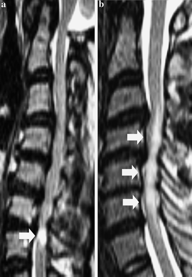
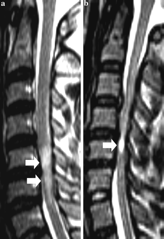
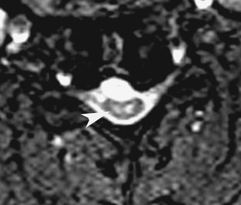
Results: Twenty-two studies fulfilled our search criteria. There were 11 prospective studies and a total of 1,508 patients were studied. The majority of studies classified ISI based on either the longitudinal extent (12 studies) or the qualitative features of the ISI (10 studies). Three studies used both parameters to classify T2W ISI. Other classifications were based on the position of ISI (1 study), presence of snake-eye appearance on axial MR images (1 study) and signal intensity ratio (SIR) (1 study). Poorer functional outcomes correlated with sharp, intense ISI (6 studies) and multisegmental ISI (5 studies) (Class II evidence). Five of ten studies reported that the regression of ISI postoperatively was associated with better neurological outcomes (Class II evidence).
Conclusions: Methodological variations in previous studies made it difficult to compare studies and results. Both multisegmental T2W ISI and sharp, intense T2W ISI are associated with poorer surgical outcome (Class II evidence). The regression of T2W ISI postoperatively correlates with better functional outcomes (Class II). Future studies on the significance of ISI should ensure use of a uniform grading system, standardized outcome measures and multivariate analyses to control for other preoperative variables.
Figures
Similar articles
-
Change in morphology of intramedullary T2-weighted increased signal intensity after anterior decompressive surgery for cervical spondylotic myelopathy.Spine (Phila Pa 1976). 2014 Aug 15;39(18):1458-62. doi: 10.1097/BRS.0000000000000440. Spine (Phila Pa 1976). 2014. PMID: 24859579
-
The evolution of T2-weighted intramedullary signal changes following ventral decompressive surgery for cervical spondylotic myelopathy: Clinical article.J Neurosurg Spine. 2014 Oct;21(4):538-46. doi: 10.3171/2014.6.SPINE13727. Epub 2014 Jul 11. J Neurosurg Spine. 2014. PMID: 25014501
-
Increased signal intensity on postoperative T2-weighted axial images in cervical spondylotic myelopathy: Patterns of changes and associated impact on outcomes.J Clin Neurosci. 2021 Aug;90:244-250. doi: 10.1016/j.jocn.2021.06.007. Epub 2021 Jun 15. J Clin Neurosci. 2021. PMID: 34275557
-
The value of preoperative magnetic resonance imaging in predicting postoperative recovery in patients with cervical spondylosis myelopathy: a meta-analysis.Clinics (Sao Paulo). 2016 Mar;71(3):179-84. doi: 10.6061/clinics/2016(03)10. Clinics (Sao Paulo). 2016. PMID: 27074180 Free PMC article. Review.
-
Does three-grade classification of T2-weighted increased signal intensity reflect the severity of myelopathy and surgical outcomes in patients with cervical compressive myelopathy? A systematic review and meta-analysis.Neurosurg Rev. 2020 Jun;43(3):967-976. doi: 10.1007/s10143-019-01106-3. Epub 2019 May 3. Neurosurg Rev. 2020. PMID: 31053986
Cited by
-
Machine-learning-based prediction by stacking ensemble strategy for surgical outcomes in patients with degenerative cervical myelopathy.J Orthop Surg Res. 2024 Sep 4;19(1):539. doi: 10.1186/s13018-024-05004-3. J Orthop Surg Res. 2024. PMID: 39227869 Free PMC article.
-
Cervical Spondylotic Myelopathy: Natural Course and the Value of Diagnostic Techniques -WFNS Spine Committee Recommendations.Neurospine. 2019 Sep;16(3):386-402. doi: 10.14245/ns.1938240.120. Epub 2019 Sep 30. Neurospine. 2019. PMID: 31607071 Free PMC article.
-
Is the "snake-eye" MRI sign correlated to anterior spinal artery occlusion on CT angiography in cervical spondylotic myelopathy and amyotrophy?Eur Spine J. 2014 Jul;23(7):1541-7. doi: 10.1007/s00586-014-3348-1. Epub 2014 May 14. Eur Spine J. 2014. PMID: 24823850
-
Role of Diffusion Tensor MR Imaging in Degenerative Cervical Spine Disease: a Review of the Literature.Clin Neuroradiol. 2016 Sep;26(3):265-76. doi: 10.1007/s00062-015-0467-y. Epub 2015 Sep 30. Clin Neuroradiol. 2016. PMID: 26423129 Review.
-
Classification of cervical disc herniation myelopathy or radiculopathy: a magnetic resonance imaging-based analysis.Quant Imaging Med Surg. 2023 Aug 1;13(8):4984-4994. doi: 10.21037/qims-22-1387. Epub 2023 May 29. Quant Imaging Med Surg. 2023. PMID: 37581078 Free PMC article.
References
-
- Mummaneni PV, Kaiser MG, Matz PG, Anderson PA, Groff M, Heary R, Holly L, Ryken T, Choudhri T, Vresilovic E, Resnick D. Preoperative patient selection with magnetic resonance imaging, computed tomography, and electroencephalography: does the test predict outcome after cervical surgery? J Neurosurg Spine. 2009;11(2):119–129. doi: 10.3171/2009.3.SPINE08717. - DOI - PubMed
Publication types
MeSH terms
LinkOut - more resources
Full Text Sources
Medical
