

The effects of image compression on quantitative measurements of digital panoramic radiographs
- PMID: 22926465
- PMCID: PMC3505705
- DOI: 10.4317/medoral.17912
The effects of image compression on quantitative measurements of digital panoramic radiographs
Abstract
Objectives: The aims of this study were to explore how image compression affects density, fractal dimension, linear and angular measurements on digital panoramic images and assess inter and intra-observer repeatability of these measurements.
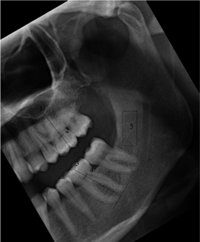
Study design: Sixty-one digital panoramic images in TIFF format (Tagged Image File Format) were compressed to JPEG (Joint Photographic Experts Group) images. Two observers measured gonial angle, antegonial angle, mandibular cortical width, coronal pulp width of maxillary and mandibular first molar, tooth length of maxillary and mandibular first molar on the left side of these images twice. Fractal dimension of the selected regions of interests were calculated and the density of each panoramic radiograph as a whole were also measured on TIFF and JPEG compressed images. Intra-observer and inter-observer consistency was evaluated with Cronbach's alpha. Paired samples t-test and Kolmogorov-Smirnov test was used to evaluate the difference between the measurements of TIFF and JPEG compressed images.
Results: The repeatability of angular measurements had the highest Cronbach's alpha value (0.997). There was statistically significant difference for both of the observers in mandibular cortical width (MCW) measurements (1st ob. p: 0.002; 2nd ob. p: 0.003), density (p<0.001) and fractal dimension (p<0.001) between TIFF and JPEG images. There was statistically significant difference for the first observer in antegonial angle (1st ob p< 0.001) and maxillary molar coronal pulp width (1st ob. p< 0.001) between JPEG and TIFF files.
Conclusions: The repeatability of angular measurements is better than linear measurements. Mandibular cortical width, fractal dimension and density are affected from compression. Observer dependent factors might also cause statistically significant differences between the measurements in TIFF and JPEG images.
Figures


Similar articles
-
The effects of compression on the image quality of digital panoramic radiographs.Clin Oral Investig. 2012 Jun;16(3):719-26. doi: 10.1007/s00784-011-0587-y. Epub 2011 Jul 6. Clin Oral Investig. 2012. PMID: 21732088
-
Effects of Image Compression on Linear Measurements of Digital Panoramic Radiographs.Braz Dent J. 2016 Oct-Dec;27(6):757-760. doi: 10.1590/0103-6440201601157. Braz Dent J. 2016. PMID: 27982191
-
Evaluation of proximal caries in images resulting from different modes of radiographic digitalization.Dentomaxillofac Radiol. 2011 Sep;40(6):338-43. doi: 10.1259/dmfr/67185962. Dentomaxillofac Radiol. 2011. PMID: 21831972 Free PMC article.
-
TIFF, GIF, and PNG: get the picture?Biomed Instrum Technol. 2007 Jul-Aug;41(4):297-300. doi: 10.2345/0899-8205(2007)41[297:TGAPGT]2.0.CO;2. Biomed Instrum Technol. 2007. PMID: 17849757 Review.
-
Influence of the digital file format on radiographic diagnostic in dentistry: a scoping review.Braz Oral Res. 2024 Sep 30;38:e100. doi: 10.1590/1807-3107bor-2024.vol38.0100. eCollection 2024. Braz Oral Res. 2024. PMID: 39356906 Free PMC article.
Cited by
-
Use of fractal analysis in dental images: a systematic review.Dentomaxillofac Radiol. 2020 Feb;49(2):20180457. doi: 10.1259/dmfr.20180457. Epub 2019 Aug 20. Dentomaxillofac Radiol. 2020. PMID: 31429597 Free PMC article.
-
Evaluation of the bone structure surrounding photofunctionalized implants using the fractal analysis method: a split-mouth randomized clinical study.BMC Oral Health. 2025 Jul 1;25(1):964. doi: 10.1186/s12903-025-06330-6. BMC Oral Health. 2025. PMID: 40598042 Free PMC article. Clinical Trial.
-
Evaluation of graft osteogenesis using fractal dimension analysis on cone-beam computed tomography images following maxillary sinus lift surgery.BMC Oral Health. 2025 Aug 21;25(1):1346. doi: 10.1186/s12903-025-06695-8. BMC Oral Health. 2025. PMID: 40841897 Free PMC article.
-
Mandibular radiomorphometric parameters of women with cemento-osseous dysplasia.Dentomaxillofac Radiol. 2020 May 1;49(4):20190359. doi: 10.1259/dmfr.20190359. Epub 2019 Dec 20. Dentomaxillofac Radiol. 2020. PMID: 31846355 Free PMC article.
-
The effects of technical factors on the fractal dimension in different dental radiographic images.Eur Oral Res. 2023 May 4;57(2):68-74. doi: 10.26650/eor.2023984422. Eur Oral Res. 2023. PMID: 37525855 Free PMC article.
References
-
- Stramotas S, Geenty JP, Petocz P, Darendeliler MA. Accuracy of linear and angular measurements on panoramic radiographs taken at various positions in vitro. Eur J Orthod. 2002;24:43–52. - PubMed
-
- Shahabi M, Ramazanzadeh BA, Mokhber N. Comparison between the external gonial angle in panoramic radiographs and lateral cephalograms of adult patients with Class I malocclusion. J Oral Sci. 2009;51:425–9. - PubMed
-
- Ghosh S, Vengal M, Pai KM, Abhishek K. Remodeling of the antegonial angle region in the human mandible: a panoramic radiographic cross-sectional study. Med Oral Patol Oral Cir Bucal. 2010;15:e802–7. - PubMed
-
- Devlin H, Allen P, Graham J, Jacobs R, Nicopoulou-Karayianni K, Lindh C et al. The role of the dental surgeon in detecting osteoporosis: the OSTEODENT study. Br Dent J. 2008;204:E16. - PubMed
-
- Kvaal SI, Kollveit KM, Thomsen IO, Solheim T. Age estimation of adults from dental radiographs. Forensic Sci Int. 1995;74:175–85. - PubMed
MeSH terms
LinkOut - more resources
Full Text Sources
Miscellaneous
