. 2013 Mar;27(3):569-77.
doi: 10.1038/leu.2012.247. Epub 2012 Aug 28.

Anti-leukemic mechanisms of pegylated arginase I in acute lymphoblastic T-cell leukemia

Affiliations

Anti-leukemic mechanisms of pegylated arginase I in acute lymphoblastic T-cell leukemia

K Morrow et al. Leukemia. 2013 Mar.

Abstract

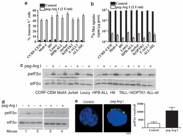
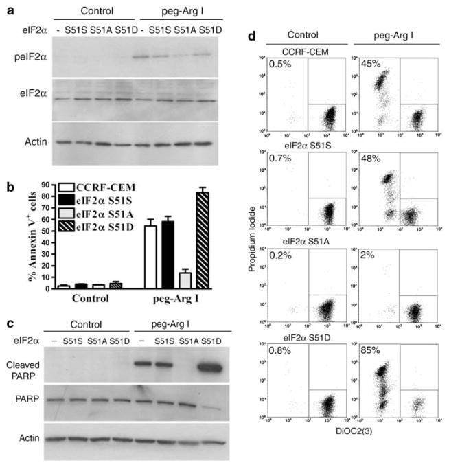
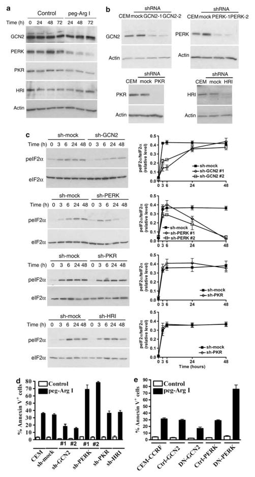
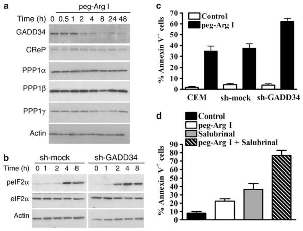
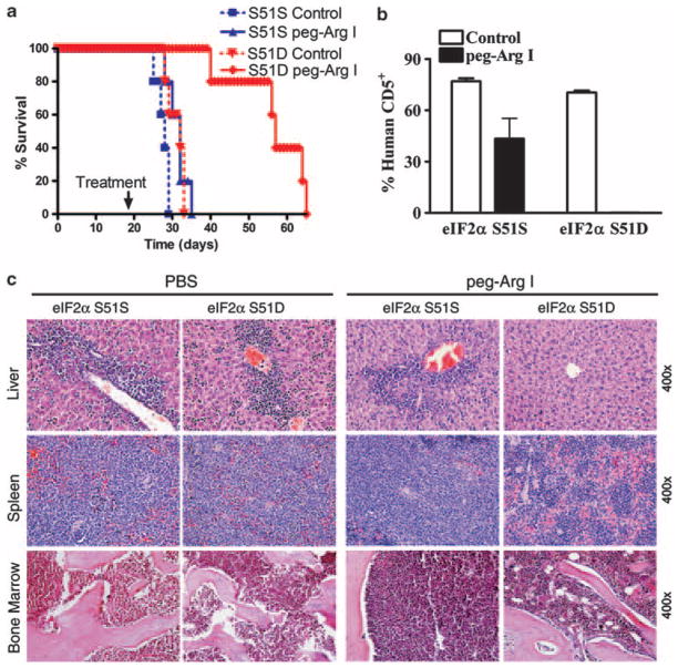
New treatments for adults with acute lymphoblastic T-cell leukemia (T-ALL) are urgently needed, as the current rate of overall remission in these patients is only about 40 percent. We recently showed the potential therapeutic benefit of the pegylated-human-arginase I (peg-Arg I) in T-ALL. However, the mechanisms by which peg-Arg I induces an anti-T-ALL effect remained unknown. Our results show the induction of T-ALL cell apoptosis by peg-Arg I, which associated with a global arrest in protein synthesis and with the phosphorylation of the eukaryotic-translation-initiation factor 2 alpha (eIF2α). Inhibition of eIF2α phosphorylation in T-ALL cells prevented the apoptosis induced by peg-Arg I, whereas the expression of a phosphomimetic eIF2α form increased the sensibility of T-ALL cells to peg-Arg I. Phosphorylation of eIF2α by peg-Arg I was mediated through kinases PERK and GCN2 and down-regulation of phosphatase GADD34. GCN2 and decreased GADD34 promoted T-ALL cell apoptosis after treatment with peg-Arg I, whereas PERK had an unexpected anti-apoptotic role. Additional results showed that phospho-eIF2α signaling further increased the anti-leukemic effects induced by peg-Arg I in T-ALL-bearing mice. These results suggest the central role of phospho-eIF2α in the anti-T-ALL effects induced by peg-Arg I and support its study as a therapeutic target.


Conflict of interest statement

CONFLICT OF INTEREST

The authors declare no conflict of interest.

Figures

Figure 1
Figure 1
Peg-Arg I induces apoptosis, an arrested translation, and phosphorylation of eIF2α in T-ALL cells. (a) Malignant T cell lines (1 × 10⁵) were cultured for 48 h in the presence or the absence of peg-Arg I (2 IU/ml) or PBS (control). Then, the levels of the apoptosis marker annexin V were determined by flow cytometry. (b) T-ALL cell lines were treated with peg-Arg I for 24 h, after which de novo translation was tested using 35S-Metionine (35S-Met) incorporation, as described in the methods. (c) Malignant T cell lines (1 × 10⁵/ml) were cultured for 8 h in the presence or the absence of peg-Arg I (2 IU/ml), after which phospho-eIF2α levels were detected by western blot. (de). A representative experiment showing the expression of phosphorylated and total eIF2α by western blot (d) and phospho-eIF2α by cytofluorescence (e) in CD5+ cells sorted from individual mice bearing CCRF-CEM cells for 30 days and treated with PBS (n = 6) or 0.5 mg per mouse peg-Arg I (n = 7) for 4 h. The experiment was repeated a minimum of 3 times obtaining similar results.
Figure 2
Figure 2
Phosphorylation of eIF2α is a central mediator in the induction of T-ALL cell apoptosis by peg-Arg I. (a) Phospho-eIF2α expression in untransfected CCRF-CEM cells (−), and CCRF-CEM cells stably transfected with eIF2α-S51S, eIF2α-S51 A or eIF2α-S51D, and treated with peg-Arg I for 8 h. (bd) Expression of the apoptosis markers annexin V (b), cleaved poly-ADP-ribose polymerase (PARP) (c), and DiOC2(3)/propidium iodide (d) in cells from a cultured with peg-Arg I for 72 h. The experiments were repeated a minimum of three times obtaining similar results.
Figure 3
Figure 3
Role of eIF2α kinases in the phosphorylation of eIF2α and the cellular apoptosis induced by peg-Arg I. (a) Expression of eIF2α kinases was detected by western blot in CCRF-CEM cells treated with peg-Arg I (2 IU/ml). (b) Stably silencing of the expression of the specific eIF2α kinases was achieved after transfection of CCRF-CEM cells with plasmids coding for specific sh-RNA or sh-controls and further selection of single clones in medium supplemented with 800 μg/ml Geneticin. (c) Cells from (b) were treated with peg-Arg I (2 IU/ml) and the levels of phospho-eIF2α (3–48 h) were detected by western blot (left column) and phospho-eIF2α/eIF2α densitometric values graphed (right column). (de) CCRF-CEM cells previously silenced for GCN2, PERK, PKR, or HRI (d), or expressing DN-GCN or DN-PERK (e) were treated with peg-Arg I (2 IU/ml) for 48 h, after which the levels annexin V were measured by flow cytometry. Experiments were repeated a minimum of three times obtaining similar results.
Figure 4
Figure 4
Inhibition of GADD34 accelerates eIF2α phosphorylation and further increases cellular apoptosis induced by peg-Arg I. (a) CCRF-CEM cells were cultured in medium with or without peg-Arg I (2 IU/ml) and the expression of eIF2α phosphatase components was tested by immunoblotting. (b) Silencing of GADD34 in CCRF-CEM cells induces an earlier phosphorylation of eIF2α after treatment with peg-Arg I. (c) CCRF-CEM cells stably transfected with plasmids coding for sh-GADD34 or sh-mock were treated with peg-Arg I for 24 h, after which annexin V expression was detected using flow cytometry. (d) Expression of annexin V in CCRF-CEM cells treated for 24 h with peg-Arg I (2 IU/ml) and/or salubrinal (20 μM). Values are from 3 similar experiments.
Figure 5
Figure 5
Phospho-eIF2α sensitizes T-ALL to the anti-leukemic effect of peg-Arg I in vivo. (a) Survival of mice bearing CCRF-CEM cells stably transduced with eIF2α-S51S or eIF2α-S51D and that were treated with PBS (Control) (n = 10) or peg-Arg I (n = 10). Peg-Arg I extended the survival of mice injected with T-ALL cells expressing a phosphomimetic form of eIF2α. (b) Percentage of human CD5+ cells was established in the spleens after 30 days of the injection of CCRF-CEM cells expressing eIF2α-S51S or eIF2α-S51D. Mice were treated with 0.5 mg/mouse peg-Arg I (n = 10) or PBS (n = 10) twice a week starting on day 19 post-T-ALL injection, as described in the methods. (c) A representative hematoxylin and eosin staining histopathology from liver, spleen and bone marrow from b.

References

    1. Inaba H, Bhojwani D, Pauley JL, Pei D, Cheng C, Metzger ML, et al. Combination chemotherapy with clofarabine, cyclophosphamide, and etoposide in children with refractory or relapsed haematological malignancies. Br J Haematol. 2012;156:275–279. - PMC - PubMed
    1. Pui CH, Sandlund JT, Pei D, Campana D, Rivera GK, Ribeiro RC, et al. Improved outcome for children with acute lymphoblastic leukemia: results of Total Therapy Study XIIIB at St Jude Children’s Research Hospital. Blood. 2004;104:2690–2696. - PubMed
    1. Tanosaki R, Tobinai K. Adult T-cell leukemia-lymphoma: current treatment strategies and novel immunological approaches. Expert Rev Hematol. 2010;3:743–753. - PubMed
    1. Laport GF, Larson RA. Treatment of adult acute lymphoblastic leukemia. Semin Oncol. 1997;24:70–82. - PubMed
    1. Hernandez CP, Morrow K, Lopez-Barcons LA, Zabaleta J, Sierra R, Velasco C, et al. Pegylated arginase I: a potential therapeutic approach in T-ALL. Blood. 2010;115:5214–5221. - PMC - PubMed

Publication types

MeSH terms