

New synaptic and molecular targets for neuroprotection in Parkinson's disease
- PMID: 22927178
- PMCID: PMC4161019
- DOI: 10.1002/mds.25096
New synaptic and molecular targets for neuroprotection in Parkinson's disease
Abstract
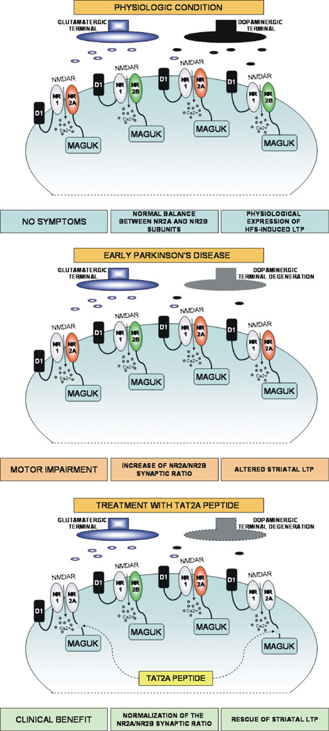
The defining anatomical feature of Parkinson's disease (PD) is the degeneration of substantia nigra pars compacta (SNc) neurons, resulting in striatal dopamine (DA) deficiency and in the subsequent alteration of basal ganglia physiology. Treatments targeting the dopaminergic system alleviate PD symptoms but are not able to slow the neurodegenerative process that underlies PD progression. The nucleus striatum comprises a complex network of projecting neurons and interneurons that integrates different neural signals to modulate the activity of the basal ganglia circuitry. In this review we describe new potential molecular and synaptic striatal targets for the development of both symptomatic and neuroprotective strategies for PD. In particular, we focus on the interaction between adenosine A2A receptors and dopamine D2 receptors, on the role of a correct assembly of NMDA receptors, and on the sGC/cGMP/PKG pathway. Moreover, we also discuss the possibility to target the cell death program parthanatos and the kinase LRRK2 in order to develop new putative neuroprotective agents for PD acting on dopaminergic nigral neurons as well as on other basal ganglia structures.
Copyright © 2013 Movement Disorders Society.
Conflict of interest statement
Full financial disclosures and author roles may be found in the online version of this article.
Figures


Similar articles
-
Synaptic dysfunction in Parkinson's disease.Adv Exp Med Biol. 2012;970:553-72. doi: 10.1007/978-3-7091-0932-8_24. Adv Exp Med Biol. 2012. PMID: 22351072 Review.
-
Dopamine D2 receptor-mediated neuroprotection in a G2019S Lrrk2 genetic model of Parkinson's disease.Cell Death Dis. 2018 Feb 12;9(2):204. doi: 10.1038/s41419-017-0221-2. Cell Death Dis. 2018. PMID: 29434188 Free PMC article.
-
The impact of reactive oxygen species and genetic mitochondrial mutations in Parkinson's disease.Gene. 2013 Dec 10;532(1):18-23. doi: 10.1016/j.gene.2013.07.085. Epub 2013 Aug 15. Gene. 2013. PMID: 23954870 Review.
-
LRRK2-G2019S mice display alterations in glutamatergic synaptic transmission in midbrain dopamine neurons.J Neurochem. 2022 Apr;161(2):158-172. doi: 10.1111/jnc.15588. Epub 2022 Feb 27. J Neurochem. 2022. PMID: 35152441 Free PMC article.
-
Subthalamic nucleus-mediated excitotoxicity in Parkinson's disease: a target for neuroprotection.Ann Neurol. 1998 Sep;44(3 Suppl 1):S175-88. doi: 10.1002/ana.410440726. Ann Neurol. 1998. PMID: 9749591 Review.
Cited by
-
Molecular Mechanisms of Parthanatos and Its Role in Diverse Diseases.Int J Mol Sci. 2022 Jun 30;23(13):7292. doi: 10.3390/ijms23137292. Int J Mol Sci. 2022. PMID: 35806303 Free PMC article. Review.
-
Pharmacological mechanism and therapeutic efficacy of Icariside II in the treatment of acute ischemic stroke: a systematic review and network pharmacological analysis.BMC Complement Med Ther. 2022 Sep 30;22(1):253. doi: 10.1186/s12906-022-03732-9. BMC Complement Med Ther. 2022. PMID: 36180911 Free PMC article.
-
Adult Conditional Knockout of PGC-1α Leads to Loss of Dopamine Neurons.eNeuro. 2016 Sep 2;3(4):ENEURO.0183-16.2016. doi: 10.1523/ENEURO.0183-16.2016. eCollection 2016 Jul-Aug. eNeuro. 2016. PMID: 27622213 Free PMC article.
-
Maternal stress programs accelerated aging of the basal ganglia motor system in offspring.Neurobiol Stress. 2020 Nov 2;13:100265. doi: 10.1016/j.ynstr.2020.100265. eCollection 2020 Nov. Neurobiol Stress. 2020. PMID: 33344718 Free PMC article.
-
Zinc in Regulating Protein Kinases and Phosphatases in Neurodegenerative Diseases.Biomolecules. 2022 Jun 4;12(6):785. doi: 10.3390/biom12060785. Biomolecules. 2022. PMID: 35740910 Free PMC article. Review.
References
-
- Calabresi P, Filippo MD, Ghiglieri V, Tambasco N, Picconi B. Levodopa-induced dyskinesias in patients with Parkinson’s disease: filling the bench-to-bedside gap. Lancet Neurol. 2010;9:1106–1117. - PubMed
-
- Lang AE, Obeso JA. Challenges in Parkinson’s disease: restoration of the nigrostriatal dopamine system is not enough. Lancet Neurol. 2004;3:309–316. - PubMed
-
- Olanow CW, Kieburtz K, Schapira AH. Why have we failed to achieve neuroprotection in Parkinson’s disease? Ann Neurol. 2008;64(Suppl 2):S101–S110. - PubMed
-
- Stephens B, Mueller AJ, Shering AF, et al. Evidence of a break-down of corticostriatal connections in Parkinson’s disease. Neuroscience. 2005;132:741–754. - PubMed
-
- Deutch AY. Striatal plasticity in parkinsonism: dystrophic changes in medium spiny neurons and progression in Parkinson’s disease. J Neural Transm Suppl. 2006:67–70. - PubMed
Publication types
MeSH terms
Substances
Grants and funding
LinkOut - more resources
Full Text Sources
Medical
Miscellaneous
