. 2012 Nov 1;120(18):3699-707.
doi: 10.1182/blood-2012-01-401364. Epub 2012 Aug 27.

Cytosolic sensing of extracellular self-DNA transported into monocytes by the antimicrobial peptide LL37

Affiliations

Cytosolic sensing of extracellular self-DNA transported into monocytes by the antimicrobial peptide LL37

Georgios Chamilos et al. Blood. .

Abstract

The intracellular location of nucleic acid sensors prevents recognition of extracellular self-DNA released by dying cells. However, on forming a complex with the endogenous antimicrobial peptide LL37, extracellular DNA is transported into endosomal compartments of plasmacytoid dendritic cells, leading to activation of Toll-like receptor-9 and induction of type I IFNs. Whether LL37 also transports self-DNA into nonplasmacytoid dendritic cells, leading to type I IFN production via other intracellular DNA receptors is unknown. Here we found that LL37 very efficiently transports self-DNA into monocytes, leading the production of type I IFNs in a Toll-like receptor-independent manner. This type I IFN induction was mediated by double-stranded B form DNA, regardless of its sequence, CpG content, or methylation status, and required signaling through the adaptor protein STING and TBK1 kinase, indicating the involvement of cytosolic DNA sensors. Thus, our study identifies a novel link between the antimicrobial peptides and type I IFN responses involving DNA-dependent activation of cytosolic sensors in monocytes.


Figures

Figure 1
Figure 1
Self-DNA-LL37 complexes induce type I IFNs in human monocytes. (A) IFN-α produced by various human blood-derived immune cells (pDCs, monocytes, myeloid DCs, T cells, B cells, and NK cells), and structural nonhematopoietic cells (fibroblasts, keratinocytes) after overnight stimulation with self-DNA-LL37 complexes (105 cells/condition). Each symbol represents an independent experiment, and horizontal bars represent the mean.*P < .0001 (unpaired Student t test). (B) Fold induction of Ifna2 and Ifnb mRNA at different time points (0, 6, 12, and 24 hours) after stimulation of human monocytes and pDCs with self-DNA-LL37 complexes. Representative results are shown from 1 of 3 independent experiments. Gene expression data are normalized to human GAPDH gene. (C) IFN-α produced by monocytes and pDCs after stimulation with increasing concentrations of huDNA either alone or premixed with LL37 (105 cells/condition). Data are representative of at least 5 experiments; error bars represent the SD of triplicate wells.
Figure 2
Figure 2
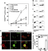
LL37 efficiently transports self-DNA into human monocytes. (A) Degree of association of Alexa488-labeled huDNA-LL37 complexes with human blood-derived immune cells (pDCs, monocytes, and myeloid DCs), and structural nonhematopoietic cells (fibroblasts, keratinocytes) at different time points (0, 1, 4, and 12 hours) after stimulation, assessed by flow cytometry. Alexa488-labeled huDNA alone showed minimal association with all cell types tested. Data are the mean ± SEM of 4 independent experiments. *P < .0001 (paired Student t test). (B) Representative FACS analysis of Alexa488 huDNA-LL37 complex association with human blood-derived immune cells (pDCs, monocytes, and myeloid DCs), and structural nonhematopoietic cells (fibroblasts, keratinocytes) 4 hours after stimulation is shown. (C) Intracellular localization of Alexa488 huDNA-LL37 complexes 4 and 24 hours after stimulation of human monocytes, assessed by confocal microscopy. Green represents Alexa488 huDNA; and red, surface staining of monocytes with Alexa647 HLA-DR. Bar represents 5 μm (D) Percentage of cytoplasmic distribution of Alexa488 huDNA-LL37 complexes 4 and 24 hours after stimulation of human pDCs, and monocytes. Data are representative of 3 independent experiments and are given as mean ± SEM of the percentage of cells with cytoplasmic DNAAlexa distribution; at least 150 cells containing Alexa488 huDNA-LL37 complexes from 10 different fields were counted for each condition. *P < .0001 (paired Student t test).
Figure 3
Figure 3
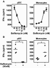
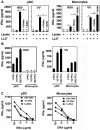
IFN-α induction in monocytes by self-DNA-LL37 complexes is TLR-independent. (A) IFN-α produced by pDCs (5 × 10⁴ cells/condition) and monocytes (2 × 10⁵ cells/condition) stimulated with huDNA (huDNA)-LL37 complexes after pretreatment with increasing concentrations of bafilomycin. Data are representative of at least 3 experiments; error bars represent the SD of triplicate wells. (B) IFN-α production by pDCs and monocytes stimulated with huDNA-LL37 complexes with or without pretreatment with 50nM bafilomycin. Each symbol represents an independent experiment, and horizontal bars represent the mean. *P < .0001 (paired Student t test).
Figure 4
Figure 4
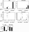
Type I IFN induction in monocytes by DNA-LL37 complexes requires dsDNA but is independent of DNA sequence and methylation. (A) IFN-α produced by pDCs (5 × 10⁴ cells/condition) and monocytes (2 × 10⁵ cells/condition) after overnight stimulation with 10 μg/mL of ssCpG DNA, or dsCpG DNA sequences of phosphodiester backbone alone or in complex with LL37 (10μM), with or without pretreatment with bafilomycin (50nM). In contrast to pDCs, in monocytes ssDNA sequences in complex with LL37 failed to induce IFN-α production. Data are representative of at least 4 independent experiments; error bars represent the SD of triplicate wells. (B) IFN-α produced by pDCs and monocytes after overnight stimulation with 10 μg/mL of a CpG-containing plasmid or a CpG-free plasmid, alone or with LL37 (10μM), with or without pretreatment with 50nM bafilomycin. Each symbol represents an independent experiment, and horizontal bars represent the mean. (C) IFN-α produced by pDCs (5 × 10⁴ cells/condition) and monocytes (2 × 10⁵ cells/condition) after overnight stimulation with 10 μg/mL of unmethylated or methylated dsCpG DNA, either alone or in complex with LL37 (10μM). Data are presented as mean ± SEM of 3 independent experiments. *P < .0001 (paired Student t test).
Figure 5
Figure 5
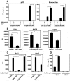
Type I IFN induction by DNA-LL37 complexes in human monocytes occurs through cytosolic DNA sensors. (A) IFN-α produced by pDCs and monocytes after overnight stimulation with 10 μg/mL of poly dA:dT or poly dC:dG DNA, either alone or in complex with LL37 (10μM), with or without pretreatment with 50nM bafilomycin. Data are representative of at least 5 independent experiments; error bars represent the SD of triplicate wells. *P < .0001 (paired Student t test). (B) Real-time PCR of TBK1, MyD88, and STING expression after transfection with siRNA targeting TBK1, MyD88, STING, or control nontargeting RNAi sequences (control siRNA [C RNAi]) in primary human monocytes. Data are representative of at least 3 independent experi-ments; error bars represent the SD of triplicate wells. *P < .01 (paired Student t test). (C) Real-time PCR of Ifna mRNA induction in primary human monocytes stimulated with huDNA-LL37 complexes 48 hours after transfection with siRNAs targeting TBK1, MyD88, or control nonsilencing siRNAs. Data are representative of at least 4 independent experiments for TBK1 and MyD88 silencing and 2 independent experiments for STING silencing; error bars represent the SD of triplicate wells. *P < .01 (paired Student t test). (D) ELISA of IFN-α in human monocytes after stimulation with poly dC:dG DNA (B-type DNA) in complex with LL37 complexes or brominated poly dC:dG DNA (Z-type DNA)-LL37. Data are representative of at least 3 independent experiments; error bars represent the SD of triplicate wells. *P < .0001 (paired Student t test).
Figure 6
Figure 6
In addition to huDNA, LL37 complexes with free microbial/viral DNA for type I IFN induction in monocytes. (A) IFN-α produced by pDCs (5 × 10⁴ cells/condition) and monocytes (2 × 10⁵ cells/condition) after overnight stimulation with lysates of VV and HSV viruses alone or in complex with LL37 (10μM) with or without pretreatment with 50nM bafilomycin. Data are representative of at least 5 experiments; error bars represent the SD of triplicate wells. *P < .0001 (paired Student t test). **P < .01 (paired Student t test). (B) IFN-α produced by pDCs and monocytes after overnight stimulation with increasing titers of VV and HSV viruses. HSV failed to trigger IFN-α release in monocytes; error bars represent the SD of triplicate wells. (C) IFN-α produced by pDCs (5 × 10⁴ cells/condition) and monocytes (2 × 10⁵ cells/condition) after overnight stimulation with increasing concentration of either HSV, or VV, or human genomic DNA from U937 cells (huDNA) in complex with LL37 (10μM). Data are representative of at least 3 experiments.

References

    1. Theofilopoulos AN, Baccala R, Beutler B, Kono DH. Type I interferons (alpha/beta) in immunity and autpimmunity. Annu Rev Immunol. 2005;23:307–336. - PubMed
    1. Stetson DB, Medzhitov R. Type I interferons in host defense. Immunity. 2006;25(3):373–381. - PubMed
    1. Santini SM, Lapenta C, Logozzi M, et al. Type I interferon as a powerful adjuvant for monocyte-derived dendritic cell development and activity in vitro and in Hu-PBL-SCID mice. J Exp Med. 2000;191(10):1777–1788. - PMC - PubMed
    1. Luft T, Pang KC, Thomas E, et al. Type I IFNs enhance the terminal differentiation of dendritic cells. J Immunol. 1998;161(4):1947–1953. - PubMed
    1. Hornung V, Latz E. Intracellular DNA recognition. Nat Rev Immunol. 2010;10(2):123–130. - PubMed

Publication types