. 2012 Nov 8;120(19):4072-81.
doi: 10.1182/blood-2012-04-420745. Epub 2012 Aug 27.

Mechanism of platelet dense granule biogenesis: study of cargo transport and function of Rab32 and Rab38 in a model system

Affiliations

Mechanism of platelet dense granule biogenesis: study of cargo transport and function of Rab32 and Rab38 in a model system

Andrea L Ambrosio et al. Blood. .

Abstract

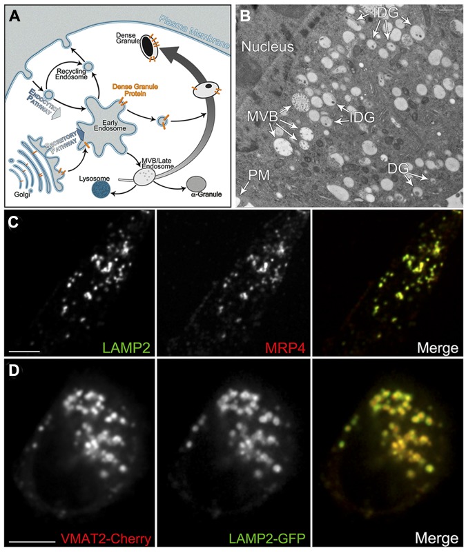
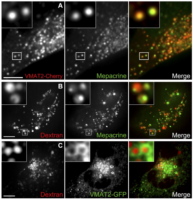
Dense granules are important in platelet aggregation to form a hemostatic plug as evidenced by the increased bleeding time in mice and humans with dense granule deficiency. Dense granules also are targeted by antiplatelet agents because of their role in thrombus formation. Therefore, the molecular understanding of the dense granule and its biogenesis is of vital importance. In this work, we establish a human megakaryocytic cell line (MEG-01) as a model system for the study of dense granule biogenesis using a variety of cell biology and biochemical approaches. Using this model system, we determine the late endocytic origin of these organelles by colocalization of the internalized fluid phase marker dextran with both mepacrine and transmembrane dense granule proteins. By mistargeting of mutant dense granule proteins, we demonstrate that sorting signals recognized by adaptor protein-3 are necessary for normal transport to dense granules. Furthermore, we show that tissue-specific Rab32 and Rab38 are crucial for the fusion of vesicles containing dense granule cargo with the maturing organelle. This work sheds light on the biogenesis of dense granules at the molecular level and opens the possibility of using this powerful model system for the investigation of new components of the biogenesis machinery.


Figures

Figure 1
Figure 1
MEG-01 cells have DGs that can be studied by different microscopy techniques. (A) Model depicting the protein traffic to DG. DG transmembrane proteins (gold) follow the secretory pathway through the Golgi complex to early endosomes where they are selectively targeted to maturing DGs originated from MVBs. (B) Thin-section transmission electron microscopy image of a MEG-01 cell subjected to HPF, fixed or embedded with glutaraldehyde-uranyl acetate-Lowicryl HM20. Original magnification, ×3900 (bar represents 500 nm). (C) Spinning-disk confocal fluorescence microscopy images of a MEG-01 cell fixed and immunostained with LAMP2 and MRP4 antibodies to label DGs (MOC = 0.63 ± 0.06, n = 5 cells). Bar represents 10 μm. (D) Spinning-disk confocal fluorescence microscopy images of a live MEG-01 cell expressing the DG markers VMAT2-Cherry and LAMP2-GFP (MOC = 0.58 ± 0.03, n = 10 cells). Bar represents 5 μm. PM indicates plasma membrane; and IDG, immature dense granule.
Figure 2
Figure 2
DGs originate from late endocytic structures. (A) DGs were labeled with the green fluorescent dye mepacrine in live MEG-01 cells expressing VMAT2-Cherry and visualized by spinning-disk confocal microscopy. The inset shows examples of colocalization between the 2 DG markers; 94% ± 2% of 171 mepacrine structures (7 cells) also contain VMAT-Cherry. (B) Live MEG-01 cells were allowed to internalize the fluid phase marker dextran Alexa Fluor 647, and DGs were subsequently labeled with mepacrine. Cells were observed by spinning-disk confocal fluorescence microscopy. Examples of structures containing both markers are presented in the magnified inset (MOC = 0.43 ± 0.04, n = 6 cells). (C) Live MEG-01 cells expressing the DG marker VMAT2-GFP were labeled with dextran Alexa Fluor 647 and imaged by spinning-disk confocal fluorescence microscopy. Inset: Examples of VMAT-GFP presence in the limiting membrane of organelles containing dextran Alexa Fluor 647 in the lumen; 66% ± 8% of 187 structures containing fluorescent dextran (7 cells) also contain VMAT-GFP. Bars represent 5 μm.
Figure 3
Figure 3
Sorting signals bound by AP-3 are crucial for the correct targeting of DG proteins in MEG-01 cells. AP-3 partially colocalizes with Rab38 in MKs and MEG-01 cells. (A-B) A single mutation in the LAMP2 cytosolic tail sorting signal (Y/A LAMP2: YEQF into AEQF) is sufficient to mistarget Y/A LAMP2-GFP to the plasma membrane in MEG-01 cells. Live cells were visualized by spinning-disk confocal fluorescence microscopy. (C-D) Similarly, mutation of the VMAT2 cytosolic tail sorting signal (IL/AA VMAT2: EEKMAIL into EEKMAAA) causes mistrafficking of the mutant protein to the plasma membrane in MEG-01 cells. Live cells were visualized by spinning-disk confocal fluorescence microscopy. (E-E′) Primary MKs were fixed and immunostained with antibodies against AP-3, Rab38, and clathrin, and imaged by spinning-disk confocal fluorescence microscopy. (E′) Close-up view of individual structures allows observation of colocalization of AP-3 and Rab38 (merge panel, MOC = 0.34 ± 0.01, n = 7 cells), whereas clathrin is also present in many of these structures (Rab38 and clathrin MOC = 0.33 ± 0.02, n = 7 cells). (F-F′) MEG-01 cells were fixed and immunostained with antibodies against AP-3, Rab38, and clathrin and imaged by spinning-disk confocal fluorescence microscopy. (F′) Similarly to the results obtained with MKs, AP-3 and Rab38 colocalize in structures that in many cases contain clathrin (Rab38 and AP-3 MOC = 0.32 ± 0.02, n = 4 cells; Rab38 and clathrin MOC = 0.33 ± 0.03, n = 4 cells). Bars represent 5 μm.
Figure 4
Figure 4
Rab32 and Rab38 are primarily present in immature DGs. (A-C) DGs were labeled with mepacrine in live MEG-01 cells expressing Cherry-Rab38 and visualized by spinning-disk confocal fluorescence microscopy; 95% ± 2% of structures containing Cherry-Rab38 (40 cells) also contain mepacrine. (A) A structure containing the highest amount of mepacrine and low Cherry-Rab38 levels is indicated with a green arrowhead, a structure with high concentration of Cherry-Rab38 and low mepacrine with a red arrowhead, and a structure with intermediate amounts of both markers with a yellow arrowhead (bar represents 5 μm). (B) Both organelles and vesicles are labeled with Cherry-Rab38, which is shown as an inset in panel C (bar represents 5 μm). (C) Although mepacrine is only present in the organelles, Cherry-Rab38 is also present in the vesicle (bar represents 500 nm). (D) Live MEG-01 cell coexpressing LAMP2-Cherry, as a DG marker, and GFP-Rab38 imaged by spinning-disk confocal fluorescence microscopy; 88 ± 4% of structures containing GFP-Rab38 (37 cells) also contain LAMP2-Cherry. Bar represents 5 μm. (E) A close-up view of structures from panel D shows a reverse correlation between the amount of Rab38 and the DG marker. (F) Fluorescence intensity line scan of the structures shown in panel E (merge panel). A.U., arbitrary units. (G-L) Immunogold electron microscopy images of immature DGs from MEG-01 cells subjected to HPF using antibodies against LAMP2 (18 nm) and Rab32 (12 nm). LAMP2 is present in 73% of the organelles label with Rab32 (n = 84 organelles). Original magnifications were ×15,000, ×20,000, ×15,000, ×20,000, ×25,000, and ×20,000, respectively. Bars represent 200 nm.
Figure 5
Figure 5
Rab32 and Rab38 colocalize with the late endocytic marker Rab7a but not with the vacuolar early endosome and recycling endosome marker Rab5a. Cherry-Rab32 and Cherry-Rab38 were cotransfected with GFP-Rab5a or GFP-Rab7a in MEG-01 cells. Confocal fluorescence microscopy images of live cells together with the corresponding fluorescence intensity line scan graphs are shown for each experiment. The white lines in the merge panels indicate the portions of the cells where fluorescence intensities for both the red and green channels were measured. (A,C) Neither Rab32 nor Rab38 colocalizes significantly with Rab5a, a marker of recycling vesicles/early endosomes (MOC = 0.16 ± 0.02, n = 13 cells and MOC = 0.14 ± 0.01, n = 7 cells, respectively). (B,D) Consistent with Rab32 and Rab38 being present in immature DGs, both proteins colocalize with Rab7a, a marker of late endosomal compartments that is also present in other LROs such as melanosomes and lamellar bodies (MOC = 0.52 ± 0.02, n = 7 cells and MOC = 0.42 ± 0.03, n = 7 cells, respectively).
Figure 6
Figure 6
Biochemical study of MEG-01 dense granules. (A) Immunoblotting analysis of fractions obtained from MEG-01 postnuclear supernatants subjected to subcellular fractionation with a 10% to 60% sucrose gradient. Markers for different cell compartments are as follow: MRP4 and LAMP2 for immature and mature DGs; PF-4 for α granules; and Rab7a and Rab32 for cytosol, vesicles, and DGs. (B) ADP (μM, solid lines) and fluorescence intensity of both infrared fluorescent-dextran (A.U., dashed line) and the fluorescent Ca2+ indicator Oregon Green BAPTA-1 dextran (A.U., dotted line) in fractions from panel A. The concentration of ADP was determined both in the untreated sucrose gradient fractions (solid circles) and in sucrose gradient fractions enriched in MRP4 structures by immunoprecipitation using an MRP4 antibody (open circles). (C) Sucrose gradient fractions were immunoprecipitated using a Rab38 antibody and the presence of MRP4 in the precipitated structures was determined by immunoblotting, confirming the occurrence of the proteins in the same structures. (D) The coexistence of both LAMP2 and MRP4 in the same organelles was confirmed by coimmnunoprecipitation using a MRP4 antibody (left) or a LAMP2 antibody (right) and immunoblotting analysis using an antibody against the other protein. SGF indicates sucrose gradient fraction; IP, immunoprecipitation; IB, immunoblotting; and IDG, immature DG.
Figure 7
Figure 7
Rab32 or Rab38 knock-down impairs normal fusion of vesicles containing dense granule proteins with the organelle. (A-F) MEG-01 cells were cotransfected with LAMP2-Cherry as a DG reporter and either control siRNA shown in panels A and B, Rab32 siRNA in panels C and D, or Rab38 siRNA in panels E and F (see supplemental Figure 11 for Rab32/38 siRNA knockdown confirmation by immunoblotting). The kymographs presented in panels B, D, and F were made by aligning on a time axis the pieces of images indicated with a white rectangle in panels A, C, and E, respectively, from each of the 60 frames of the corresponding movies (1 frame/second). (A) Control siRNA cells present diffraction-limited LAMP2 structures consistent in size with organelles. (B) The LAMP2 structures in Control siRNA cells present a limited range of motion. (C,E) Both Rab32 and Rab38 siRNA cells present LAMP2 structures that are more consistent in size with vesicles or small organelles. (D,F) The smaller LAMP2 structures in Rab32 and Rab38 siRNA cells are more dynamic and move faster than structures in Control siRNA cells. (G) The diameter of LAMP2 structures present in the representative cells shown in panels A, C, and E was measured using the Ruler function in Slidebook. (H) The average speed of LAMP2 structures present in the representative cells shown in panels A, C, and E was measured using the Manual Particle Tracking function in Slidebook. (I) Extracts from control, Rab32, and Rab38 siRNA-treated cells were fractionated in sucrose gradients. For each treatment, the amount of LAMP2 in the first fraction of the gradient, which corresponds to the vesicular LAMP2, was analyzed by immunoblotting. The levels of tubulin in the same blot were used to confirm equal loading. Bars represent 5 μm (*P < .05; **P < .001). (J) DG membrane proteins are sorted in early endosomal compartments by adaptor protein complexes, such as AP-3, which recognize sorting signals present in their cytosolic tails. Rab32 and Rab38 are recruited to the nascent clathrin-coated vesicle and through interactions with so far unknown effectors target the vesicle to the maturing DG. The DG precursor is a MVB that on receiving Rab32/Rab38 vesicles containing DG proteins, such as the ADP transporter MRP4 or the serotonin transporter VMAT2, matures into a DG.

Comment in

Similar articles

Cited by

References

    1. Huizing M, Helip-Wooley A, Westbroek W, Gunay-Aygun M, Gahl WA. Disorders of lysosome-related organelle biogenesis: clinical and molecular genetics. Annu Rev Genomics Hum Genet. 2008;9:359–386. - PMC - PubMed
    1. McNicol A, Israels SJ. Platelet dense granules: structure, function and implications for haemostasis. Thromb Res. 1999;95(1):1–18. - PubMed
    1. Kim S, Kunapuli SP. P2Y12 receptor in platelet activation. Platelets. 2011;22(1):54–58. - PubMed
    1. Konopatskaya O, Matthews SA, Harper MT, et al. Protein kinase C mediates platelet secretion and thrombus formation through protein kinase D2. Blood. 2011;118(2):416–424. - PMC - PubMed
    1. Graham GJ, Ren Q, Dilks JR, Blair P, Whiteheart SW, Flaumenhaft R. Endobrevin/VAMP-8-dependent dense granule release mediates thrombus formation in vivo. Blood. 2009;114(5):1083–1090. - PMC - PubMed

Publication types

MeSH terms