. 2012 Oct 11;120(15):3069-79.
doi: 10.1182/blood-2012-01-403493. Epub 2012 Aug 27.

Fluvastatin inhibits FLT3 glycosylation in human and murine cells and prolongs survival of mice with FLT3/ITD leukemia

Affiliations

Fluvastatin inhibits FLT3 glycosylation in human and murine cells and prolongs survival of mice with FLT3/ITD leukemia

Allen B Williams et al. Blood. .

Abstract

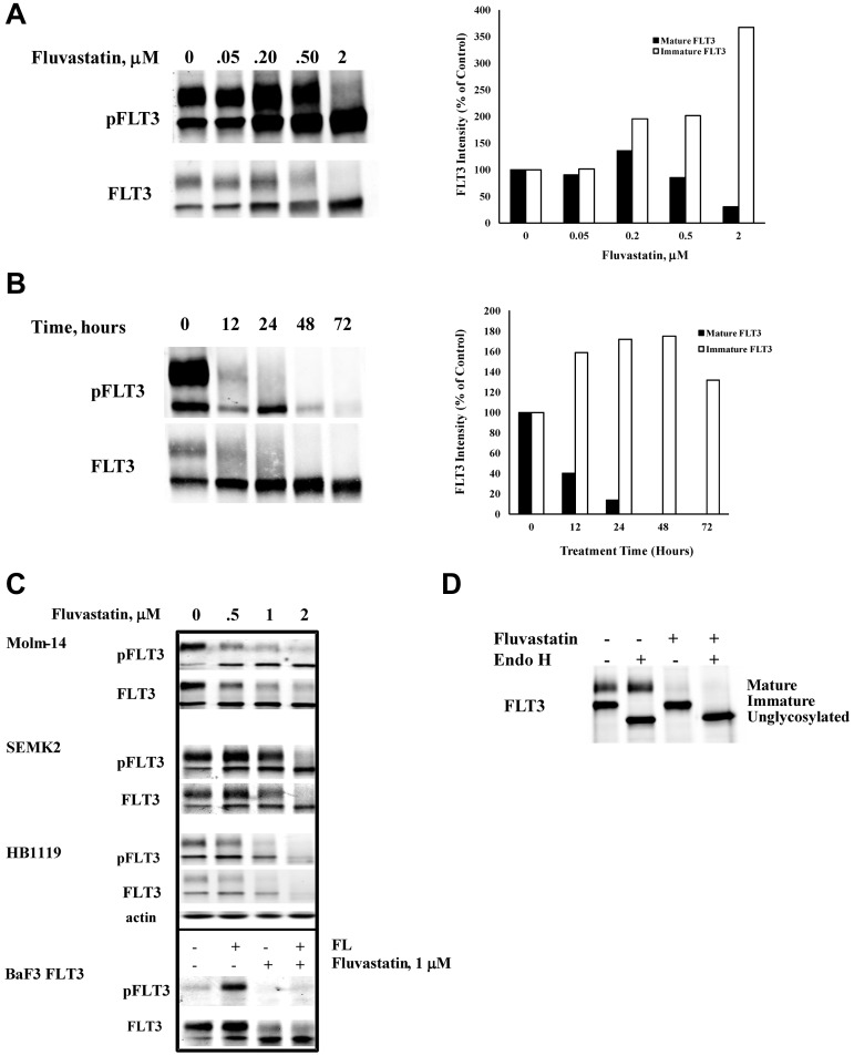
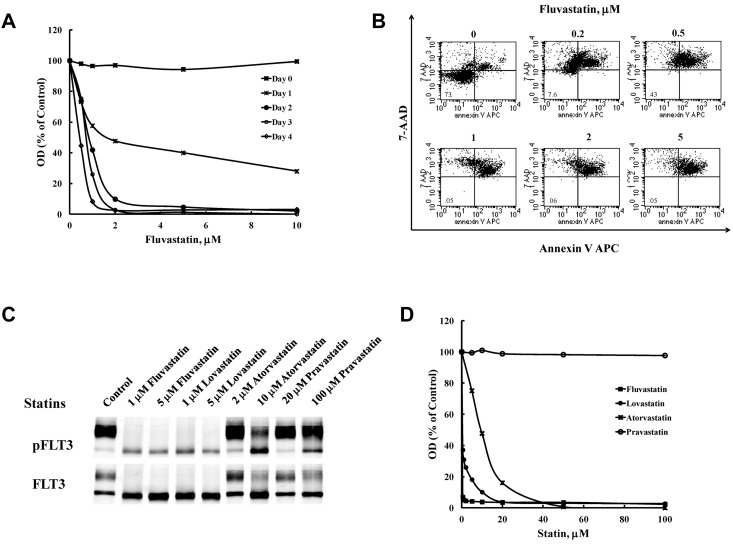
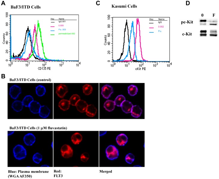
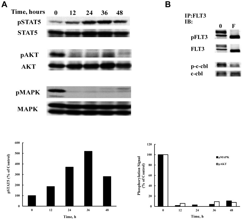
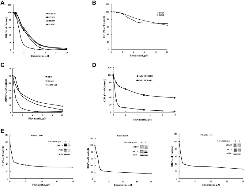
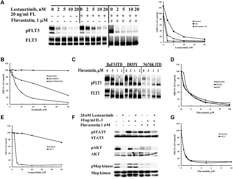
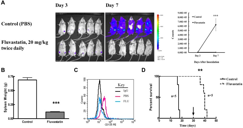
FLT3 is frequently mutated in acute myeloid leukemia (AML), but resistance has limited the benefit of tyrosine kinase inhibitors (TKI). We demonstrate that statins can impair FLT3 glycosylation, thus leading to loss of surface expression and induction of cell death, as well as mitigation of TKI resistance. Immunofluorescence microscopy confirms a reduction in surface localization and an increase in intracellular FLT3/internal tandem duplication (ITD) accumulation. This aberrant localization was associated with increased STAT5 activation but inhibition of both MAPK and AKT phosphorylation. Growth inhibition studies indicate that FLT3/ITD-expressing cells were killed with an IC(50) within a range of 0.2-2μM fluvastatin. Several mechanisms of resistance could be circumvented by fluvastatin treatment. An increase in the IC(50) for inhibition of phosphorylated FLT3/ITD by lestaurtinib caused by exogenous FLT3 ligand, resistance to sorafenib caused by the D835Y or FLT3/ITD N676K mutations, and activation of the IL-3 compensatory pathway were all negated by fluvastatin treatment. Finally, fluvastatin treatment in vivo reduced engraftment of BaF3 FLT3/ITD cells in Balb/c mice. These results demonstrate that statins, a class of drugs already approved by the US Food and Drug Administration, might be repurposed for the management of FLT3 mutant acute myeloid leukemia cases either alone or in conjunction with FLT3 TKI.


Figures

Figure 1
Figure 1
Fluvastatin prevents glycosylation of FLT3 and impedes receptor maturation. (A) BaF3 FLT3/ITD cells were treated with increasing concentrations of fluvastatin for 24 hours. Lysates were immunoprecipitated with anti-FLT3 antibody and resolved by SDS-PAGE and then probed with FLT3 or 4G10 antiphosphotyrosine antibodies. Quantitation of the upper and lower FLT3 bands is shown in the graph. (B) Cells were treated with 1μM fluvastatin for the indicated time points and analyzed for FLT3 expression as described. Quantitation of the upper and lower FLT3 bands is shown in the graph. (C) The effect of increasing fluvastatin concentration treatment for up to 24 hours on the FLT3-expressing cell lines HB1119, Molm-14, and SEMK-2 cells. In the bottom panel, wild-type FLT3-expressing BaF3 cells were treated ± 1μM fluvastatin for 24 hours followed by ± stimulation with 20 ng/mL FLT3 ligand for 5 minutes. (D) BaF3 FLT3/ITD cells were treated with 1μM fluvastatin for 24 hours after which immunoprecipitated FLT3/ITD was digested with endoglycosidase H for 1 hour. Results are typical of at least 3 independent experiments.
Figure 2
Figure 2
Treatment with statins inhibits proliferation in BaF3 FLT3/ITD cells. (A) BaF3 FLT3/ITD cells were treated with fluvastatin for the indicated times and processed using the MTT assay or (B) by measuring binding of annexin V and 7-AAD after 48 hours to assess induction of apoptosis. (C) Western blotting and (D) the MTT assay were also used to measure the effect of fluvastatin, lovastatin, atorvastatin, or pravastatin on FLT3 glycosylation and growth inhibition. Results are representative of 3 independent experiments.
Figure 3
Figure 3
Fluvastatin prevents translocation of FLT3/ITD to the cell surface leading to intracellular accumulation of the immature form. (A) BaF3 FLT3/ITD cells were treated with 1μM fluvastatin for 24 hours. Afterward, surface or total FLT3/ITD was measured by incubating with CD135-PE for 30 minutes and analyzed by flow cytometry. The untreated cells are shown in red, fluvastatin-treated cells in blue, and total FLT3 from cells treated with fluvastatin and then permeabilized is shown in green. Cell-surface staining with IgG is shown in black for both the permeabilized and nonpermeabilized cells. (B) Cells treated with or without fluvastatin were adhered to poly-L-lysine slides and stained for plasma membrane (WGA-A350, blue) or FLT3 (Cy3, red). Slides were visualized by immunofluorescence microscopy using a Nikon TE2000 Eclipse microscope and 60 × 1.4 oil-immersion lens. (C) Kasumi cells were treated with 1μM fluvastatin for 24 hours and assessed for surface c-Kit expression. Cells stained with IgG are shown in black, untreated cells in red, and fluvastatin-treated cells in blue. (D) Western blot results show fluvastatin treatment led to a reduction in the amount of mature c-Kit expression in Kasumi-1 cells. Results are representative of at least 3 independent experiments.
Figure 4
Figure 4
Fluvastatin alters FLT3/ITD signaling. (A) BaF3 FLT3/ITD cells were treated with 1μM fluvastatin for the indicated times and 50 μg of lysate from each time point was analyzed by SDS-PAGE and Western blotting. (B) Cells were treated with 1μM fluvastatin for 24 hours and lysates were immunoprecipitated with a FLT3 antibody (S-18). Blots were then probed for FLT3, phosphotyrosine, c-cbl, or phospho c-cbl. Typical results of 3 experiments are shown.
Figure 5
Figure 5
Fluvastatin inhibits proliferation that is driven by glycosylated transmembrane receptors. Inhibition of growth was assessed after a 48-hour incubation period using the MTT assay in cell lines driven by either: (A) activated FLT3 including FLT3/ITD (MV4-11, Molm-14 cells), FLT3 (SEMK2 cells), or a D835H kinase domain mutant (HB1119 cells); (B) the nonglycosylated oncogenic intracellular kinases BCR-ABL (K562 cells) or Fes kinase (U937 cells); or (C) other glycosylated transmembrane receptors, such as insulin (HL60 cells), c-Kit (MO7E cells), or mutant c-Kit (Kasumi cells). (D) The effect of fluvastatin was assessed on proliferation of BaF3 cells expressing either FLT3/ITD or BCR-ABL after treatment for 48 hours by MTT. (E) Primary patient AML samples harboring a FLT3/ITD mutation were treated with the indicated concentrations of fluvastatin for 72 hours and analyzed by MTT assay or Western blotting. Typical results are shown for at least 2 separate experiments.
Figure 6
Figure 6
Fluvastatin reverses resistance to FLT3 tyrosine kinase inhibitors mediated by multiple mechanisms. (A) Resistance to lestaurtinib induced by stimulation with FLT3 ligand (FL) is overcome by prior treatment with fluvastatin. BaF3 FLT3/ITD cells were treated with or without 1μM fluvastatin for 24 hours followed by incubation in lestaurtinib for 1 hour and then stimulated with 20 ng/mL FL for 5 minutes. Densitometry of the pFLT3 bands plotted in response to treatment is shown. (B) BaF3 cells expressing a FLT3/ITD, D835Y, or FLT3/ITD N676K resistance mutation were treated with sorafenib for 48 hours and viability was assessed by MTT assay. (C) The same cells were treated for 24 hours with increasing concentrations of fluvastatin to study FLT3 glycosylation or (D) for 48-96 hours and analyzed by MTT assay. Cells were simultaneously treated with either (E) lestaurtinib or (G) fluvastatin and 10 ng/mL interleukin-3 for 48 hours after which cell growth was evaluated by MTT. (F) Activation of IL-3 receptor signaling pathways was assessed by treating cells with or without 1μM fluvastatin for 24 hours followed by incubation with or without lestaurtinib for 1 hour and a 10-minute stimulation with or without 10 ng/mL IL-3 followed by SDS-PAGE Western blotting analysis of STAT5, AKT, and MAPK activation states. Results are representative of at least 3 experiments.
Figure 7
Figure 7
Fluvastatin reduces tumor burden in mice engrafted with FLT3/ITD cells. (A) Balb/c mice were injected via tail vein with 2 × 10⁵ luciferase-positive BaF3 FLT3/ITD cells and treated with PBS (control) or fluvastatin at 20 mg/kg twice daily starting 3 days after injection. Mice were injected with luciferin and analyzed on an IVIS Spectrum imager on days 3 and 7. The mean (+ SEM) bioluminescence for the 5 mice in each group at days 3 and 7 is plotted on the right. (B) Twice-daily treatments were continued as described in panel A until mice became moribund (control group) at which point both groups of mice were euthanized and spleen weights (mean + SEM) were measured. (C) Peripheral blood from transplanted mice treated with or without 20 mg/kg fluvastatin twice daily on days 10 and 11 after transplantation was assayed for surface FLT3 expression. IgG-stained cells are shown in black, cells from PBS-treated mice in red, and cells from fluvastatin-treated mice in blue. (D) Survival of transplanted mice was assessed by twice-daily PBS or fluvastatin treatments from day 3 until day 30 (or until mice became moribund) at which point treatment was stopped (arrow). Five mice were used in each group and the experiment was conducted twice.

References

    1. Small D, Levenstein M, Kim E, et al. STK-1, the human homolog of Flk-2/Flt-3, is selectively expressed in CD34+ human bone marrow cells and is involved in the proliferation of early progenitor/stem cells. Proc Natl Acad Sci U S A. 1994;91(2):459–463. - PMC - PubMed
    1. Reilly JT. FLT3 and its role in the pathogenesis of acute myeloid leukaemia. Leuk Lymphoma. 2003;44(1):1–7. - PubMed
    1. Chan PM. Differential signaling of Flt3 activating mutations in acute myeloid leukemia: a working model. Protein Cell. 2011;2(2):108–115. - PMC - PubMed
    1. Lyman SD, James L, Vanden Bos T, et al. Molecular cloning of a ligand for the flt3/flk-2 tyrosine kinase receptor: a proliferative factor for primitive hematopoietic cells. Cell. 1993;75(6):1157–1167. - PubMed
    1. Lavagna-Sevenier C, Marchetto S, Birnbaum D, Rosnet O. FLT3 signaling in hematopoietic cells involves CBL, SHC and an unknown P115 as prominent tyrosine-phosphorylated substrates. Leukemia. 1998;12(3):301–310. - PubMed

Publication types

MeSH terms