. 2012 Oct 26;111(10):1274-85.
doi: 10.1161/CIRCRESAHA.112.277525. Epub 2012 Aug 27.

Interleukin-27 receptor limits atherosclerosis in Ldlr-/- mice

Affiliations

Interleukin-27 receptor limits atherosclerosis in Ldlr-/- mice

Ekaterina K Koltsova et al. Circ Res. .

Abstract

Rationale: Atherosclerosis is a chronic inflammatory disease of the arterial wall. Several proinflammatory cytokines are known to promote atherosclerosis, but less is known about the physiological role of anti-inflammatory cytokines. Interleukin (IL)-27 is a recently discovered member of the IL-6/IL-12 family. The IL-27 receptor is composed of IL-27 receptor A (WSX-1) and gp130 and is required for all established IL-27 signaling pathways. The expression of the IL-27 subunit Ebi3 is elevated in human atheromas, yet its function in atherosclerosis remains unknown.

Objective: The aim of this study was to test the role of IL-27 receptor signaling in immune cells in atherosclerosis development.

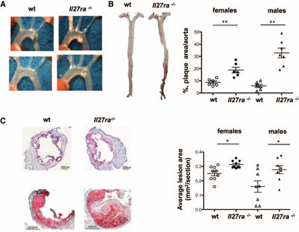
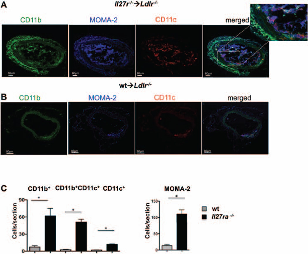
Methods and results: Atherosclerosis-prone Ldlr(-/-) mice transplanted with Il27ra(-/-) bone marrow and fed Western diet for 16 weeks developed significantly larger atherosclerotic lesions in aortic roots, aortic arches, and abdominal aortas. Augmented disease correlated with increased accumulation of CD45(+) leukocytes and CD4(+) T cells in the aorta, which produced increased amounts of IL-17A and tumor necrosis factor. Several chemokines, including CCL2, were upregulated in the aortas of Ldlr(-/-) mice receiving Il27ra(-/-) bone marrow, resulting in accumulation of CD11b(+) and CD11c(+) macrophages and dendritic cells in atherosclerotic aortas.

Conclusions: The absence of anti-inflammatory IL-27 signaling skews immune responses toward T-helper 17, resulting in increased production of IL-17A and tumor necrosis factor, which in turn enhances chemokine expression and drives the accumulation of proatherogenic myeloid cells in atherosclerotic aortas. These findings establish a novel antiatherogenic role for IL-27 receptor signaling, which acts to suppress the production of proinflammatory cytokines and chemokines and to curb the recruitment of inflammatory myeloid cells into atherosclerotic aortas.


Figures

Figure 1
Figure 1. Increased atherosclerotic plaque area inIl27ra−/−bone marrow–transplanted mice
Ldlr−/− mice were lethally irradiated and reconstituted with 5×10⁶ unfractioned bone marrow cells from C57BL/6 (wild-type [wt]) or Il27ra−/− mice. After 4 weeks of reconstitution, mice were fed with western diet for 16 weeks. A, Images of aortic arch of Ldlr−/− mice receiving Il27ra−/− bone marrow or C57BL/6 (wt) control. B, Representative en face images of Sudan IV–stained whole aortas of Ldlr−/− mice receiving Il27ra−/− or wt bone marrow. B, right, Quantification of plaque area as percentage of aortic surface in Ldlr−/− mice receiving Il27ra−/− or wt bone marrow (n=6–7 females or males per group, respectively). C, left, Representative images of aortic roots (top) and single valve (bottom) of Ldlr−/− mice receiving Il27ra−/− or C57BL/6 (wt) bone marrow. C, right, Aortic root lesions were quantified on frozen sections stained with Oil-red-O in the 300 µm following the aortic valve (wt [n=8–9 males or females] mice and Il27ra−/− [n=8–9 males or females] mice from 3 experiments).
Figure 2
Figure 2. Leukocyte composition of mouse aorta
A, Aortas from bone marrow–transplanted mice were made into single-cell suspensions by enzymatic digestion and stained. Live CD45+ leukocytes were counted in aortas of 16-week Western diet–fed Ldlr−/− mice transplanted with Il27ra−/− (n=7) or C57BL/6 (wild-type [wt]; n=7) bone marrow. Left panels show representative dot plots. Right panel shows mean±SEM, *P<0.05. B, left, Live CD45+ cells from aortas were stained for TCRβ and CD4. Numbers indicate percentage in indicated gates. B, right, Absolute number of live CD45+, TCRβ+, CD4+TCRβ+, and CD8+TCRβ+ cells in aortas from Ldlr−/− mice transplanted with Il27ra−/− or wt bone marrow (n=7) each. Mean±SEM *P<0.05. Localization of CD3+ cells in aortic roots (C) and abdominal aortas (D) of Ldlr−/− mice transplanted with Il27ra−/− or C57BL/6 (wt) bone marrow as seen by immunofluorescence. L indicates lumen; P, plaque; A, adventitia. Representative images from 1 of 3 independent experiments. Mean±SEM number of CD3+ cells per section in aortic roots (C, right) and abdominal aortas (D, right) of Ldlr−/− mice transplanted with Il27ra−/− (n=6) or C57BL/6 (wt; n=6) bone marrow are shown.
Figure 3
Figure 3. Enhanced production of T-helper 17 cytokines and transcription factors in mice receivingIl27ra−/− bone marrow
A, Relative gene expression in Il27ra−/−Ldlr−/− (n=6) mice normalized to β-actin and then normalized to gene expression in wild-type (wt)→Ldlr−/− (n=6) mice in aortic arches, abdominal aortas (Ab. aorta), paraaortic lymph nodes (paLNs), and spleens after 16 weeks of Western diet. B and C, Interleukin (IL)-17A, tumor necrosis factor (TNF), IL-6, and interferon (IFN) γ were measured by bead array in supernatants of aortic (n=8; B) and splenic (n=8; C) cell suspensions, stimulated with anti-CD3/anti-CD28 for 48 h. Mean±SEM; *P<0.05.
Figure 4
Figure 4. Enhanced production of T-helper 17 cytokines in aortas of mice receiving Il27ra−/− bone marrow
Percent of CD4 T cells expressing tumor necrosis factor (TNF), interleukin (IL)-17A, and interferon (IFN) γ by intracellular staining in aorta (A, left), spleen (B, left), or paraaortic lymph nodes (paLNs) (C, left) of Ldlr−/− mice transplanted with Il27ra−/− (n=6) or wild-type (wt; n=6) bone marrow fed 16 weeks with Western diet. A–C, right, Representative fluorescence-activated cell sorter plot of CD4+TCRβ+ T cells stained for IL-17A and TNF in aortas (A), spleens (B), and paLNs (C) of Ldlr−/− mice transplanted with Il27ra−/− or wt bone marrow fed 16 weeks with Western diet. Mean±SEM; *P<0.05.
Figure 5
Figure 5. Regulatory T cells in spleen and lymph nodes inIl27ra−/− bone marrow–transplanted mice
A, Percentage of live CD4+TCRβ+Foxp3+ T regulatory cells (Treg) in spleens and paraaortic lymph nodes (paLNs) from Ldlr−/− mice transplanted with Il27ra−/− (n=10) or wild-type (wt) bone marrow (n=10). B, Percentage of live CD4+TCRβ+, Foxp3IL10+ Tr1 cells in spleens and paLNs of Ldlr−/− mice transplanted with Il27ra−/− (n=10) or wt bone marrow (n=10). Mean±SEM; *P<0.05. LN indicates lymph node.
Figure 6
Figure 6. Analysis of interleukin (IL)-17–dependent chemokine expression in mouse aortas and lymphoid organs
A, Relative chemokine gene expression in Il27ra−/−→Ldlr−/− (n=6) normalized to β-actin and then normalized to gene expression in wild-type (wt)→Ldlr−/− (n=6) mice in aortic arches, abdominal aortas (Ab. aortas), paraaortic lymph nodes (LNs), and spleens after 16 weeks of Western diet. CCL2 protein in supernatants of aortic cell suspensions unstimulated (n=6; B) or stimulated with anti-CD3/anti-CD28 for 48 h (n=8; C). Mean±SEM; *P<0.05.
Figure 7
Figure 7. Interleukin-27 receptor (IL-27R) controls the recruitment of myeloid cells to atherosclerotic aortas
A, left, Live CD45+ cells from aortas of Ldlr−/− mice transplanted with bone marrow from Il27ra−/− or wild-type (wt) mice fed with Western diet for 16 weeks were stained for CD11b and CD11c. Numbers indicate percentage in each quadrant. A, right, Absolute number of live CD45+, CD11b+CD11c, CD11b+CD11c+, and CD11bCD11c+ cells per aorta of Ldlr−/− transplanted with Il27ra−/− (n=8) or wt (n=7) bone marrow was determined based on flow cytometry data. Mean±SEM; *P<0.05. B and C, Localization and abundance of CD11b+CD11c(green), CD11b+CD11c+ (yellow), CD11bCD11c+(red), and MOMA-2+ (blue) cells in aortic roots of Ldlr−/− mice transplanted with Il27ra−/− (B) or C57BL/6 (wt; C) bone marrow characterized by immunofluorescence. Dotted white lines indicate border of lamina muscularis. L indicates lumen; P, plaque; A, adventitia. Representative images from 1 of 6 independent experiments. D, Quantification of CD11b+CD11c, CD11b+CD11c+, CD11bCD11c+, and MOMA-2+ cells in the aortic roots of Ldlr−/− mice transplanted with Il27ra−/− (n=6) or C57BL/6 (wt) (n=6) bone marrow as in B and C. Mean±SEM; *P<0.05.
Figure 8
Figure 8. Interleukin-27 receptor (IL-27R) controls the recruitment of myeloid cells to abdominal aortas of atherosclerotic mice
Localization and abundance of CD11b+CD11c(green), CD11b+CD11c+ (yellow), CD11bCD11c+(red), and MOMA-2+ (blue) cells in abdominal aortas of Ldlr−/− mice transplanted with Il27ra−/− (A) or C57BL/6 (wild-type [wt]) (B) bone marrow characterized by immunofluorescence. Dotted white lines indicate border of lamina muscularis. L indicates lumen; P, plaque; A, adventitia. Representative images from 1 of 6 independent experiments. C, Quantification of immunofluoresecent staining for CD11b+CD11c, CD11b+CD11c+, CD11bCD11c+, and MOMA-2+ cells in the abdominal aortas of Ldlr−/− mice transplanted with Il27ra−/− (n=6) or C57BL/6 (wt; n=6) bone marrow as shown in A and B. Mean±SEM; *P<0.05.

References

    1. Ait-Oufella H, Taleb S, Mallat Z, Tedgui A. Recent advances on the role of cytokines in atherosclerosis. Arterioscler Thromb Vasc Biol. 2011;31:969–979. - PubMed
    1. Zernecke A, Weber C. Chemokines in the vascular inflammatory response of atherosclerosis. Cardiovasc Res. 2010;86:192–201. - PubMed
    1. Ley K, Miller YI, Hedrick CC. Monocyte and macrophage dynamics during atherogenesis. Arterioscler Thromb Vasc Biol. 2011;31:1506–1516. - PMC - PubMed
    1. Galkina E, Ley K. Immune and inflammatory mechanisms of atherosclerosis (*) Annu Rev Immunol. 2009;27:165–197. - PMC - PubMed
    1. Buono C, Come CE, Stavrakis G, Maguire GF, Connelly PW, Lichtman AH. Influence of interferon-gamma on the extent and phenotype of diet-induced atherosclerosis in the LDLR-deficient mouse. Arterioscler Thromb Vasc Biol. 2003;23:454–460. - PubMed

Publication types

MeSH terms