. 2012 Oct 2;109(40):16184-9.
doi: 10.1073/pnas.1213343109. Epub 2012 Aug 27.

Insulin stimulation of SREBP-1c processing in transgenic rat hepatocytes requires p70 S6-kinase

Affiliations

Insulin stimulation of SREBP-1c processing in transgenic rat hepatocytes requires p70 S6-kinase

Joshua L Owen et al. Proc Natl Acad Sci U S A. .

Abstract

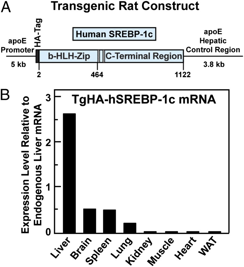
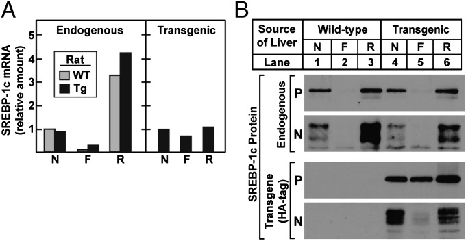
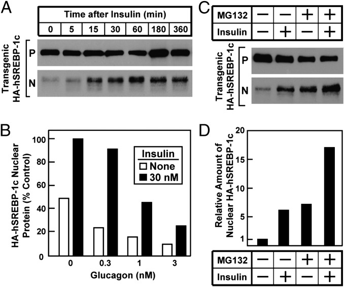
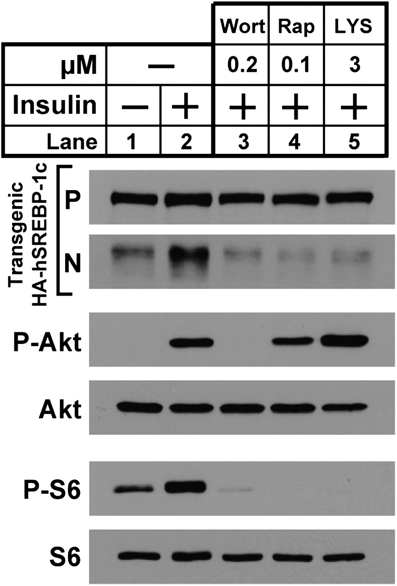
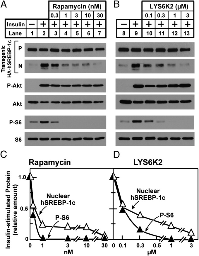
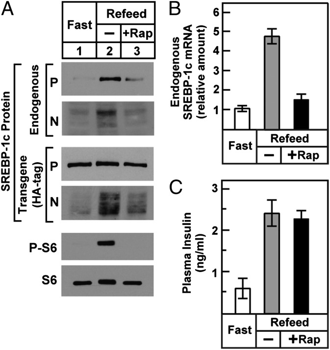
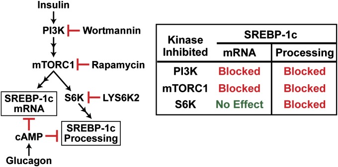
Insulin activates sterol regulatory element-binding protein-1c (SREBP-1c) in liver, thereby increasing fatty acid and triglyceride synthesis. We created a line of transgenic rats that produce epitope-tagged human SREBP-1c in liver under control of the constitutive apolipoprotein E promoter/enhancer. This system allows us to dissect the pathway by which insulin stimulates SREBP-1c processing without interference by the insulin-mediated increase in SREBP-1c mRNA. Rats are used because freshly isolated rat hepatocytes respond much more robustly to insulin than do mouse hepatocytes. The data reveal that insulin-mediated stimulation of SREBP-1c processing requires the mechanistic target of rapamycin complex 1 (mTORC1), which also is required for insulin-mediated SREBP-1c mRNA induction. However, in contrast to mRNA induction, insulin stimulation of SREBP-1c processing is blocked by an inhibitor of p70 S6-kinase. The data indicate that the pathways for insulin enhancement of SREBP-1c mRNA and proteolytic processing diverge after mTORC1. Stimulation of processing requires the mTORC1 target p70 S6-kinase, whereas induction of mRNA bypasses this enzyme. Insulin stimulation of both processes is blocked by glucagon. The transgenic rat system will be useful in further defining the molecular mechanism for insulin stimulation of lipid synthesis in liver in normal and diabetic states.


Conflict of interest statement

The authors declare no conflict of interest.

Figures

Fig. 1.
Fig. 1.
Generation of transgenic rats expressing HA-tagged full-length human SREBP-1c in liver. (A) The transgene construct contains a cDNA fragment encoding HA-tagged full-length human SREBP-1c (amino acids 2–1122) under control of human apoE promoter and its hepatic control region. (B) Tissue distribution of human HA-hSREBP-1c transgene. Male TgHA-hSREBP-1c rats (2–3 mo old) that were fed a chow diet ad libitum were killed 6 h into the dark cycle. Equal amounts of total RNA from the indicated tissues of four transgenic rats were pooled and subjected to real-time PCR. Each value represents the amount of transgenic human SREBP-1c mRNA in the indicated tissue relative to that of endogenous rat SREBP-1c mRNA in the liver, which is arbitrarily defined as 1. The cycle threshold (Ct) values for endogenous and transgenic SREBP-1c in the liver were 24.4 and 23.0, respectively.
Fig. 2.
Fig. 2.
Effect of fasting and refeeding on the hepatic levels of endogenous and transgenic SREBP-1c mRNAs and proteins in WT and TgHA-hSREBP-1c rats. Male WT and transgenic TgHA-hSREBP-1c (Tg) littermates (2–3 mo old, four rats per group) were subjected to fasting and refeeding as described in Materials and Methods. The nonfasted group (N) was fed a chow diet ad libitum, the fasted group (F) was fasted for 48 h, and the re-fed group (R) was fasted for 48 h and re-fed a high-carbohydrate diet for 6 h before study. (A) Equal amounts of total RNA from livers of four rats were pooled and subjected to real-time PCR. For the endogenous rat SREBP-1c mRNA, each value represents the amount relative to the amount in the nonfasted WT rats, which was arbitrarily defined as 1 (Ct value, 24.2). For the transgenic human SREBP-1c mRNA, the level in the nonfasted transgenic rats was arbitrarily defined as 1 (Ct value, 23.2). (B) Liver membrane and nuclear extract fractions were prepared individually and pooled from four rats per group. The mean values for the total amount of protein in the 24 membrane and nuclear extract fractions were 2,459 and 458 μg, respectively. Aliquots of membrane (30 μg) and nuclear extract (30 μg) fractions were subjected to 8% SDS/PAGE and immunoblot analysis with both anti-rat SREBP-1 and anti-HA antibodies to detect membrane-bound precursor (P) and cleaved nuclear (N) forms of endogenous rat SREBP-1 and HA-tagged transgenic human SREBP-1c, respectively. The exposure times for the autoradiograms were 1 s for all precursors, 10 s for transgenic nuclear extract, and 60 s for endogenous nuclear extracts.
Fig. 3.
Fig. 3.
Insulin-mediated stimulation of proteolytic processing of transgenic SREBP-1c in primary rat hepatocytes. Hepatocytes from nonfasted TgHA-hSREBP-1c rats were prepared and plated on day 0 as described in Materials and Methods. (A) Time course of insulin effect. On day 1, the cells were left untreated or treated with 100 nM insulin for the indicated time, harvested, and then pooled (three dishes of cells per sample) for immunoblot analysis of precursor (P) and nuclear (N) forms of transgenic HA-hSREBP-1c protein. The gels were exposed to film for 5 s. (B) Effect of glucagon. On day 1, cells were left untreated or were pretreated for 2 h with the indicated concentration of glucagon, after which the cells were left untreated or were treated with 30 nM insulin for 1 h before harvest as above. Immunoblot analysis of nuclear transgenic HA-hSREBP-1c was carried out and then was scanned and quantified by densitometry as described in SI Materials and Methods. The amount of the cleaved nuclear HA-hSREBP-1c protein in the cells without any treatment was arbitrarily set at 1. (C and D) Effect of proteasomal inhibitor. (C) On day 1, hepatocytes were left untreated or were treated 10 nM insulin for 1 h in the absence or presence of 10 μM MG132 (proteasomal inhibitor) and then were harvested for immunoblot analysis as in A. The gels were exposed to film for 5–10 s. (D) The gel of nuclear extract fractions (N) in C was scanned and quantified by densitometry as in B, with the amount of nuclear HA-hSREBP-1c protein in cells without any treatment arbitrarily set at 1.
Fig. 4.
Fig. 4.
Effects of protein kinase inhibitors on insulin-stimulated proteolytic processing of transgenic SREBP-1c in rat hepatocytes. Hepatocytes from nonfasted TgHA-hSREBP-1c rats were prepared and plated on day 0. On day 1, cells were left untreated or were pretreated for 3 h with the indicated protein kinase inhibitor: 0.2 μM wortmannin (lane 3), 0.1 μM rapamycin (lane 4), or 3 μM LYS6K2 (lane 5). After this preincubation, the cells were left untreated or were treated with 10 nM insulin for 30 min, harvested, and then pooled (three dishes of cells per sample). Each sample was subjected to immunoblot analyses of the following proteins: precursor (P) and nuclear (N) forms of transgenic HA-hSREBP-1c, phosphorylated Akt (P-Akt), total Akt, phosphorylated ribosomal protein S6 (P-S6), and total S6. Gels were exposed to film for 3–25 s.
Fig. 5.
Fig. 5.
mTORC1 and S6K are required for insulin to stimulate proteolytic processing of transgenic SREBP-1c in rat hepatocytes. Hepatocytes from nonfasted TgHA-hSREBP-1c rats were prepared and plated on day 0. On day 1, cells were left untreated or were pretreated for 2 h with the indicated concentration of rapamycin or LYS6K2, after which the cells were left untreated or were treated with 30 nM insulin for 1 h, harvested, and then pooled (three dishes of cells per sample) for immunoblot analysis. (A and B) Samples were subjected to immunoblot analysis as in Fig. 4. (C and D) Quantification of immunoblots. The gels of nuclear extract fractions (N) of transgenic HA-hSREBP-1c protein and cytosol fractions of phosphorylated ribosomal protein S6 (P-S6) in A and B were scanned and quantified by densitometry. The amount of nuclear HA-hSREBP-1c or cytosol P-S6 protein in cells treated with insulin alone (lanes 2 and 9) was arbitrarily set at 1.
Fig. 6.
Fig. 6.
Effect of mTORC1 inhibition on SREBP-1c processing (A) and SREBP-1c transcription (B) in livers of TgHA-hSREBP-1c rats subjected to fasting and refeeding. Male transgenic rats (2–3 mo old, four rats per group) were fasted for 48 h or were fasted for 48 h and then re-fed a high-carbohydrate diet for 3 h before study. Animals in the re-fed group were injected i.p. with vehicle or 20 mg/kg rapamycin (+Rap) 1 h before refeeding. (A) Liver membrane, nuclear extract, and cytosol fractions were prepared individually and pooled (four rats per group). Membrane and nuclear extract fractions were subjected to immunoblot analysis to detect precursor (P) and nuclear (N) forms of HA-tagged transgenic human SREBP-1c and endogenous rat SREBP-1 as in Fig. 2. Immunoblot analysis of cytosolic phosphorylated ribosomal protein S6 (P-S6) and total S6 was carried out as described in SI Materials and Methods. Gels were exposed to film for 3–60 s. (B) Total liver RNA was prepared individually and subjected to real-time PCR. Each bar (mean ± SEM of data from four rats) represents the amount of endogenous SREBP-1c mRNA relative to that in the fasted group, which was arbitrarily defined as 1. (C) Plasma insulin levels. Insulin was measured in plasma obtained immediately after administration of anesthesia as described in Materials and Methods. Each bar represents mean ± SEM of data from four rats.
Fig. 7.
Fig. 7.
Pathways for insulin stimulation of SREBP-1c mRNA and proteolytic processing diverge after mTORC1. Both processes are inhibited by glucagon acting through cAMP.

Comment in

Similar articles

Cited by

References

    1. Browning JD, Horton JD. Molecular mediators of hepatic steatosis and liver injury. J Clin Invest. 2004;114:147–152. - PMC - PubMed
    1. Leavens KF, Birnbaum MJ. Insulin signaling to hepatic lipid metabolism in health and disease. Crit Rev Biochem Mol Biol. 2011;46:200–215. - PubMed
    1. Horton JD, Goldstein JL, Brown MS. SREBPs: Activators of the complete program of cholesterol and fatty acid synthesis in the liver. J Clin Invest. 2002;109:1125–1131. - PMC - PubMed
    1. Krycer JR, Sharpe LJ, Luu W, Brown AJ. The Akt-SREBP nexus: Cell signaling meets lipid metabolism. Trends Endocrinol Metab. 2010;21:268–276. - PubMed
    1. Foretz M, Guichard C, Ferré P, Foufelle F. Sterol regulatory element binding protein-1c is a major mediator of insulin action on the hepatic expression of glucokinase and lipogenesis-related genes. Proc Natl Acad Sci USA. 1999;96:12737–12742. - PMC - PubMed

Publication types

MeSH terms