. 2012 Sep 11;109(37):14995-5000.
doi: 10.1073/pnas.1207516109. Epub 2012 Aug 27.

Interactions between the termini of lumen enzymes and shell proteins mediate enzyme encapsulation into bacterial microcompartments

Affiliations

Interactions between the termini of lumen enzymes and shell proteins mediate enzyme encapsulation into bacterial microcompartments

Chenguang Fan et al. Proc Natl Acad Sci U S A. .

Abstract

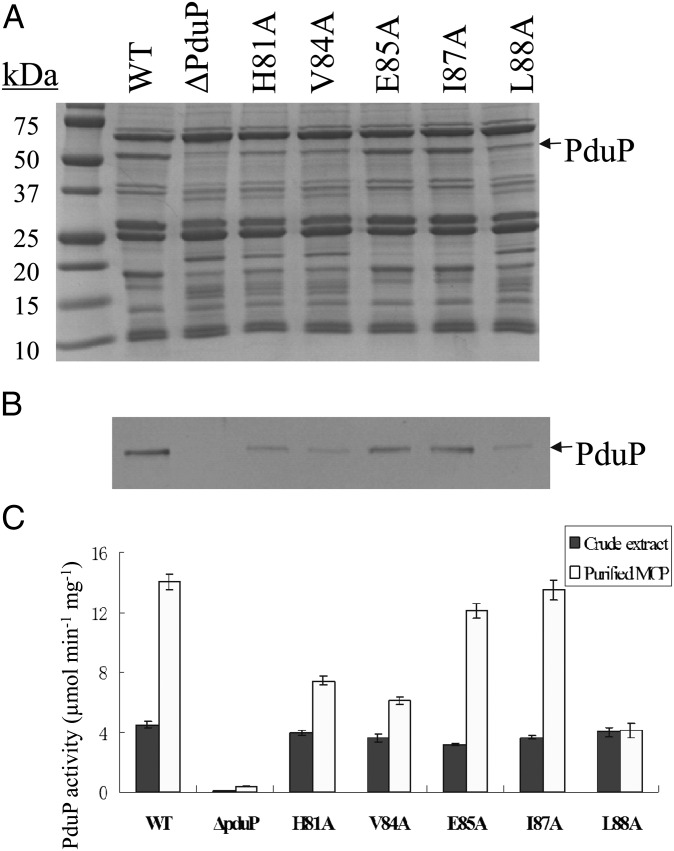
Bacterial microcompartments (MCPs) are a widespread family of proteinaceous organelles that consist of metabolic enzymes encapsulated within a protein shell. For MCPs to function specific enzymes must be encapsulated. We recently reported that a short N-terminal targeting sequence of propionaldehyde dehydrogenase (PduP) is necessary and sufficient for the packaging of enzymes into a MCP that functions in 1,2-propanediol (1,2-PD) utilization (Pdu) by Salmonella enterica. Here we show that encapsulation is mediated by binding of the PduP targeting sequence to a short C-terminal helix of the PduA shell protein. In vitro studies indicated binding between PduP and PduA (and PduJ) but not other MCP shell proteins. Alanine scanning mutagenesis determined that the key residues involved in binding are E7, I10, and L14 of PduP and H81, V84, and L88 of PduA. In vivo targeting studies indicated that the binding between the N terminus of PduP and the C terminus of PduA is critical for encapsulation of PduP within the Pdu MCP. Structural models suggest that the N terminus of PduP and C terminus of PduA both form helical structures that bind one another via the key residues identified by mutagenesis. Cumulatively, these results show that the N-terminal targeting sequence of PduP promotes its encapsulation by binding to MCP shell proteins. This is a unique report determining the mechanism by which a MCP targeting sequence functions. We propose that specific interactions between the termini of shell proteins and lumen enzymes have general importance for guiding the assembly and the higher level organization of bacterial MCPs.


Conflict of interest statement

The authors declare no conflict of interest.

Figures

Fig. 1.
Fig. 1.
Model for Pdu microcompartment. The hexagonal frame indicates the shell of the Pdu microcompartment, which is composed of nine different polypeptides (PduABB'JKMNTU). Encapsulated within the shell are enzymes for the activation of cob(III)alamin to coenzyme B12 (PduGH-S-O) as well as three 1,2-PD degradative enzymes: coenzyme B12-dependent diol dehydratase (PduCDE), propionaldehyde dehydrogenase (PduP), and 1-propanol dehydrogenase (PduQ). The proposed function of the Pdu MCP is to sequester propionaldehyde and channel it to downstream enzymes to prevent toxicity and DNA damage. A key requirement of this model is that specific enzymes are encapsulated within the protein shell. Enzymes not reported to be components of purified MCPs are positioned outside the MCP in the cytoplasm of the cell: phosphotransacylase (PduL) and propionate kinase (PduW).
Fig. 2.
Fig. 2.
SDS-PAGE analysis of His-tag pull downs used to test binding of PduA to PduP. Fractions that eluted from a Ni-NTA column at high imidazole concentrations are shown. Coelution of an untagged protein with a His-tagged bait indicates binding. M, molecular markers. Lane 1: PduP-His6 and PduA; lane 2: truncated PduP-His6 and PduA; lane 3: PduP-His6 and truncated PduA; lane 4: PduP and His6-PduA; lane 5: truncated PduP and His6-PduA; lane 6: PduP and truncated His6-PduA. A total of 5 μg of proteins was loaded in each lane. P, PduP; P′, truncated PduP, which has residues 2–18 deleted. A,PduA; A′, truncated PduA, which has 14 C-terminal amino acids deleted.
Fig. 3.
Fig. 3.
Results for the alanine scanning mutagenesis of the N terminus of PduP. (A) SDS-PAGE and (B) Western blotting of purified MCPs. M, molecular markers; no insert, ΔpduP/plac22; WT, ΔpduP/plac22-pduP. Substitutions introduced into the targeting sequence of PduP are labeled for each lane. SDS-PAGE gel and Western blotting are aligned for each variant. A total of 10 μg of purified MCPs was loaded in each lane. (C) PduP enzyme activities for the cell extracts and purified MCPs. Dark columns represent the activities in crude extracts; light columns represent the activities in purified MCPs. Means and SEs are based on three replicates. (D) Multiple sequence alignment of the N-terminal regions of representative PduP homologs from different organisms. The National Center for Biotechnology Information gene accession identifications, beginning from the top entry, are 16765381, 365906594, 283832579, 311279056, 218549353, 238784333, 317491711, 365834525, and 237808453. *Three key amino acids for targeting PduP into the lumen of MCPs.
Fig. 4.
Fig. 4.
Results for the site-directed mutagenesis of the C terminus of PduA. (A) SDS-PAGE. (B) Western blotting of purified MCPs. M, molecular markers. Substitutions are labeled for each lane. SDS-PAGE gel and Western blotting are aligned for each variant. Ten milligrams of purified MCPs was loaded in each lane. (C) PduP enzyme activities for the cell extracts and purified MCPs. Dark columns represent the activities in crude extracts; light columns represent the activities in purified MCPs. Means and SEs are based on three replicates.
Fig. 5.
Fig. 5.
Structure model of N terminus of PduP and its interaction with PduA. (A) Structural model of N terminus of PduP predicted by the PEPstr server. (B) Structure model of N terminus of PduP binding with the hexamer of PduA predicted by the RosettaDock server. Cyan color indicates the N terminus of PduP; gray, red, orange, golden, yellow, and blue color indicate the subunits of the PduA hexamer. (C) Structure model of N terminus of PduP binding with the monomer of PduA predicted by the RosettaDock server. Six key residues for binding are labeled.

Similar articles

Cited by

References

    1. Kerfeld CA, Heinhorst S, Cannon GC. Bacterial microcompartments. Annu Rev Microbiol. 2010;64:391–408. - PubMed
    1. Yeates TO, Kerfeld CA, Heinhorst S, Cannon GC, Shively JM. Protein-based organelles in bacteria: Carboxysomes and related microcompartments. Nat Rev Microbiol. 2008;6:681–691. - PubMed
    1. Price GD, Badger MR, Woodger FJ, Long BM. Advances in understanding the cyanobacterial CO2-concentrating-mechanism (CCM): Functional components, Ci transporters, diversity, genetic regulation and prospects for engineering into plants. J Exp Bot. 2008;59:1441–1461. - PubMed
    1. Cheng S, Liu Y, Crowley CS, Yeates TO, Bobik TA. Bacterial microcompartments: their properties and paradoxes. Bioessays. 2008;30:1084–1095. - PMC - PubMed
    1. Bobik TA. Polyhedral organelles compartmenting bacterial metabolic processes. Appl Microbiol Biotechnol. 2006;70:517–525. - PubMed

Publication types

LinkOut - more resources