. 2012 Sep 18;109(38):E2543-50.
doi: 10.1073/pnas.1205664109. Epub 2012 Aug 27.

Pathogenic bacterium Helicobacter pylori alters the expression profile of p53 protein isoforms and p53 response to cellular stresses

Affiliations

Pathogenic bacterium Helicobacter pylori alters the expression profile of p53 protein isoforms and p53 response to cellular stresses

Jinxiong Wei et al. Proc Natl Acad Sci U S A. .

Abstract

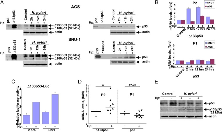
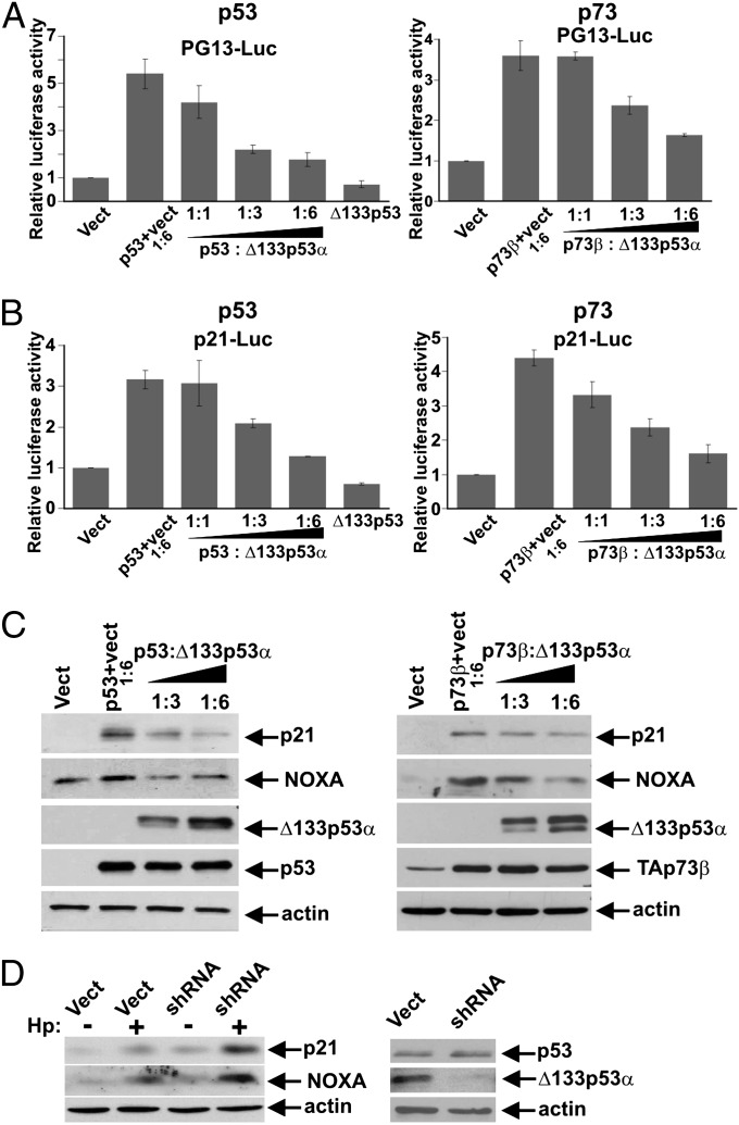
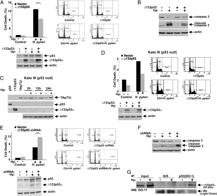
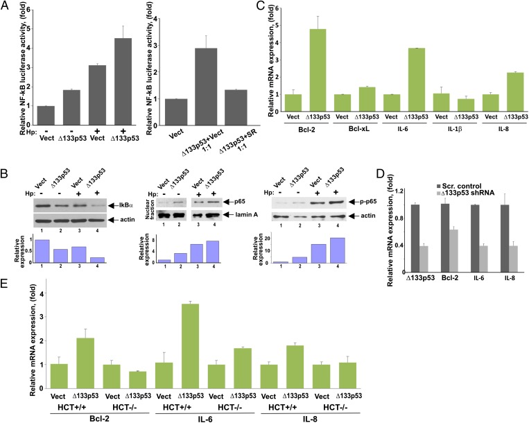
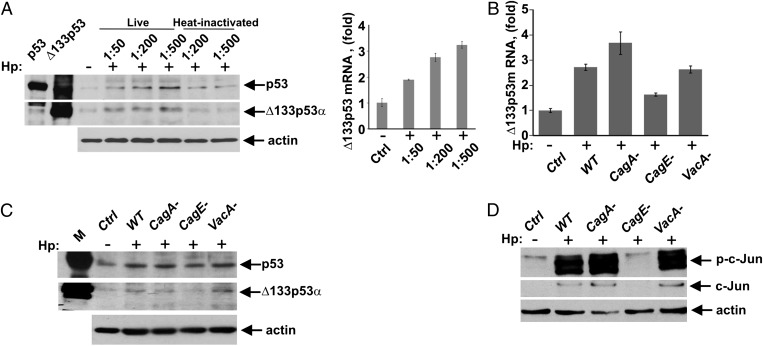
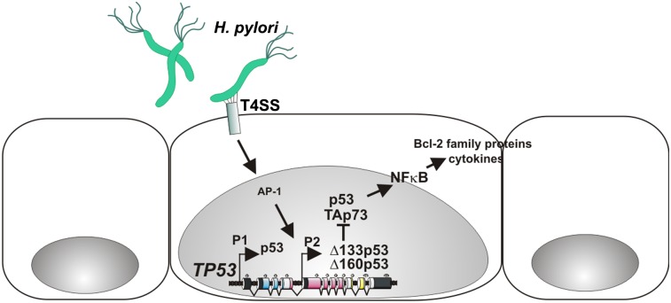
The p53 protein plays a central role in the prevention of tumorigenesis. Cellular stresses, such as DNA damage and aberrant oncogene activation, trigger induction of p53 that halts cellular proliferation and allows cells to be repaired. If cellular damage is beyond the capability of the repair mechanisms, p53 induces apoptosis or cell cycle arrest, preventing damaged cells from becoming cancerous. However, emerging evidence suggests that the function of p53 needs to be considered as isoform-specific. Here, we report that the expression profile of p53 can be shifted toward inhibitory p53 isoforms by the pathogenic bacterium Helicobacter pylori, which is known for its strong association with gastric cancer and gastric mucosa-associated lymphoid tissue lymphoma. We found that interaction of H. pylori with gastric epithelial cells, mediated via the cag pathogenicity island, induces N-terminally truncated Δ133p53 and Δ160p53 isoforms in human cells. Induction of an orthologous p53 isoform, Δ153p53, was also found in H. pylori-infected Mongolian gerbils. The p53 isoforms inhibit p53 and p73 activities, induce NF-κB, and increase survival of infected cells. Expression of Δ133p53, in response to H. pylori infection, is regulated by phosphorylation of c-Jun and activation of activator protein-1-dependent transcription. Together, these results provide unique insights into the regulation of p53 protein and may contribute to the understanding of tumorigenesis associated with H. pylori.


Conflict of interest statement

The authors declare no conflict of interest.

Figures

Fig. 1.
Fig. 1.
Truncated p53 isoforms are up-regulated by H. pylori. (A) Protein lysates were prepared from control (−) AGS cells (Upper) and SNU-1 cells (Lower) or from those co-cultured (+) with H. pylori strain J166 for the indicated time and analyzed for expression of p53 and Δ133p53 (Δ160p53) isoforms by Western blotting using DO-1 (Right) and DO-11 (Left) antibodies, respectively. (B) Bar graph represents qRT-PCR analyses of p53 transcripts produced from the P2 (Upper) and P1 (Lower) promoters in SNU-1 and AGS cells co-cultured with H. pylori for the indicated time. Data are normalized to HPRT1 mRNA expression. The mRNA expression of p53 isoforms in uninfected cells is arbitrarily set at 1. (C) SNU-1 cells transfected with a luciferase reporter (Δ133p53-Luc) were co-cultured with H. pylori (Hp) for the indicated time and analyzed using a dual-luciferase reporter assay. (D) Expression of p53 transcripts derived from the P1 and P2 promoters in the gastric mucosa harvested from gerbils infected with H. pylori strain 7.13 for 3 d. H. pylori infection leads to increased expression of Δ153p53 mRNA. *P < 0.05 vs. uninfected control. (E) Expression analysis of p53 and Δ153p53 proteins in gerbils infected with H. pylori strain 7.13 for 3 d using Western blotting with DO-1 and CM1 antibodies, respectively. p53 and Δ153p53 protein migrated as bands with molecular masses of 52 kDa and 32 kDa.
Fig. 2.
Fig. 2.
Δ133p53 suppresses p53 and p73 transcriptional activities. (A) Transcriptional activities of p53 (Left) and p73 (Right) were determined using PG13-Luc reporter in SNU-1 cells following cotransfection with Δ133p53 at the indicated molar ratios. Luciferase activity in cells transfected with an empty vector (Vect) is arbitrarily set at 1. (B) Same as in A, but p21-pWWP-Luc reporter was used. (C) p53-null cells, Kato III, were cotransfected with Δ133p53 and p53 (Left) or p73 (Right) at the indicated ratios and analyzed for expression of p21 and NOXA proteins by Western blotting. (D) Expression of p21 and NOXA proteins was analyzed in SNU-1 cells, in which levels of endogenous Δ133p53 isoform were suppressed with shRNA. (Right) shRNA plasmid selectively targets the ∆133p53 isoform but not p53.
Fig. 3.
Fig. 3.
Δ133p53 protein increases cell survival of H. pylori-infected cells. (A) SNU-1 cells stably transfected with Δ133p53 or empty vector were co-cultured with H. pylori (Hp) strain J166 for 24 h and analyzed for cell death by flow cytometry using propidium iodide (PI) staining. The percentage of cells in sub-G1 is shown. The Δ133p53 transfection significantly increases survival of H. pylori-infected cells (**P = 0.002; n = 3). (Lower) Expression of Δ133p53 protein in analyzed cells is shown. (B) Same as in A, but apoptosis was analyzed by Western blotting with antibodies recognizing uncleaved and cleaved caspase 3. (C) p73 is strongly up-regulated by H. pylori in p53-null Kato III cells. Cells were co-cultured with H. pylori strain J166 and analyzed for expression of TAp73β, p53, and Δ133p53 proteins at the indicated time using p73, DO-1, and DO-11 antibodies. (D) Same as in A, but Kato III cells were analyzed. (Lower) Expression of exogenous Δ133p53 protein. (E) SNU-1 cells, stably transfected with either shRNA against Δ133p53 or scrambled control vector, were co-cultured with H. pylori strain J166 for 24 h and analyzed as described in A. Down-regulation of Δ133p53 significantly increases cell death induced by H. pylori (**P = 0.009; n = 3). (Lower) Expression of endogenous Δ133p53 protein is shown. (F) Same as in E, but apoptosis was analyzed by Western blotting with antibodies recognizing uncleaved and cleaved caspase 3. (G) Δ133p53 protein physically interacts with p53. Protein lysates from SNU-1 cells co-cultured with H. pylori (+) for 12 h or left uninfected (−) were immunoprecipitated with either p53 (DO-1) antibody or nonspecific mouse IgG (N/S). The p53–Δ133p53 protein complexes were analyzed by Western blotting with the p53 (DO-11) antibody, which recognizes the Δ133p53 isoform. Equal antibody loading was verified by detecting light chains of antibodies.
Fig. 4.
Fig. 4.
Δ133p53 protein regulates NF-κB activity. (A) NF-κB activity was assessed in SNU-1 cells transfected with either Δ133p53 expression vector (Δ133p53) or empty control vector (Vect) and co-cultured with H. pylori (Hp) (+) for 8 h or left uninfected (−). NF-κB activity was assessed using the pNFκB-Luc reporter. (Right) Reporter analysis was repeated in the presence of the NF-κB inhibitor, IκB SR. SNU-1 cells were cotransfected with Δ133p53 vector and SR (or empty vector) at a 1:1 ratio. (B) SNU-1 cells stably transfected with Δ133p53 or empty control vector were co-cultured with H. pylori for 3 h and analyzed for IκBα protein (Left) as well as nuclear localization of p65 (RelA) protein (Center) and its phosphorylation at serine 536 (Right) by Western blotting. The graphs represent the results of densitometric analysis of immunoblots and depict actin (or lamin A)-normalized protein expression. (C) SNU-1 cells were transfected with either Δ133p53 or empty control vector for 48 h and analyzed for mRNA expression of the indicated NF-κB target genes by qPCR. (D) AGS cells were stably transfected with a specific shRNA against Δ133p53 or scrambled (Scr.) control vector and were analyzed for mRNA expression of Bcl-2, IL-6, and IL-8 by qPCR. (E) Same as in C, but isogenic cell lines HCT116+/+ (WT p53) and HCT116−/− (p53-null) were used.
Fig. 5.
Fig. 5.
AP-1 transcription factor is involved in the regulation of p53 isoforms. (A) Location of AP-1 binding site in the p53 P2 promoter are shown. The activity of luciferase reporters controlled by the 1.5-kb P2 promoter or its AP-1 binding site mutant was analyzed 24 h after transfection of c-Jun. Luciferase activity in cells transfected with empty vector (Vect) is arbitrarily set at 1. (Lower) Four nucleotides were mutated to inactivate the AP-1 binding site. (B) Protein and mRNA levels of Δ133p53 were determined in SNU-1 cells transfected with c-Jun or empty control vector. (C) DNA binding of c-Jun to the native P2 p53 gene promoter was analyzed by ChIP in SNU-1 cells co-cultured with H. pylori strain J166 for 12 h (+) or left uninfected (−). An antibody recognizing phospho-c-Jun (Ser63) was used for detection of the c-Jun binding. (D) Protein and mRNA levels of Δ133p53 were analyzed in SNU-1 cells transfected with either c-Jun siRNA or scrambled control siRNA for 48 h and co-cultured with H. pylori strain J166 for an additional 12 h.
Fig. 6.
Fig. 6.
Induction of p53 isoforms is mediated by the T4SS. (A) Protein lysates were prepared from control SNU-1 cells (−) or those co-cultured with H. pylori (Hp) (+) strain J166 at the indicated cell/bacteria ratios or with heat-inactivated bacteria and analyzed for expression of Δ133p53 protein by Western blotting. (Right) qPCR analysis of Δ133p53 transcripts in SNU-1 cells co-cultured with H. pylori is shown. Data are normalized to HPRT1 mRNA expression. The Δ133p53 mRNA level in uninfected cells is arbitrarily set at 1. (B) SNU-1 cells were co-cultured with WT H. pylori strain J166 or its isogenic cagA-, cagE-, or vacA-null mutants and analyzed for Δ133p53 mRNA by qPCR. SNU-1 cells were co-cultured with H. pylori as described in B for 12 h and analyzed for expression of p53 and Δ133p53 proteins (C), as well as c-Jun protein levels and its phosphorylation at position Ser63 (D), by Western blotting.
Fig. P1.
Fig. P1.
Regulation of p53 isoforms in H. pylori-infected cells. The p53 gene has the main P1 and alternative P2 promoters. Interaction of H. pylori with gastric epithelial cells, mediated by the cag-PAI, activates transcription from the P2 promoter and induces synthesis of N-terminally truncated ∆133p53 and ∆160p53 isoforms. The p53 isoforms inhibit the activities of p53 and p73, induce NF-κB activity, and increase survival of infected cells. The expression of ∆133p53 in response to H. pylori infection is regulated by activation of AP-1–dependent transcription. The transactivation, DNA binding, and oligomerization domains of p53 protein are shown in blue, red, and yellow, respectively.

Similar articles

Cited by

References

    1. Polk DB, Peek RM, Jr. Helicobacter pylori: Gastric cancer and beyond. Nat Rev Cancer 10:403–414. - PMC - PubMed
    1. Levine AJ, Hu W, Feng Z. The P53 pathway: What questions remain to be explored? Cell Death Differ. 2006;13:1027–1036. - PubMed
    1. Vousden KH, Prives C. Blinded by the Light: The Growing Complexity of p53. Cell. 2009;137:413–431. - PubMed
    1. Matsumoto Y, et al. Helicobacter pylori infection triggers aberrant expression of activation-induced cytidine deaminase in gastric epithelium. Nat Med. 2007;13:470–476. - PubMed
    1. Wei J, et al. Regulation of p53 tumor suppressor by Helicobacter pylori in gastric epithelial cells. Gastroenterology. 2010;139:1333–1343. - PMC - PubMed

Publication types