. 2012 Sep 11;109(37):14906-11.
doi: 10.1073/pnas.1211976109. Epub 2012 Aug 27.

Heritable yeast prions have a highly organized three-dimensional architecture with interfiber structures

Affiliations

Heritable yeast prions have a highly organized three-dimensional architecture with interfiber structures

Helen R Saibil et al. Proc Natl Acad Sci U S A. .

Abstract

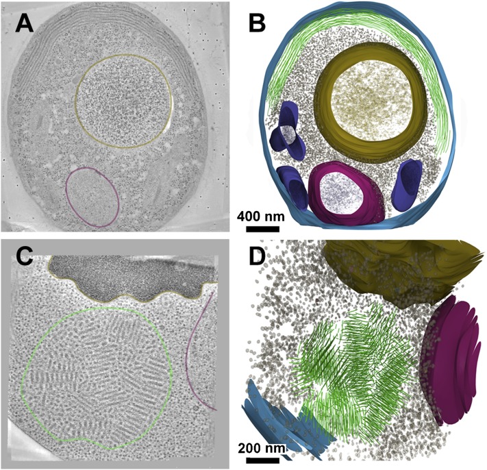
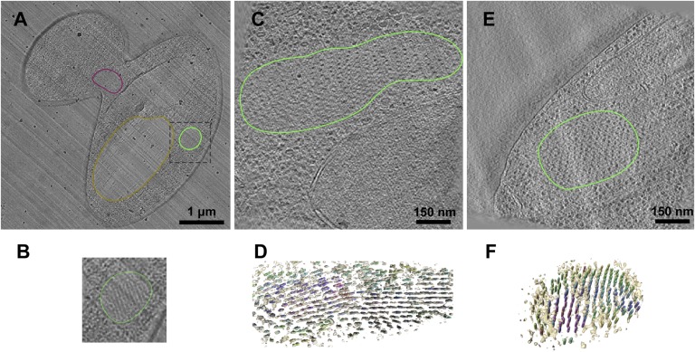
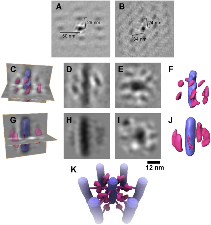
Yeast prions constitute a "protein-only" mechanism of inheritance that is widely deployed by wild yeast to create diverse phenotypes. One of the best-characterized prions, [PSI(+)], is governed by a conformational change in the prion domain of Sup35, a translation-termination factor. When this domain switches from its normal soluble form to an insoluble amyloid, the ensuing change in protein synthesis creates new traits. Two factors make these traits heritable: (i) the amyloid conformation is self-templating; and (ii) the protein-remodeling factor heat-shock protein (Hsp)104 (acting together with Hsp70 chaperones) partitions the template to daughter cells with high fidelity. Prions formed by several other yeast proteins create their own phenotypes but share the same mechanistic basis of inheritance. Except for the amyloid fibril itself, the cellular architecture underlying these protein-based elements of inheritance is unknown. To study the 3D arrangement of prion assemblies in their cellular context, we examined yeast [PSI(+)] prions in the native, hydrated state in situ, taking advantage of recently developed methods for cryosectioning of vitrified cells. Cryo-electron tomography of the vitrified sections revealed the prion assemblies as aligned bundles of regularly spaced fibrils in the cytoplasm with no bounding structures. Although the fibers were widely spaced, other cellular complexes, such as ribosomes, were excluded from the fibril arrays. Subtomogram image averaging, made possible by the organized nature of the assemblies, uncovered the presence of an additional array of densities between the fibers. We suggest these structures constitute a self-organizing mechanism that coordinates fiber deposition and the regulation of prion inheritance.


Conflict of interest statement

The authors declare no conflict of interest.

Figures

Fig. 1.
Fig. 1.
Three-dimensional analysis of ring and dot structures in freeze-substituted yeast cells. (A) Section through a tomogram of a ring cell, with about half of the ring in the plane of the section. The nucleus is outlined in magenta and the vacuole in brown. (B) Rendered 3D model from serial tomograms of this ring cell. Ring fibers are in green; membrane, blue; nucleus, magenta; vacuole, brown; mitochondria, purple; and large complexes (presumably ribosomes) as gray dots. The large complexes in the cytoplasm are excluded from the fiber area. (C) Section through a dot cell tomogram, colored as above. (D) Rendered 3D model of the dot from serial tomograms, showing the mosaic arrangement of fiber bundles. (Coloring as in B.)
Fig. 2.
Fig. 2.
Cryosections of dot and ring cells. (A) Overview section of a budding yeast cell with a dot aggregate, outlined as in Fig. 1. The cell is compressed along the direction of sectioning, indicated by the knife marks (diagonal streaks). (B) Enlargement of the dot region. (C) Tomogram slice of a ring, adjacent to the nucleus. (D) Rendered views of the fibers in the ring, colored by their cross-correlation values, with the most ordered regions in purple and least ordered in yellow. (E) Tomogram slice of a dot cell. (F) Rendered view of the fibril array in the dot, colored as in D.
Fig. 3.
Fig. 3.
Subtomogram averages of the fiber arrays. Subregions of the cryotomograms were aligned and averaged to reveal additional structures surrounding the fibers. (A) Section through the array of ring fibers, with the region in the center showing the averaged density surrounded by the array of fibers. (B) Equivalent view of the dot fiber array. The variable spacing might arise from sectioning artifacts and/or oblique fibril orientations. (C) Rendered views of the ring average superposed on density sections. (D and E) Long- and cross-sections of the averaged ring fiber. (F) Rendered view of the averaged fiber with the surrounding structures. The fiber is shown in purple and the additional structures in pink. (G) Rendered view of the dot average superposed on density sections as above. (H and I) Long- and cross-sections of the dot fiber average. (J) Rendered view of the averaged fiber with the surrounding structures, colored as in F. The differences in dot and ring structures most likely arise from the lower resolution obtained for the dot average. The defocus needed to obtain sufficient contrast in cryosections causes a white halo around high-density (dark) features; therefore, obscuring regions of contact between the central fiber and surrounding structures. (K) Illustration of the deduced arrangement of cross-bridges between the fibers. Fibers are in purple, and cross-bridge structures are in pink.
Fig. 4.
Fig. 4.
Fluorescence microscopy showing colocalization of chaperones with NM-YFP in ring and dot structures. DIC, differential interference contrast images. The prion fibers are labeled with YFP (NM-YFP), and the chaperones are labeled with CFP. The results show that Hsp104, the Hsp70 proteins Ssa1/2 and Sse1, and the Hsp40 protein Sis1 are all found in dots and rings.

References

    1. Collinge J, Clarke AR. A general model of prion strains and their pathogenicity. Science. 2007;318:930–936. - PubMed
    1. Polymenidou M, Cleveland DW. The seeds of neurodegeneration: Prion-like spreading in ALS. Cell. 2011;147:498–508. - PMC - PubMed
    1. Halfmann R, Lindquist S. Epigenetics in the extreme: Prions and the inheritance of environmentally acquired traits. Science. 2010;330:629–632. - PubMed
    1. Tuite MF, Serio TR. The prion hypothesis: From biological anomaly to basic regulatory mechanism. Nat Rev Mol Cell Biol. 2010;11:823–833. - PMC - PubMed
    1. Shorter J, Lindquist S. Prions as adaptive conduits of memory and inheritance. Nat Rev Genet. 2005;6:435–450. - PubMed

Publication types

LinkOut - more resources