. 2012 Sep 18;109(38):15502-7.
doi: 10.1073/pnas.1206446109. Epub 2012 Aug 27.

Low-density lipoprotein receptor overexpression enhances the rate of brain-to-blood Aβ clearance in a mouse model of β-amyloidosis

Affiliations

Low-density lipoprotein receptor overexpression enhances the rate of brain-to-blood Aβ clearance in a mouse model of β-amyloidosis

Joseph M Castellano et al. Proc Natl Acad Sci U S A. .

Abstract

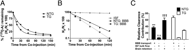
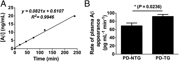
The apolipoprotein E (APOE)-ε4 allele is the strongest genetic risk factor for late-onset, sporadic Alzheimer's disease, likely increasing risk by altering amyloid-β (Aβ) accumulation. We recently demonstrated that the low-density lipoprotein receptor (LDLR) is a major apoE receptor in the brain that strongly regulates amyloid plaque deposition. In the current study, we sought to understand the mechanism by which LDLR regulates Aβ accumulation by altering Aβ clearance from brain interstitial fluid. We hypothesized that increasing LDLR levels enhances blood-brain barrier-mediated Aβ clearance, thus leading to reduced Aβ accumulation. Using the brain Aβ efflux index method, we found that blood-brain barrier-mediated clearance of exogenously administered Aβ is enhanced with LDLR overexpression. We next developed a method to directly assess the elimination of centrally derived, endogenous Aβ into the plasma of mice using an anti-Aβ antibody that prevents degradation of plasma Aβ, allowing its rate of appearance from the brain to be measured. Using this plasma Aβ accumulation technique, we found that LDLR overexpression enhances brain-to-blood Aβ transport. Together, our results suggest a unique mechanism by which LDLR regulates brain-to-blood Aβ clearance, which may serve as a useful therapeutic avenue in targeting Aβ clearance from the brain.


Conflict of interest statement

Conflict of interest statement: D.M.H. cofounded C2N Diagnostics. Some measurements of samples were assessed by employees of C2N Diagnostics. D.M.H. is on the scientific advisory boards of Satori and En Vivo, and consults for Pfizer, Bristol-Myers Squibb, and Innogenetics.

Figures

Fig. 1.
Fig. 1.
LDLR enhances clearance of radiolabeled Aβ from brain. (A) Percentage remaining for 12 nM [125I]Aβ40 microinjected in ISF of caudate-putamen in NTG (■) and TG (○) mice killed at various time points. Percentage recovery was calculated from Eq. S1 (SI Materials and Methods). (B) Time-dependent clearance of [125I]Aβ40 by passive ISF bulk flow (♦) and across the BBB after correction for degradation within brain by TCA precipitation method (NTG, ■; TG, ○) calculated from data in Fig. 1A and Eq. S4 (SI Materials and Methods). (C) Using fractional rate constants calculated in Table S1, relative contributions of degradation-corrected clearance of [125I]Aβ40 by the BBB and ISF bulk flow, as well as retention within brain, were calculated for NTG (black bars) and TG (white bars) mice. Each component is indicated with a plus sign (+). Complete time course includes 32–41 mice (n = 4–6 mice per time point for each group). Values in A and C are represented as mean ± SEM. When two-way ANOVA was significant (with genotype and component as factors), differences among clearance components were assessed using Tukey’s post hoc test for multiple comparisons. ***P < 0.001, % BBB for NTG vs. TG. †††P < 0.001, % brain retention for NTG vs. TG. n.s., no significant difference between ISF bulk flow components between NTG and TG.
Fig. 2.
Fig. 2.
LDLR overexpression in PDAPP mice markedly decreases brain Aβ/amyloid deposition and apoE levels. (A) Representative coronal brain sections from 10-mo-old, sex-matched PDAPP+/− mice expressing normal levels of LDLR (PD-NTG), and PDAPP+/− mice overexpressing LDLR (PD-TG). Aβ immunostaining was performed using anti-Aβ antibody (biotinylated 3D6). (Scale bars, 300 μm.) (B) Quantification of the area of the hippocampus or cortex occupied by Aβ immunostaining (n = 9 mice per group). (C) Representative amyloid burden in coronal brain sections from 10-mo-old, sex-matched PD-NTG mice and PD-TG mice. Amyloid was visualized using the congophilic fluorescent dye, X-34. (Scale bars, 100 μm.) (D) Quantification of the area of hippocampus or cortex occupied by X-34 staining (n = 9–10 mice per group). In B and D, groups were compared using the Mann–Whitney U test. *P < 0.05, **P < 0.01, ***P < 0.001. (E) ApoE protein levels measured by sensitive sandwich ELISA in hippocampal and cortical homogenates from PD-NTG and PD-TG mice (at 3–4 mo of age to avoid confounding effects from amyloid plaque deposition; n = 9 mice per group). Differences between groups were assessed using two-tailed Student’s t test (with Welch's correction for E). ***P < 0.001. Values represent means ± SEM.
Fig. 3.
Fig. 3.
Intravenous HJ5.1 administration results in stable antibody steady-state levels in plasma without altering brain ISF Aβ metabolism. (A) Concentration of biotinylated HJ5.1 (HJ5.1B) in plasma collected by serial retro-orbital bleeds following intrajugular injection of HJ5.1B in PDAPP+/− mice (n = 4; 3–4 mo old). (B) In vivo microdialysis was performed in PDAPP+/− mice expressing normal levels of LDLR (PD-NTG) to monitor ISF Aβ1x levels during baseline sampling as well as the period following intrajugular administration of 250 μg HJ5.1 (n = 5; 3–4 mo old). (C) Mean effect of HJ5.1 treatment on ISF Aβ1x levels compared with mean baseline period preceding treatment. (D and E) Experiments in B and C were repeated in PDAPP+/− mice overexpressing LDLR (PD-TG) (n = 5; 3–4 mo old). (F) ISF Aβ1-x levels during the baseline period of microdialysis were compared between PD-NTG and PD-TG mice. Differences between groups were assessed by paired Student’s t test in C and E and Student’s t test in F. **P < 0.01. Values represent mean ± SEM.
Fig. 4.
Fig. 4.
Antibody-assisted plasma accumulation of brain Aβ reveals faster brain-to-blood appearance rate in PDAPP mice overexpressing LDLR. (A) Representative plasma accumulation experiment illustrating kinetics of brain-derived Aβ appearance in plasma collected by serial retro-orbital bleeds following HJ5.1 treatment. Appearance rates were calculated from the slopes of individual linear regressions, e.g., for A, 82.1 pg⋅mL−1⋅min−1. (B) Mean rate of Aβ appearance in PDAPP+/− mice expressing normal levels of LDLR (PD-NTG) or PDAPP+/− mice overexpressing LDLR (PD-TG) (n = 6–7 per group; 3.5–4.5 mo old). Difference between groups was analyzed using two-tailed Student’s t test. *P < 0.05. Values in B represent mean ± SEM.

Similar articles

Cited by

References

    1. Hardy J. A hundred years of Alzheimer’s disease research. Neuron. 2006;52:3–13. - PubMed
    1. Golde TE, Schneider LS, Koo EH. Anti-aβ therapeutics in Alzheimer’s disease: The need for a paradigm shift. Neuron. 2011;69:203–213. - PMC - PubMed
    1. Benilova I, Karran E, De Strooper B. The toxic Aβ oligomer and Alzheimer’s disease: An emperor in need of clothes. Nat Neurosci. 2012;15:349–357. - PubMed
    1. Mawuenyega KG, et al. Decreased clearance of CNS beta-amyloid in Alzheimer’s disease. Science. 2010;330:1774. - PMC - PubMed
    1. Verghese PB, Castellano JM, Holtzman DM. Apolipoprotein E in Alzheimer’s disease and other neurological disorders. Lancet Neurol. 2011;10:241–252. - PMC - PubMed

Publication types