. 2012 Sep 11;109(37):14983-8.
doi: 10.1073/pnas.1206641109. Epub 2012 Aug 27.

Resolvin E1 promotes phagocytosis-induced neutrophil apoptosis and accelerates resolution of pulmonary inflammation

Affiliations

Resolvin E1 promotes phagocytosis-induced neutrophil apoptosis and accelerates resolution of pulmonary inflammation

Driss El Kebir et al. Proc Natl Acad Sci U S A. .

Abstract

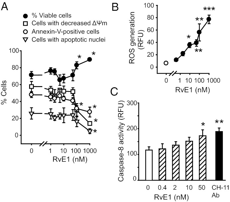
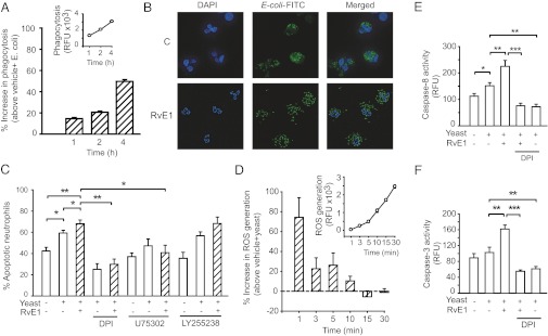
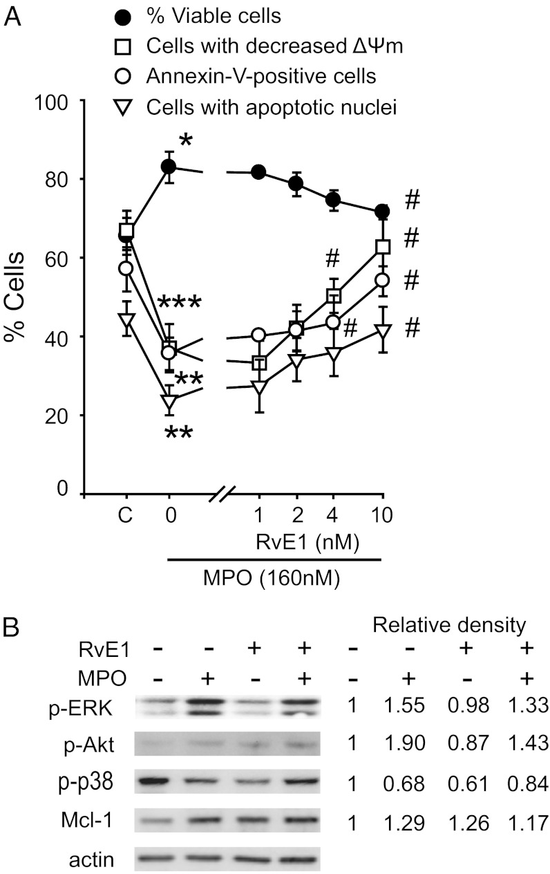
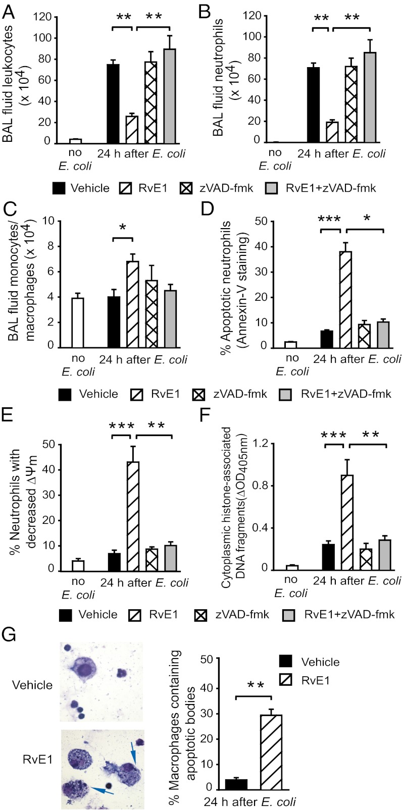
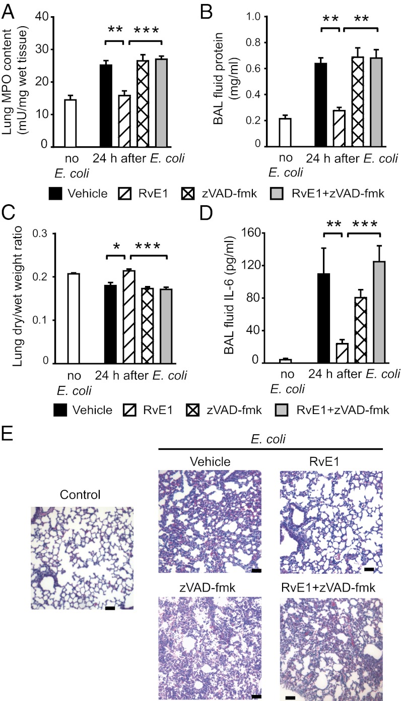
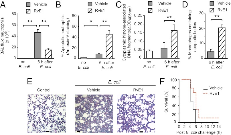
Inappropriate neutrophil activation contributes to the pathogenesis of acute lung injury (ALI). Apoptosis is essential for removal of neutrophils from inflamed tissues and timely resolution of inflammation. Resolvin E1 (RvE1) is an endogenous lipid mediator derived from the ω-3 polyunsaturated fatty acid eicosapentaenoic acid that displays proresolving actions. Because the balance of prosurvival and proapoptosis signals determines the fate of neutrophils, we investigated the impact of RvE1 on neutrophil apoptosis and the outcome of neutrophil-mediated pulmonary inflammation in mice. Culture of human neutrophils with RvE1 accelerated apoptosis evoked by phagocytosis of opsonized Escherichia coli or yeast. RvE1 through the leukotriene B(4) receptor BLT1 enhanced NADPH oxidase-derived reactive oxygen species generation and subsequent activation of caspase-8 and caspase-3. RvE1 also attenuated ERK and Akt-mediated apoptosis-suppressing signals from myeloperoxidase, serum amyloid A, and bacterial DNA, shifting the balance of pro- and anti-survival signals toward apoptosis via induction of mitochondrial dysfunction. In mice, RvE1 treatment enhanced the resolution of established neutrophil-mediated pulmonary injury evoked by intratracheal instillation or i.p. administration of live E. coli or intratracheal instillation of carrageenan plus myeloperoxidase via facilitating neutrophil apoptosis and their removal by macrophages. The actions of RvE1 were prevented by the pan-caspase inhibitor zVAD-fmk. These results identify a mechanism, promotion of phagocytosis-induced neutrophil apoptosis and mitigation of potent anti-apoptosis signals, by which RvE1 could enhance resolution of acute lung inflammation.


Conflict of interest statement

Conflict of interest statement: P.G. is president of Resolvyx Pharmaceuticals and retains founder stock in the company.

Figures

Fig. 1.
Fig. 1.
Effects of RvE1 on neutrophil apoptosis. Human neutrophils (5 × 10⁶ cells/mL) were cultured for 24 h with RvE1 and viability, mitochondrial transmembrane potential (ΔΨm) [chloromethyl-X-rosamine (CMXRos) staining] and apoptosis (annexin-V–FITC binding and nuclear DNA content) were assessed. (A) RVE1 at high concentrations suppresses intrinsic apoptosis. Data are means ± SEM (n = 4–11). *P < 0.05 vs. untreated. (B) To monitor ROS production, neutrophils were loaded with H2DCFDA (5 μM) and then left untreated (C, control) or challenged with RvE1 for 15 min. Data are means ± SEM (n = 4–7). *P < 0.05; **P < 0.01; ***P < 0.001 vs. untreated. (C) Caspase-8 activity was assessed at 4 h post-RvE1 with flow cytometry using FITC-labeled z-Ile-Glu(Ome)-Thr-Asp(Ome)-fluoromethylketone (z-IETD-fmk) as a substrate. The effect of the Fas-activating antibody (CH-11 Ab) is shown for comparison. (n = 4–7). *P < 0.05; **P < 0.01 vs. untreated.
Fig. 2.
Fig. 2.
RvE1 enhances phagocytosis-induced neutrophil apoptosis. (A and B) Human neutrophils (5 × 10⁶ cells/mL) were mixed with opsonized FITC-labeled E. coli at a ratio of 1:10 for 1, 2, and 4 h. Extracellular fluorescence was quenched with 0.2% trypan blue and intracellular fluorescence was analyzed with flow cytometry (A) or fluorescence microscopy (B). Inset: representative time course of neutrophil phagocytosis of bacteria. Results are expressed as percentage increase above vehicle plus E. coli (n = 4). (C) Neutrophils were cultured for 24 h with yeast (five yeast particles/neutrophil) with or without RvE1 (10 nM), U75302 (1 μM), LY255238 (1 μM), or DPI (20 μM), stained with acridine orange (10 μg/mL), and apoptosis was assessed by nuclear morphology (condensed or fragmented chromatin) under a fluorescence microscope. Data are means ± SEM (n = 5–6). *P < 0.05; **P < 0.01. (D) ROS generation. The effects of RvE1 are expressed as percentage increase above vehicle plus yeast. Inset: Time course of ROS production by phagocytosing neutrophils. Data are means ± SEM (n = 5). Caspase-8 (E) and caspase-3 (F) activity was assessed at 4 h post-RvE1 with flow cytometry using FITC-labeled z-IETD-fmk and acetyl-Asp-Glu-Val-Asp-fluoromethylketone (Ac-DEVD-fmk), respectively, as substrates. Data are means ± SEM (n = 6). *P < 0.05; **P < 0.01; ***P < 0.001.
Fig. 3.
Fig. 3.
RvE1 attenuates MPO suppression of neutrophil apoptosis. (A) Human neutrophils (5 × 10⁶ cells/mL) were cultured for 20 min with RvE1 then with MPO (160 nM) for 24 h. Viability, mitochondrial transmembrane potential (ΔΨm) (CMXRos staining), and apoptosis (annexin-V–FITC binding and nuclear DNA content) were then assessed. Data are means ± SEM (n = 4–7). *P < 0.05; **P < 0.01; ***P < 0.001 vs. untreated (C, control). #P < 0.01 vs. MPO-treated. (B) Impact on MAP kinases and Mcl-1. Neutrophils were lysed after culture with MPO (160 nM) ± RvE1 (10 nM) for 30 min (MAP kinases) or 1 h (for Mcl-1). Proteins were subjected to immunoblotting with antibodies to phosphorylated kinases, Mcl-1, or actin. Numerical values indicate relative density of bands normalized with density of actin bands. Results are representative of three separate experiments.
Fig. 4.
Fig. 4.
RvE1 enhances resolution of E. coli–evoked pneumonia. Six hours after intratracheal instillation of 107 live E. coli, mice were treated with vehicle or RvE1 (25 μg/kg, i.p.) and/or the pan-caspase inhibitor zVAD-fmk (10 μg/kg, i.p. three times at 4-h intervals). Mice were killed 24 h later and BAL fluid total leukocyte (A), neutrophil (B), and monocyte/macrophage counts (C), the percentage of annexin-V–positive (apoptotic) neutrophils (D), the percentage of neutrophils with decreased mitochondrial transmembrane potential (ΔΨm) (E), BAL cell cytoplasmic histone-associated DNA fragments (F), and the number of BAL fluid macrophages containing apoptotic bodies (arrows, Left) (G) were determined. Data are means ± SEM (n = 6–8 mice per group). *P < 0.05, **P < 0.01; ***P < 0.001.
Fig. 5.
Fig. 5.
RvE1 enhances resolution of E. coli–evoked pneumonia. Six hours after intratracheal instillation of 107 live E. coli, mice were treated with vehicle or RvE1 (25 μg/kg, i.p.) and/or the pan-caspase inhibitor zVAD-fmk (10 μg/kg, i.p. three times at 4-h intervals). Mice were killed 24 h later and lung myeloproxidase content (A), BAL fluid protein (B), lung dry-to-wet weight ratio (C), and BAL fluid IL-6 level (D) were determined. Data are means ± SEM (n = 6–8 mice per group). *P < 0.05, **P < 0.01; ***P < 0.001. (E) Lung tissue sections from naive mice (control), mice with inflammation evoked by E. coli, or mice treated with vehicle, RvE1, and/or zVAD-fmk for 24 h. Hematoxylin and eosin stain (H&E); scale bars: 100 μm.
Fig. 6.
Fig. 6.
RvE1 attenuates lung inflammation and prolongs survival of mice with E. coli peritonitis. One hour after i.p. injection of 2 × 10⁸ live E. coli, mice were treated with vehicle or RvE1 (25 μg/kg, i.p.). Mice were killed at 6-h post–E. coli and BAL fluid neutrophil counts (A), the percentage of annexin-V–positive (apoptotic) neutrophils (B), BAL fluid cell cytoplasmic histone-associated DNA fragments (C), and the percentage of macrophages containing apoptotic bodies (D) were determined. Data are means ± SEM (n = 6 mice per group). *P < 0.05, **P < 0.01. (E) Lung tissue sections from naive mice (control) and mice challenged with E. coli and treated with RvE1 or vehicle. H&E stain; scale bars: 100 μm. (F) Kaplan-Meier survival plots for mice challenged intraperitoneally with 109 live E. coli and treated with RvE1 or vehicle (n = 10 per group). P = 0.0315 by the log-rank test.

References

    1. Nathan C, Ding A. Nonresolving inflammation. Cell. 2010;140:871–882. - PubMed
    1. Serhan CN, Chiang N, Van Dyke TE. Resolving inflammation: Dual anti-inflammatory and pro-resolution lipid mediators. Nat Rev Immunol. 2008;8:349–361. - PMC - PubMed
    1. Ware LB, Matthay MA. The acute respiratory distress syndrome. N Engl J Med. 2000;342:1334–1349. - PubMed
    1. Savill JS, et al. Macrophage phagocytosis of aging neutrophils in inflammation. Programmed cell death in the neutrophil leads to its recognition by macrophages. J Clin Invest. 1989;83:865–875. - PMC - PubMed
    1. Savill J, Dransfield I, Gregory C, Haslett C. A blast from the past: Clearance of apoptotic cells regulates immune responses. Nat Rev Immunol. 2002;2:965–975. - PubMed

Publication types

MeSH terms

Substances