. 2012 Oct 19;287(43):36593-608.
doi: 10.1074/jbc.M112.365999. Epub 2012 Aug 27.

CCL2/CCR2 chemokine signaling coordinates survival and motility of breast cancer cells through Smad3 protein- and p42/44 mitogen-activated protein kinase (MAPK)-dependent mechanisms

Affiliations

CCL2/CCR2 chemokine signaling coordinates survival and motility of breast cancer cells through Smad3 protein- and p42/44 mitogen-activated protein kinase (MAPK)-dependent mechanisms

Wei Bin Fang et al. J Biol Chem. .

Abstract

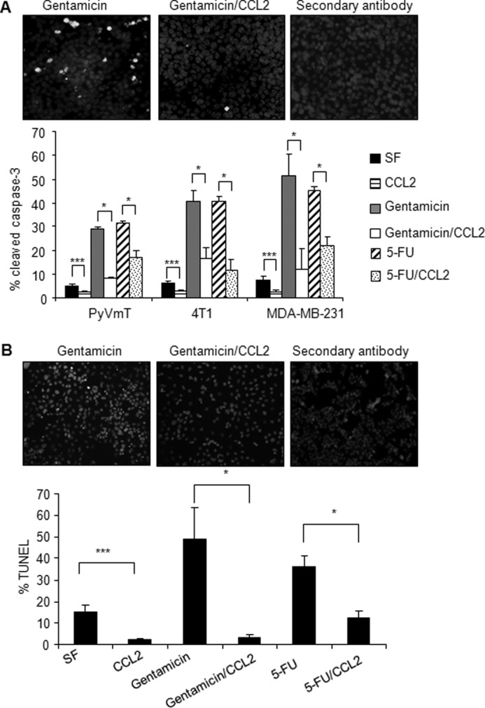
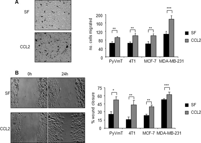
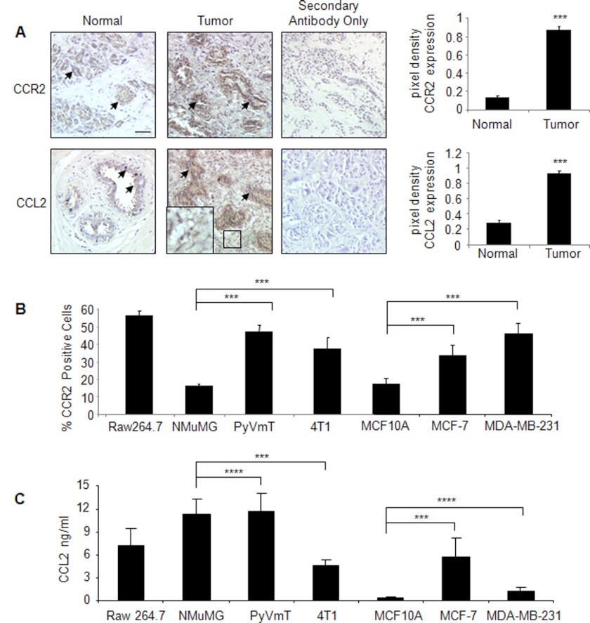
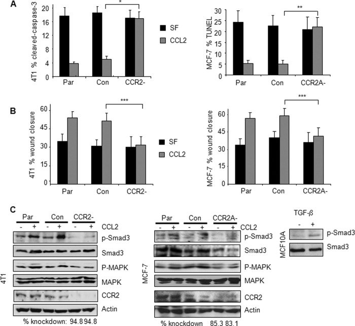
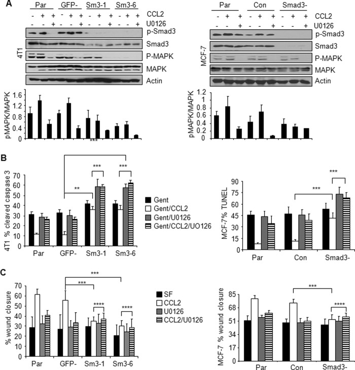
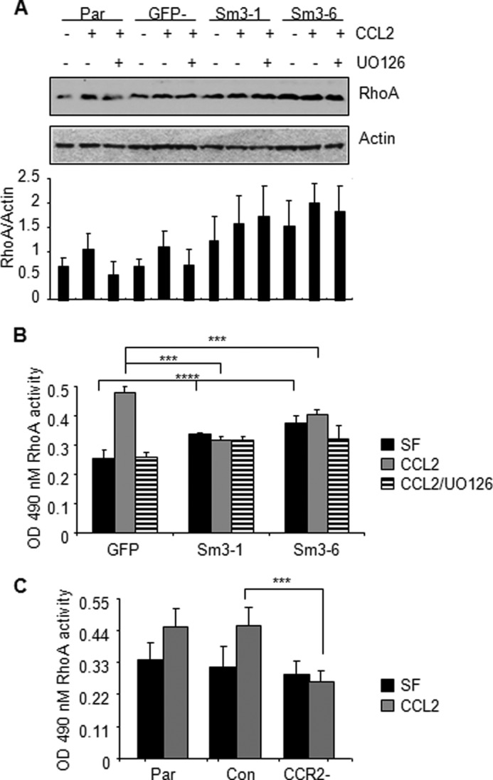
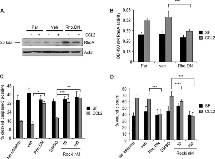
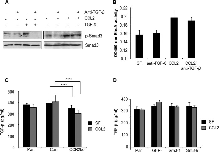
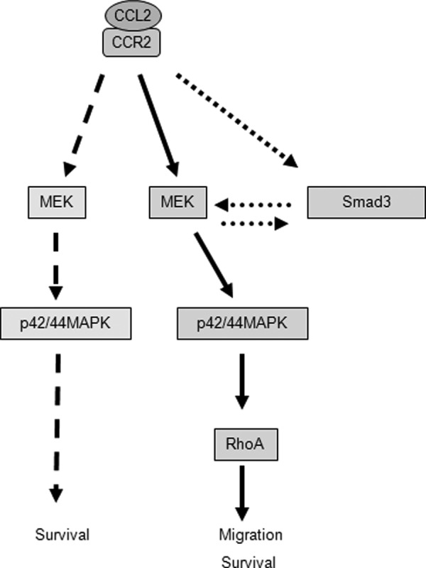
Increased cell motility and survival are important hallmarks of metastatic tumor cells. However, the mechanisms that regulate the interplay between these cellular processes remain poorly understood. In these studies, we demonstrate that CCL2, a chemokine well known for regulating immune cell migration, plays an important role in signaling to breast cancer cells. We report that in a panel of mouse and human breast cancer cell lines CCL2 enhanced cell migration and survival associated with increased phosphorylation of Smad3 and p42/44MAPK proteins. The G protein-coupled receptor CCR2 was found to be elevated in breast cancers, correlating with CCL2 expression. RNA interference of CCR2 expression in breast cancer cells significantly inhibited CCL2-induced migration, survival, and phosphorylation of Smad3 and p42/44MAPK proteins. Disruption of Smad3 expression in mammary carcinoma cells blocked CCL2-induced cell survival and migration and partially reduced p42/44MAPK phosphorylation. Ablation of MAPK phosphorylation in Smad3-deficient cells with the MEK inhibitor U0126 further reduced cell survival but not migration. These data indicate that Smad3 signaling through MEK-p42/44MAPK regulates CCL2-induced cell motility and survival, whereas CCL2 induction of MEK-p42/44MAPK signaling independent of Smad3 functions as an alternative mechanism for cell survival. Furthermore, we show that CCL2-induced Smad3 signaling through MEK-p42/44MAPK regulates expression and activity of Rho GTPase to mediate CCL2-induced breast cancer cell motility and survival. With these studies, we characterize an important role for CCL2/CCR2 chemokine signaling in regulating the intrinsic relationships between breast cancer cell motility and survival with implications on the metastatic process.


Figures

FIGURE 1.
FIGURE 1.
CCL2 blocks serum-deprived and gentamicin-induced apoptosis of mouse and human mammary carcinoma cells. A, mammary carcinoma cells were incubated in serum-free medium in the presence or absence of 20 ng/ml CCL2, 250 μg/ml gentamicin, or 250 μg/ml 5-FU for 24 h and analyzed for apoptosis by immunofluorescence staining for cleaved caspase-3 expression. Representative images of cleaved caspase-3 staining with DAPI overlay are shown for 4T1 cells. B, MCF-7 breast cancer cells were incubated in serum-free medium (SF) in the presence or absence of 250 μg/ml gentamicin and 20 ng/ml CCL2 for 24 h and analyzed for apoptosis by immunofluorescence staining for TUNEL-positive nuclei. Representative images of TUNEL staining with propidium iodide overlay are shown. Statistical analysis was determined by two-tailed t test: *, p < 0.05; ***, p < 0.001. Values are expressed as mean ± S.E. Error bars represent S.E.
FIGURE 2.
FIGURE 2.
CCL2 enhances migration of mammary carcinoma cells. Mouse and human mammary carcinoma cell lines were incubated in serum-free medium (SF) in the presence or absence of 20 ng/ml CCL2 and analyzed for changes in migration by Transwell assay (A) and wound closure assay (B). Representative images of 4T1 mammary carcinoma cells that migrated across a Transwell membrane after 8-h incubation with CCL2 are shown (A). Representative images of wound closure of 4T1 cells after 24-h incubation with CCL2 are shown (B). Statistical analysis was determined by two-tailed t test: *, p < 0.001; **, p < 0.01; ***, p < 0.05. Values are expressed as mean ± S.E. Error bars represent S.E.
FIGURE 3.
FIGURE 3.
CCR2 expression is up-regulated in breast cancer tissues and corresponds to increased CCL2 expression. A, CCL2 and CCR2 staining was performed on array slides containing de-identified cores of normal (n = 14) and invasive breast ductal carcinoma tissues (n = 20) sectioned at 5-μm thickness (catalog number 8022, US Biomax). Additional studies were performed on de-identified normal and breast cancer tissues (n = 5 per group) obtained from the Biospecimen Shared Resource at the University of Kansas Medical Center. Representative images of staining for CCL2 and CCR2 are from the tissue arrays. Arrowheads point to staining in ductal epithelial cells. The magnified inset shows CCL2 staining in the stroma. Tumor scale bar, 50 μm. Levels of CCR2 and CCL2 expression were measured by pixel density using NIH ImageJ software. Arbitrary units are shown. Statistical analysis was performed by two-tailed t test: ***, p < 0.05 compared with normal tissue. B, expression of CCR2 was analyzed in mouse and human mammary carcinoma cells by flow cytometry analysis with the Raw 264.7 murine macrophage cell line as a positive control. C, expression of CCL2 was analyzed in mouse and human mammary carcinoma cells by ELISA of conditioned medium. For B and C, statistical analysis was performed by analysis of variance test with Bonferroni's post-test of comparisons: ***, p < 0.05; ****, p > 0.05. Values are expressed as mean ± S.E. Error bars represent S.E.
FIGURE 4.
FIGURE 4.
CCR2 is required for CCL2 signaling in mouse and human breast carcinoma cells. 4T1 or MCF-7 parental (Par) cells, control (Con) siRNA-transfected cells, or cells transfected with CCR2 siRNAs were stimulated with 20 ng/ml CCL2 and analyzed for changes in gentamicin-induced apoptosis by cleaved caspase-3 or TUNEL assay as indicated (A), changes in migration by wound closure assay (B), and expression of the indicated proteins by Western blot after 8-h stimulation (C). TGF-β treatment of MCF10A cells is shown as a comparison of phospho-Smad3 expression between cell lines. The level of CCR2 knockdown was determined by densitometry analysis using NIH ImageJ software. CCR2 knockdown was compared with control siRNA samples. Values are normalized to actin. Statistical analysis was determined by two-tailed t test: *, p < 0.001; **, p < 0.01; ***, p < 0.05. Values are expressed as mean ± S.E. Error bars represent S.E.
FIGURE 5.
FIGURE 5.
Effect of Smad3 and MAPK inhibition on CCL2-induced cell survival and migration of mammary carcinoma cells. Parental cells (Par), 4T1 cells stably expressing shRNAs to GFP (GFP−) or Smad3 (Sm3-1 and Sm3-6), and MCF-7 cells transiently expressing control siRNAs (Con) or Smad3 siRNAs (Smad3−) were treated with CCL2 (20 ng/ml) in the presence or absence of 1 μm U0126 and analyzed by Western blot for changes in phosphorylation of Smad3 and p42/44MAPK (A). Densitometry analysis was performed on three independent experiments. Values are expressed as mean ± S.E. B, gentamicin (Gent)-induced apoptosis. C, migration by wound closure assay. Statistical analysis was determined by two-tailed t test: **, p < 0.01; ***, p < 0.05; ****, p > 0.05. Values are expressed as mean ± S.E. Error bars represent S.E. SF, serum-free medium.
FIGURE 6.
FIGURE 6.
CCL2/CCR2 signaling mediates RhoA activity through Smad3- and MAPK-dependent mechanisms. Parental (Par) 4T1 cells and 4T1 cells stably expressing control shRNAs (GFP−) or Smad3 shRNAs (Sm3-1 and Sm3-6) were treated with 20 ng/ml CCL2 in the presence or absence of 1 μm U0126. A, samples were analyzed for expression of RhoA by Western blot analysis after 4 h of treatment. RhoA expression was determined by densitometry analysis of three independent Western blot experiments using NIH ImageJ software. Values are expressed as mean ± S.E. B, RhoA GTPase activity was analyzed by G-LISA after 8-h incubation. Statistical analysis was determined by analysis of variance with Bonferroni's post-test of comparisons. C, parental (Par) 4T1 or cells expressing control siRNA (Con) or CCR2 siRNAs were analyzed for RhoA activity by G-LISA. Statistical analysis was determined by two-tailed t test: ***, p < 0.05; ****, p > 0.05. Values are expressed as mean ± S.E. Error bars represent S.E. SF, serum-free medium.
FIGURE 7.
FIGURE 7.
CCL2 mediation of RhoA function is important for mammary carcinoma cell survival and migration. 4T1 mammary carcinoma cells were infected with control vehicle retrovirus (veh) or retrovirus overexpressing dominant negative RhoA (Rho.DN) or treated with Rho kinase inhibitor II (Rocki) and analyzed for changes. A, expression of RhoA by immunoblot analysis. The protein band shown is at the predicted molecular mass for RhoA (25 kDa). B, RhoA activity by G-LISA. C, gentamicin-induced apoptosis. D, migration by wound closure assay. Statistical analysis was determined by two-tailed t test: *, p < 0.001; **, p < 0.01; ***, p < 0.05; ****, p > 0.05. Values are expressed as mean ± S.E. Error bars represent S.E. Par, parental; SF, serum-free medium.
FIGURE 8.
FIGURE 8.
CCL2/CCR2 signaling is not significantly dependent on TGF-β expression. A, 4T1 cells were treated with CCL2 (20 ng/ml) or TGF-β as a positive control (5 ng/ml) with or without 10 μg/ml neutralizing antibodies to TGF-β and analyzed for changes in Smad3 phosphorylation by immunoblot analysis. B, 4T1 cells were treated with CCL2 with or without 10 μg/ml neutralizing antibodies to TGF-β and analyzed for changes in RhoA activity. C, CCR2 was transiently knocked down (CCR2kd) in 4T1 cells, and cells were analyzed for expression of TGF-β by ELISA. D, Smad3-deficient 4T1 cells were treated with CCL2 and analyzed for changes in TGF-β expression by ELISA. Statistical analysis was determined by two-tailed t test: ****, p > 0.05. Values are expressed as mean ± S.E. Error bars represent S.E. Con, control; Par, parental; SF, serum-free medium.
FIGURE 9.
FIGURE 9.
Proposed model for how CCL2 signaling through CCR2 signaling regulates survival and motility of breast cancer cells. CCL2 binds to CCR2 to stimulate Smad3 and p42/44MAPK pathways. In one pathway, CCL2 induces MEK signaling through p42/44MAPK independently of Smad3 to promote cell survival. In addition, CCL2 activates Smad3, which cooperates with the MEK-p42/44MAPK pathway to regulate cell motility and survival through RhoA-dependent mechanisms.

References

    1. Gillitzer R., Goebeler M. (2001) Chemokines in cutaneous wound healing. J. Leukoc. Biol. 69, 513–521 - PubMed
    1. Laing K. J., Secombes C. J. (2004) Chemokines. Dev. Comp. Immunol. 28, 443–460 - PubMed
    1. Lentsch A. B. (2002) The Duffy antigen/receptor for chemokines (DARC) and prostate cancer. A role as clear as black and white? FASEB J. 16, 1093–1095 - PubMed
    1. Lazennec G., Richmond A. (2010) Chemokines and chemokine receptors: new insights into cancer-related inflammation. Trends Mol. Med. 16, 133–144 - PMC - PubMed
    1. Ernst C. A., Zhang Y. J., Hancock P. R., Rutledge B. J., Corless C. L., Rollins B. J. (1994) Biochemical and biologic characterization of murine monocyte chemoattractant protein-1. Identification of two functional domains. J. Immunol. 152, 3541–3549 - PubMed

Publication types

MeSH terms