Clinical Trial
. 2012 Oct 12;287(42):35244-35250.
doi: 10.1074/jbc.M111.305441. Epub 2012 Aug 26.

Identification of a novel role for dematin in regulating red cell membrane function by modulating spectrin-actin interaction

Affiliations
Clinical Trial

Identification of a novel role for dematin in regulating red cell membrane function by modulating spectrin-actin interaction

Ichiro Koshino et al. J Biol Chem. .

Abstract

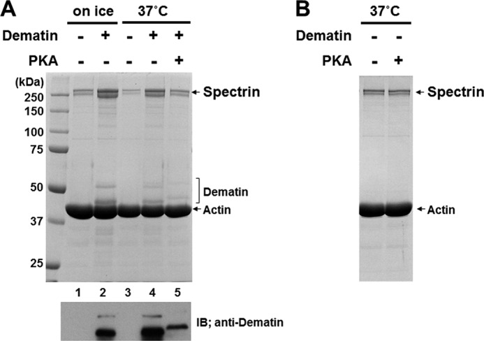
The membrane skeleton plays a central role in maintaining the elasticity and stability of the erythrocyte membrane, two biophysical features critical for optimal functioning and survival of red cells. Many constituent proteins of the membrane skeleton are phosphorylated by various kinases, and phosphorylation of β-spectrin by casein kinase and of protein 4.1R by PKC has been documented to modulate erythrocyte membrane mechanical stability. In this study, we show that activation of endogenous PKA by cAMP decreases membrane mechanical stability and that this effect is mediated primarily by phosphorylation of dematin. Co-sedimentation assay showed that dematin facilitated interaction between spectrin and F-actin, and phosphorylation of dematin by PKA markedly diminished this activity. Quartz crystal microbalance measurement revealed that purified dematin specifically bound the tail region of the spectrin dimer in a saturable manner with a submicromolar affinity. Pulldown assay using recombinant spectrin fragments showed that dematin, but not phospho-dematin, bound to the tail region of the spectrin dimer. These findings imply that dematin contributes to the maintenance of erythrocyte membrane mechanical stability by facilitating spectrin-actin interaction and that phosphorylation of dematin by PKA can modulate these effects. In this study, we have uncovered a novel functional role for dematin in regulating erythrocyte membrane function.


Figures

FIGURE 1.
FIGURE 1.
PKA phosphorylation of membrane skeletal proteins and its effect on the membrane mechanical stability. A, autoradiogram of 32P-labeled erythrocyte membrane proteins phosphorylated in the presence or absence of 0.2 μm calyculin A (CA) and/or 100 μm cAMP. Major membrane skeletal proteins are indicated. CBB, Coomassie Brilliant Blue. B, band intensities in the autoradiogram were determined by densitometric scanning and normalized by the copy number of each protein (see “Experimental Procedures” for details). White bars, control (without calyculin A); hatched bars, calyculin A alone; black bars, calyculin A + cAMP. Data are means ± S.D. (n = 3). *, p < 0.05; **, p < 0.01. C, erythrocyte membrane mechanical stability was examined using an ektacytometer. A decrease in DI reflects membrane fragmentation by applied shear stress. Representative results from five independent experiments are shown. D, erythrocyte membrane mechanical stability is expressed as T90 values, which is the time required for the DI to reach 90% of DImax. Data are means ± S.D. (n = 5). *, p < 0.05.
FIGURE 2.
FIGURE 2.
Effects of dematin and its phosphorylation on spectrin-actin interaction. A, dematin purified as described under “Experimental Procedures” was >95% pure as determined by densitometric scanning of the Coomassie Brilliant Blue (CBB)-stained gel. B, purified dematin and purified dematin phosphorylated in vitro were probed with anti-dematin monoclonal antibody (upper panel) and anti-phospho-dematin polyclonal antibody raised against the synthetic peptide CNELKKKA(pS)403LF (middle panel). The ghosts prepared from PKA-activated erythrocytes were subjected to SDS-PAGE and probed with anti-phospho-dematin polyclonal antibody (lower panel). C, upper panel, purified spectrin (1.13 μm) was incubated with F-actin (9.3 μm) on ice for 1 in the presence or absence of dematin or phospho-dematin (pDematin; ∼0.6 μm). Proteins co-sedimenting with F-actin were resolved on SDS gel. Spectrin, actin, and dematin are indicated. Lower panel, an immunoblot (IB) using anti-dematin mAb to unambiguously indicate co-sedimenting dematin is shown. D, the band intensity of spectrin co-sedimenting with F-actin was determined by densitometric scanning of a Coomassie Brilliant Blue-stained gel and is shown as the relative amount to the control (without dematin; None). Data are means ± S.D. (n = 3). **, p < 0.01. E, upper panel, spectrin was incubated with F-actin on ice for 1 h in the presence of increasing concentrations of dematin or phospho-dematin (0–0.6 μm). Proteins co-sedimenting with F-actin were resolved by SDS-PAGE. Lower panel, an immunoblot using anti-dematin mAb to unambiguously indicate dematin is shown. F, the band intensity of spectrin shown in C was determined by densitometric scanning and is shown as the relative amount to that obtained without dematin.
FIGURE 3.
FIGURE 3.
Phosphorylation of dematin in the preformed complex. A, dematin was incubated with spectrin and F-actin to form a ternary complex. Then, 1 mm ATP and 2.4 mm MgCl2 were added to the reaction mixture and further incubated in the presence or absence of 133 units/ml bovine PKA catalytic subunit for 1.5 h at 37 °C. Proteins co-sedimenting with F-actin were analyzed by SDS-PAGE. An immunoblot (IB) using anti-dematin mAb to unambiguously indicate co-sedimenting dematin is shown. B, PKA treatment of the preformed spectrin-actin complex as described above did not affect spectrin binding to F-actin.
FIGURE 4.
FIGURE 4.
Specific interaction between dematin and spectrin. A, dematin or phospho-dematin was incubated with mini-spectrin immobilized on glutathione-Sepharose beads. Bound dematin and phospho-dematin (pDematin) were detected by immunoblotting using anti-dematin monoclonal antibody, which recognizes both phosphorylated and unphosphorylated dematin. The upper panel shows that equal amounts of dematin and phospho-dematin were used as input. CBB, Coomassie Brilliant Blue. B, increasing concentrations of purified dematin was pulled down by the mini-spectrin dimer (GST-α20-C + βN-2) immobilized on glutathione-Sepharose beads. C, increasing concentrations of the mini-spectrin dimer (GST-α20-C + βN-2) was added to a sensor chip on which purified dematin had been immobilized. The amount of mini-spectrin bound was quantified as change in frequency (ΔF). Representative results from three independent experiments are shown. All three experiments showed saturable and sigmoidal binding curves. A dissociation constant of 14.8 ± 1.6 nm and a Hill coefficient of 1.75 ± 0.19 were derived from analysis of the binding curves.

References

    1. Mohandas N., Chasis J. A. (1993) Red blood cell deformability, membrane material properties, and shape: regulation by transmembrane, skeletal, and cytosolic proteins and lipids. Semin. Hematol. 30, 171–192 - PubMed
    1. Mohandas N., Gallagher P. G. (2008) Red cell membrane: past, present, and future. Blood 112, 3939–3948 - PMC - PubMed
    1. Ungewickell E., Bennett P. M., Calvert R., Ohanian V., Gratzer W. B. (1979) In vitro formation of a complex between cytoskeletal proteins of the human erythrocyte. Nature 280, 811–814 - PubMed
    1. Gardner K., Bennett V. (1987) Modulation of spectrin-actin assembly by erythrocyte adducin. Nature 328, 359–362 - PubMed
    1. Mische S. M., Mooseker M. S., Morrow J. S. (1987) Erythrocyte adducin: a calmodulin-regulated actin-bundling protein that stimulates spectrin-actin binding. J. Cell Biol. 105, 2837–2845 - PMC - PubMed

Publication types