. 2012 Oct 12;287(42):34895-34903.
doi: 10.1074/jbc.M112.362293. Epub 2012 Aug 27.

A viral, transporter associated with antigen processing (TAP)-independent, high affinity ligand with alternative interactions endogenously presented by the nonclassical human leukocyte antigen E class I molecule

Affiliations

A viral, transporter associated with antigen processing (TAP)-independent, high affinity ligand with alternative interactions endogenously presented by the nonclassical human leukocyte antigen E class I molecule

Elena Lorente et al. J Biol Chem. .

Abstract

The transporter associated with antigen processing (TAP) enables the flow of viral peptides generated in the cytosol by the proteasome and other proteases to the endoplasmic reticulum, where they complex with nascent human leukocyte antigen (HLA) class I. Later, these peptide-HLA class I complexes can be recognized by CD8(+) lymphocytes. Cancerous cells and infected cells in which TAP is blocked, as well as individuals with unusable TAP complexes, are able to present peptides on HLA class I by generating them through TAP-independent processing pathways. Here, we identify a physiologically processed HLA-E ligand derived from the D8L protein in TAP-deficient vaccinia virus-infected cells. This natural high affinity HLA-E class I ligand uses alternative interactions to the anchor motifs previously described to be presented on nonclassical HLA class I molecules. This octameric peptide was also presented on HLA-Cw1 with similar binding affinity on both classical and nonclassical class I molecules. In addition, this viral peptide inhibits HLA-E-mediated cytolysis by natural killer cells. Comparison between the amino acid sequences of the presenting HLA-E and HLA-Cw1 alleles revealed a shared structural motif in both HLA class molecules, which could be related to their observed similar cross-reactivity affinities. This motif consists of several residues located on the floor of the peptide-binding site. These data expand the role of HLA-E as an antigen-presenting molecule.


Figures
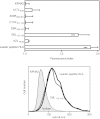
FIGURE 1.
FIGURE 1.
HLA-E stabilization with synthetic VACV ligands. The stability of HLA-E-peptide complexes on the cell surface of T2 TAP-deficient cells was measured by flow cytometry. The indicated peptides were used at 200 μm. The KPNA2 peptide and the leader peptide of HLA were used as negative and positive controls, respectively. The mAb 3D12 was used for staining. The results, calculated as fluorescence index values ± S.D., are the means of four to five independent experiments. ***, significant p values (p < 0.001). A representative experiment was depicted in the bottom panel. Shaded histogram, isotypic control; thin line, KPNA2 peptide; medium line, D8L 112–119; thick line, leader peptide of HLA.
FIGURE 2.
FIGURE 2.
Binding affinity to HLA-E and -Cw1 of VACV D8L112–119 synthetic peptide. The synthetic peptide VACV D8L112–119 (circles) was titrated bound to HLA-E (left panel) or HLA-Cw1 (right panel) on T2 TAP-deficient cells, and stabilization of HLA was measured by flow cytometry. The KPNA2 peptide was used as a negative control (solid line). The leader peptide of HLA and CMV pp657–15 were used as positive controls (squares) for binding to the HLA-E and -Cw1 alleles, respectively. The Abs used were monoclonal 3D12 (anti-HLA-E, left panel), and polyclonal SC-19438 (anti-HLA-C class I molecules, right panel). The data calculated as EC50 values ± S.D. are shown below and are the means of three to five independent experiments.
FIGURE 3.
FIGURE 3.
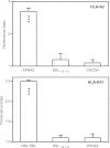
HLA-A2 and -B51 stabilization assay with synthetic VACV D8L112–119 ligand. The stability of HLA-A2-peptide (upper panel) or HLA-B51-peptide (lower panel) complexes on the surface of T2 TAP-deficient cells were measured by flow cytometry. The indicated peptides were used at 200 μm. The KPNA2 and the HBV HBc peptides were used as positive controls for binding to the HLA-A2 and -B51 alleles, respectively. The C4CON and the KPNA2 peptides were used as negative controls for binding to the HLA-A2 and -B51 alleles, respectively. The Abs used were monoclonal PA2.1 (anti-HLA-A2, upper panel) and polyclonal H00003106-B01P (anti-HLA-B class I molecules, lower panel). The results, calculated as in Fig. 1, are the mean of four independent experiments. ***, significant p values (p < 0.001).
FIGURE 4.
FIGURE 4.
VACV D8L112–119 synthetic peptide inhibits HLA-E-mediated cytotoxicity by natural killer cells. 721.221 target cells prepulsed with 100 μm of the indicated synthetic peptides were tested in a standard cytolytic assay for natural killer cytotoxicity with NK3.3 cells. The leader peptide of HLA (squares) and an irrelevant peptide (diamonds) were used as positive and negative controls, respectively. The data are the means of three independent experiments ± S.D. ***, significant p values (p < 0.001) were found between no peptide or irrelevant peptide versus leader peptide of HLA or D8L112–119.
FIGURE 5.
FIGURE 5.
Modeling of HLA-E-bound conformations of VACV D8L112–119 peptide. The backbone atoms of the indicated HLA-E-bound peptides are displayed as ribbon tubes (A, VMAPRALLL; B and C, DGLIIISI in two possible conformations: A model and B model, respectively). The atoms are represented by sticks using the following color scheme: blue, nitrogen; red, oxygen; and green, carbon. The peptide residues that interact with the indicated HLA-E pockets are designated. The HLA-E protein is not displayed. The figure was prepared using the PyMOL program.
FIGURE 6.
FIGURE 6.
HLA stabilization assay with monosubstituted Ala analogues of VACV D8L112–119 synthetic peptide. Stability at the cell surface of HLA-E on the cell surface of T2 TAP-deficient cells was measured by flow cytometry. The indicated peptides were used at 200 μm. The KPNA2 peptide was used as negative control. The mAb 3D12 was used for staining. The data, calculated as in Fig. 1, are the means of three or four independent experiments. ***, significant p values (p < 0.001).
FIGURE 7.
FIGURE 7.
Structural similarities between HLA-E and HLA-Cw1 but not with HLA-A2 or-B51. The amino acid sequence of the α1 and α2 domains of HLA-E (white backbone) was compared with the sequence of the equivalent domains of HLA-A2, -B51, and -Cw1 class I molecules using the same alignment used by Bjorkmann et al. (53, 54). The identical residues identified between HLA-E and HLA-Cw1 but not HLA-A2 or -B51 molecules (minimum desetope) and proposed as contributing to a shared structural motif that could confer peptide presenting similarities between HLA-E and HLA-Cw1 are depicted. The atoms of these four residues are represented by sticks using the following color scheme: blue, nitrogen; red, oxygen; and green, carbon. The viral peptide is not displayed. The figure was prepared using the PyMOL program.

References

    1. Kloetzel P. M., Ossendorp F. (2004) Proteasome and peptidase function in MHC-class-I-mediated antigen presentation. Curr. Opin. Immunol. 16, 76–81 - PubMed
    1. Cerundolo V., de la Salle H. (2006) Description of HLA class I- and CD8-deficient patients. Insights into the function of cytotoxic T lymphocytes and NK cells in host defense. Semin. Immunol. 18, 330–336 - PubMed
    1. Van Kaer L., Ashton-Rickardt P. G., Ploegh H. L., Tonegawa S. (1992) TAP1 mutant mice are deficient in antigen presentation, surface class I molecules, and CD4–8+ T cells. Cell 71, 1205–1214 - PubMed
    1. Voldby Larsen M., Nielsen M., Weinzierl A., Lund O. (2006) TAP-independent MHC class I presentation. Curr. Immunol. Rev. 2, 233–245
    1. Alzhanova D., Edwards D. M., Hammarlund E., Scholz I. G., Horst D., Wagner M. J., Upton C., Wiertz E. J., Slifka M. K., Früh K. (2009) Cowpox virus inhibits the transporter associated with antigen processing to evade T cell recognition. Cell Host. Microbe 6, 433–445 - PMC - PubMed

Publication types

MeSH terms