. 2012 Dec;343(3):763-78.
doi: 10.1124/jpet.112.197590. Epub 2012 Aug 27.

The autophagy-senescence connection in chemotherapy: must tumor cells (self) eat before they sleep?

Affiliations

The autophagy-senescence connection in chemotherapy: must tumor cells (self) eat before they sleep?

Rachel W Goehe et al. J Pharmacol Exp Ther. 2012 Dec.

Abstract

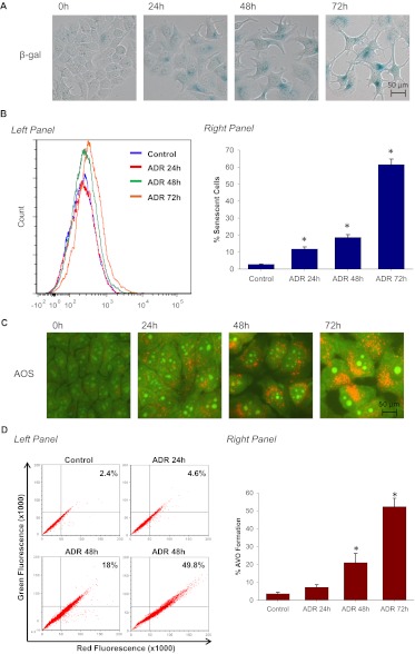
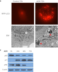
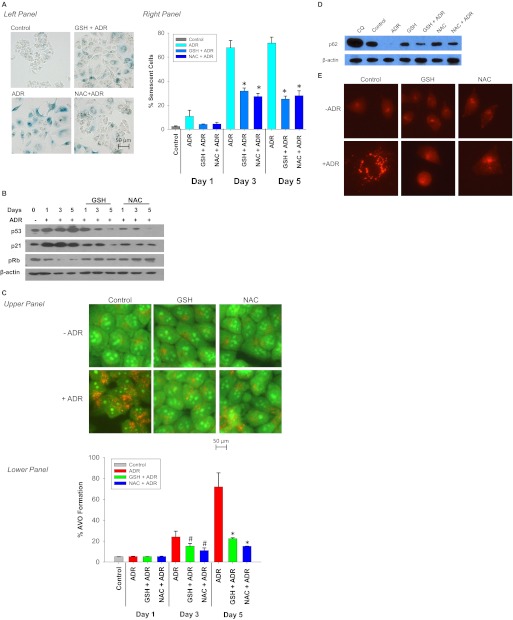
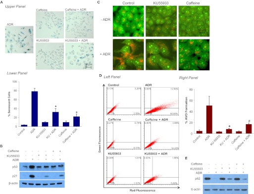
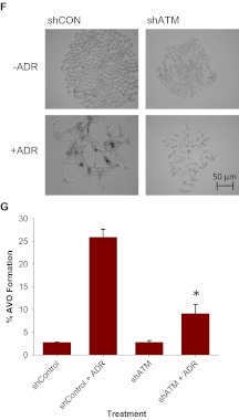
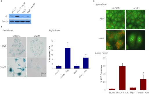
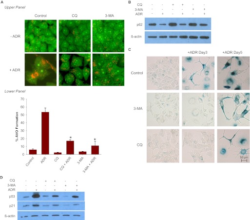
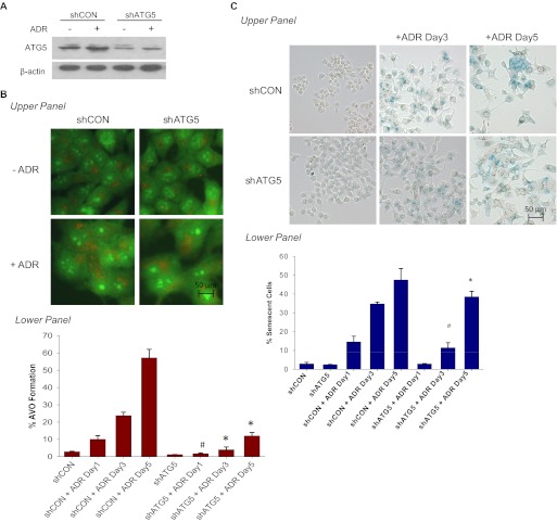
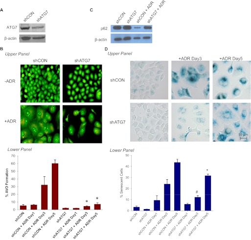
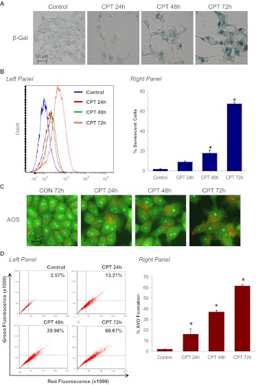
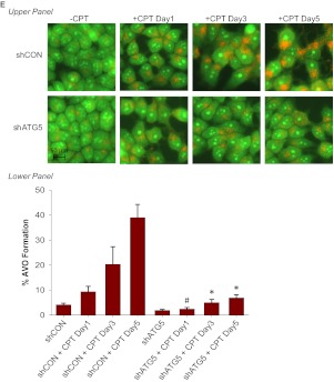
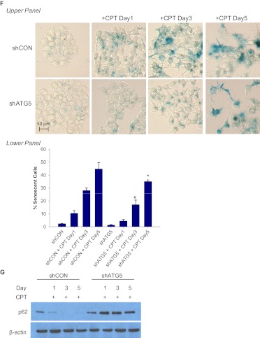
Exposure of MCF-7 breast tumor cells or HCT-116 colon carcinoma cells to clinically relevant concentrations of doxorubicin (Adriamycin; Farmitalia Research Laboratories, Milan, Italy) or camptothecin results in both autophagy and senescence. To determine whether autophagy is required for chemotherapy-induced senescence, reactive oxygen generation induced by Adriamycin was suppressed by N-acetyl cysteine and glutathione, and the induction of ataxia telangiectasia mutated, p53, and p21 was modulated pharmacologically and/or genetically. In all cases, autophagy and senescence were collaterally suppressed. The close association between autophagy and senescence indicated by these experiments reflects their collateral regulation via common signaling pathways. The potential relationship between autophagy and senescence was further examined through pharmacologic inhibition of autophagy with chloroquine and 3-methyl-adenine and genetic ablation of the autophagy-related genes ATG5 and ATG7. However, inhibition of autophagy by pharmacological and genetic approaches could not entirely abrogate the senescence response, which was only reduced and/or delayed. Taken together, our findings suggest that autophagy and senescence tend to occur in parallel, and furthermore that autophagy accelerates the development of the senescent phenotype. However, these responses are not inexorably linked or interdependent, as senescence can occur when autophagy is abrogated.


Figures

Fig. 1.
Fig. 1.
ADR induces both senescence and autophagy in MCF-7 cells. MCF-7 cells were treated with 1 μM ADR for 2 h followed by drug removal and replacement with fresh medium. A, β-galactosidase staining. Control cells generally show minimal staining. B, left, representative histogram of positive C12FDG fluorescent cells for each treatment. Right, flow cytometric detection of SA-β-gal activity as measured by C12FDG-fluorescin fluorescence. *, p < 0.001 compared with control. C, cells were stained with acridine orange and imaged by fluorescence microscopy. D, left, representative dot plots of positive acridine orange staining for each treatment. Right, cells were quantified for AVO formation produced by ADR treatment by FACS analysis. *, p < 0.001 compared with control. E, punctate signal of RFP-LC3 and transmission electron microscopy imaging 72 h after ADR treatment. Arrowheads in red indicate autophagosome formation. F, immunoblot analysis of p62, p21, pRb, and β-actin. Data are representative of an average of three independent experiments.
Fig. 1.
Fig. 1.
ADR induces both senescence and autophagy in MCF-7 cells. MCF-7 cells were treated with 1 μM ADR for 2 h followed by drug removal and replacement with fresh medium. A, β-galactosidase staining. Control cells generally show minimal staining. B, left, representative histogram of positive C12FDG fluorescent cells for each treatment. Right, flow cytometric detection of SA-β-gal activity as measured by C12FDG-fluorescin fluorescence. *, p < 0.001 compared with control. C, cells were stained with acridine orange and imaged by fluorescence microscopy. D, left, representative dot plots of positive acridine orange staining for each treatment. Right, cells were quantified for AVO formation produced by ADR treatment by FACS analysis. *, p < 0.001 compared with control. E, punctate signal of RFP-LC3 and transmission electron microscopy imaging 72 h after ADR treatment. Arrowheads in red indicate autophagosome formation. F, immunoblot analysis of p62, p21, pRb, and β-actin. Data are representative of an average of three independent experiments.
Fig. 2.
Fig. 2.
Involvement of free radicals in ADR-induced senescence and autophagy. MCF-7 cells were pretreated with 20 μM GSH or 20 μM NAC for 1 h followed by 1 μM ADR for 2 h. The drugs were removed, and fresh media were restored. A, senescence was evaluated by β-galactosidase staining 72 h after treatment (left) and quantified for C12FDG-fluorescin fluorescence by FACS analysis (right). *, p < 0.001 compared with control. B, immunoblot analysis of p53, p21, pRb, and β-actin. C, acridine orange staining visualized by fluorescence microscopy (top) and quantified by FACS analysis (bottom). #, p < 0.05 compared with ADR treatment on day 3; *, p < 0.001 compared with ADR treatment on day 5. D, immunoblot analysis of p62 and β-actin were evaluated 72 h after treatment. E, fluorescence microscopy for RFP-LC3 at 72 h after initiation of the indicated treatments.
Fig. 3.
Fig. 3.
Influence of ATM activity on ADR-induced senescence and autophagy. MCF-7 cells were treated with 1 μM ADR for 2 h preceded by a 2-h exposure to 20 μM KU55933 or 2 mM caffeine. A, top, MCF-7 cells were fixed and stained for β-galactosidase. Bottom, flow cytometric detection of SA-β-gal activity as measured by C12FDG-fluorescin fluorescence. *, p < 0.001 compared with ADR treatment. B, immunoblot analysis of p53, p21, and β-actin. C, acridine orange staining visualized by fluorescence microscopy. D, left, representative dot plots of positive acridine orange staining for each of the indicated treatment. Right, quantification of acridine orange staining by FACS analysis. *, p < 0.001 compared with ADR; #, p < 0.003 compared with ADR. E, immunoblot analyses of p62 and β-actin. F and G, MCF-7 cells were transfected with either shRNA against a scramble control (shCON) or shRNA against ATM (shATM). F, shCON and shATM MCF-7 cells were assessed for β-galactosidase staining 72 h after ADR treatment. G, cells with AVO formation were monitored by FACS analysis 72 h after treatment. *, p < 0.001 compared with shCON + ADR.
Fig. 3.
Fig. 3.
Influence of ATM activity on ADR-induced senescence and autophagy. MCF-7 cells were treated with 1 μM ADR for 2 h preceded by a 2-h exposure to 20 μM KU55933 or 2 mM caffeine. A, top, MCF-7 cells were fixed and stained for β-galactosidase. Bottom, flow cytometric detection of SA-β-gal activity as measured by C12FDG-fluorescin fluorescence. *, p < 0.001 compared with ADR treatment. B, immunoblot analysis of p53, p21, and β-actin. C, acridine orange staining visualized by fluorescence microscopy. D, left, representative dot plots of positive acridine orange staining for each of the indicated treatment. Right, quantification of acridine orange staining by FACS analysis. *, p < 0.001 compared with ADR; #, p < 0.003 compared with ADR. E, immunoblot analyses of p62 and β-actin. F and G, MCF-7 cells were transfected with either shRNA against a scramble control (shCON) or shRNA against ATM (shATM). F, shCON and shATM MCF-7 cells were assessed for β-galactosidase staining 72 h after ADR treatment. G, cells with AVO formation were monitored by FACS analysis 72 h after treatment. *, p < 0.001 compared with shCON + ADR.
Fig. 4.
Fig. 4.
Down-regulation of p21 attenuates ADR-induced autophagy. MCF-7 cells were stably infected with either shRNA against a scramble control (shCON) or shRNA against p21 (shp21). Cell lines were treated with 1 μM ADR for 2 h, which was then replaced with complete medium. A, immunoblot analyses of lysates from shCON and shp21 cells treated with or without ADR for 72 h. B, cells were stained with β-galactosidase, and images were taken 72 h after ADR treatment (left) and quantified for C12FDG-fluorescin fluorescence by FACS analysis (right). *, p < 0.05 compared with shCON + ADR. C, top, acridine orange images were taken at 72 h after ADR treatment. Bottom, cells with AVO formation were monitored by FACS analysis 72 h after treatment. *, p < 0.003 compared with shCON + ADR.
Fig. 5.
Fig. 5.
Impact of autophagic inhibitors on ADR-induced senescence. MCF-7 cells were pretreated with 5 mM 3-MA or 5 μM chloroquine for 1 h followed by 1 μM ADR for 2 h. ADR was removed, and fresh media were restored with or without 3-MA or chloroquine. A, top, acridine orange images were taken at 72 h after ADR treatment. Bottom, cells with AVO formation were monitored by FACS analysis 72 h after treatment. *, p < 0.001 compared with ADR. B, immunoblot analysis of p62 and β-actin. C, cells were stained with β-galactosidase, and images were taken at the indicated times after ADR treatment. D, immunoblot analysis of p21, p53, and β-actin.
Fig. 6.
Fig. 6.
Genetic silencing of ATG5 temporarily delayed ADR-induced senescence. MCF-7 cells were stably infected with either shRNA against a scramble control (shCON) or shRNA against ATG5 (shATG5). Cell lines were treated with 1 μM ADR for 2 h, which was then replaced with complete medium. A, immunoblot analyses of lysates from shCON and shATG5 cells treated with or without ADR for 72 h for ATG5 and β-actin. B, acridine orange staining visualized by fluorescence microscopy (top) and quantified by FACS analysis (bottom). *, p < 0.001 compared with shCON + ADR day 3 and shCON + ADR day 5; #, p < 0.05 compared with ADR day 1. C, β-galactosidase staining (top) and flow cytometric detection of SA-β-gal activity as measured by C12FDG-fluorescin fluorescence (bottom). #, p < 0.05 compared with shATG5; *, p < 0.001 compared with shATG5.
Fig. 7.
Fig. 7.
Genetic silencing of ATG7 temporarily delayed ADR-induced senescence. MCF-7 cells were stably infected with either shRNA against a scramble control (shCON) or shRNA against ATG7 (shATG7). Cell lines were treated with 1 μM ADR for 2 h, which was then replaced with complete medium. A, immunoblot analyses of lysates from shCON and shATG7 cells treated with or without ADR for 72 h for ATG7 and β-actin. B, acridine orange staining visualized by fluorescence microscopy (top) and quantified by FACS analysis (bottom). *, p < 0.001 compared with shCON + ADR day 3 and shCON + ADR day 5. C, immunoblot analyses of lysates from shCON and shATG7 cells treated with or without ADR for 72 h for p62 and β-actin. D, β-galactosidase staining (top) and flow cytometric detection of SA-β-gal activity as measured by C12FDG-fluorescin fluorescence (bottom). #, p < 0.05 compared with shATG7; *, p < 0.001 compared with shATG7. E, immunoblot analysis of p21 and β-actin.
Fig. 8.
Fig. 8.
Silencing autophagy temporarily delayed CPT-induced senescence. MCF-7 cells were treated with 5 μM CPT for 2 h followed by drug removal and replacement with fresh medium. A, β-galactosidase staining. B, left, representative histograms of positive C12FDG fluorescent cells for each treatment. Right, flow cytometric detection of SA-β-gal activity as measured by C12FDG-fluorescin fluorescence. *, p < 0.001 compared with control. C, cells were stained with acridine orange and imaged by fluorescence microscopy. D, left, representative dot plots of positive acridine orange staining for each treatment. Right, cells were quantified for AVO formation produced by CPT treatment by FACS analysis. *, p < 0.001 compared with control. E, acridine orange staining of CPT-treated MCF-7 cells at the indicated time points (top) and quantification of AVO formation by FACS analysis (bottom). *, p < 0.001 compared with shCON + CPT day 3 and shCON + CPT day 5; #, p < 0.05 compared with shCON + CPT day 1. F, β-galactosidase staining of CPT-treated MCF-7 cells at the indicated time points (top) and quantification of C12FDG-fluorescin fluorescence by FACS analysis (bottom). #, p < 0.05 compared with shATG5; *, p < 0.001 compared with shATG5. G, immunoblot analysis of p62 and β-actin.
Fig. 8.
Fig. 8.
Silencing autophagy temporarily delayed CPT-induced senescence. MCF-7 cells were treated with 5 μM CPT for 2 h followed by drug removal and replacement with fresh medium. A, β-galactosidase staining. B, left, representative histograms of positive C12FDG fluorescent cells for each treatment. Right, flow cytometric detection of SA-β-gal activity as measured by C12FDG-fluorescin fluorescence. *, p < 0.001 compared with control. C, cells were stained with acridine orange and imaged by fluorescence microscopy. D, left, representative dot plots of positive acridine orange staining for each treatment. Right, cells were quantified for AVO formation produced by CPT treatment by FACS analysis. *, p < 0.001 compared with control. E, acridine orange staining of CPT-treated MCF-7 cells at the indicated time points (top) and quantification of AVO formation by FACS analysis (bottom). *, p < 0.001 compared with shCON + CPT day 3 and shCON + CPT day 5; #, p < 0.05 compared with shCON + CPT day 1. F, β-galactosidase staining of CPT-treated MCF-7 cells at the indicated time points (top) and quantification of C12FDG-fluorescin fluorescence by FACS analysis (bottom). #, p < 0.05 compared with shATG5; *, p < 0.001 compared with shATG5. G, immunoblot analysis of p62 and β-actin.

References

    1. Akbas SH, Timur M, Ozben T. (2005) The effect of quercetin on topotecan cytotoxicity in MCF-7 and MDA-MB 231 human breast cancer cells. J Surg Res 125:49–55 - PubMed
    1. Alexander A, Kim J, Walker CL. (2010) ATM engages the TSC2/mTORC1 signaling node to regulate autophagy. Autophagy 6:672–673 - PMC - PubMed
    1. Amaravadi RK, Yu D, Lum JJ, Bui T, Christophorou MA, Evan GI, Thomas-Tikhonenko A, Thompson CB. (2007) Autophagy inhibition enhances therapy-induced apoptosis in a Myc-induced model of lymphoma. J Clin Invest 117:326–336 - PMC - PubMed
    1. Azad MB, Chen Y, Gibson SB. (2009) Regulation of autophagy by reactive oxygen species (ROS): implications for cancer progression and treatment. Antioxid Redox Signal 11:777–790 - PubMed
    1. Bandhakavi S, Kim YM, Ro SH, Xie H, Onsongo G, Jun CB, Kim DH, Griffin TJ. (2010) Quantitative nuclear proteomics identifies mTOR regulation of DNA damage response. Mol Cell Proteomics 9:403–414 - PMC - PubMed

Publication types