. 2012 Nov;32(21):4400-9.
doi: 10.1128/MCB.00441-12. Epub 2012 Aug 27.

Late-stage maturation of the Rieske Fe/S protein: Mzm1 stabilizes Rip1 but does not facilitate its translocation by the AAA ATPase Bcs1

Affiliations

Late-stage maturation of the Rieske Fe/S protein: Mzm1 stabilizes Rip1 but does not facilitate its translocation by the AAA ATPase Bcs1

Tie-Zhong Cui et al. Mol Cell Biol. 2012 Nov.

Abstract

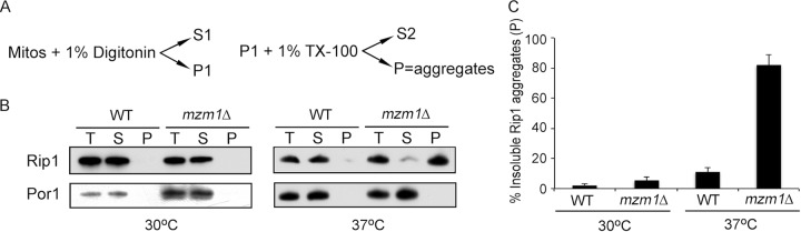
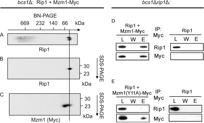
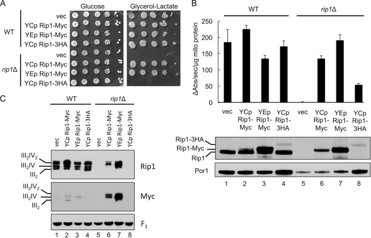
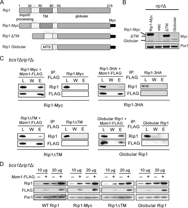
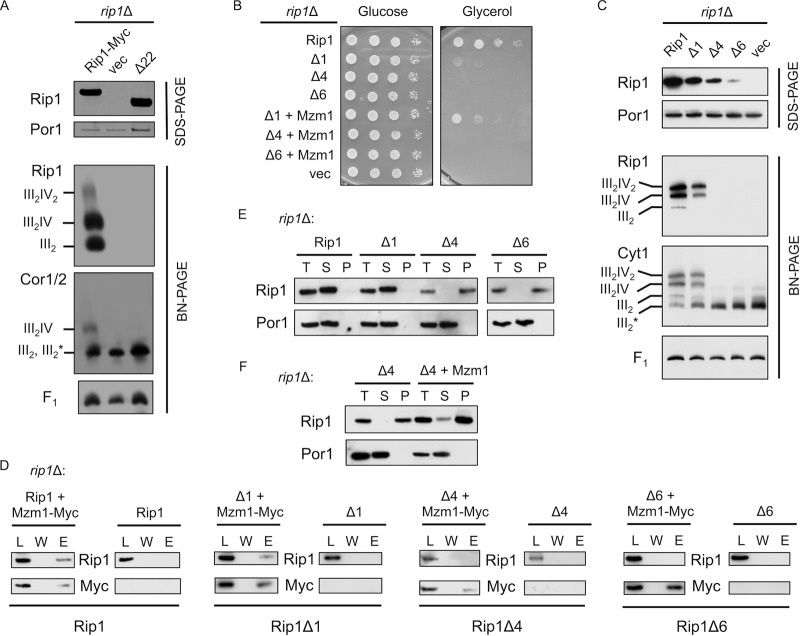
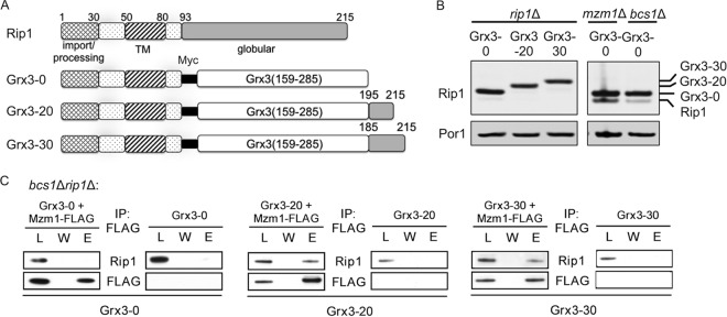
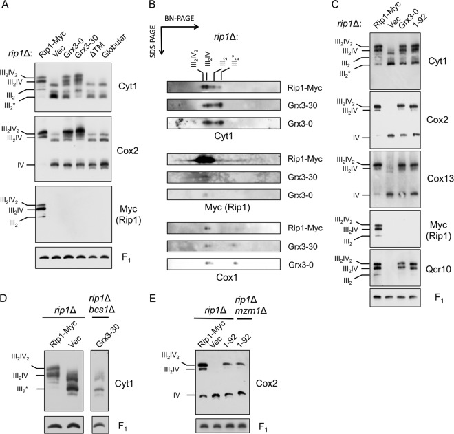
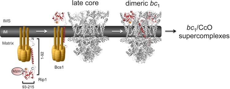
The final step in the assembly of the ubiquinol-cytochrome c reductase or bc(1) complex involves the insertion of the Rieske Fe/S cluster protein, Rip1. Maturation of Rip1 occurs within the mitochondrial matrix prior to its translocation across the inner membrane (IM) in a process mediated by the Bcs1 ATPase and subsequent insertion into the bc(1) complex. Here we show that the matrix protein Mzm1 functions as a Rip1 chaperone, stabilizing Rip1 prior to the translocation step. In the absence of Mzm1, Rip1 is prone to either proteolytic degradation or temperature-induced aggregation. A series of Rip1 truncations were engineered to probe motifs necessary for Mzm1 interaction and Bcs1-mediated translocation of Rip1. The Mzm1 interaction with Rip1 persists in Rip1 variants lacking its transmembrane domain or containing only its C-terminal globular Fe/S domain. Replacement of the globular domain of Rip1 with that of the heterologous folded protein Grx3 abrogated Mzm1 interaction; however, appending the C-terminal 30 residues of Rip1 to the Rip1-Grx3 chimera restored Mzm1 interaction. The Rip1-Grx3 chimera and a Rip1 truncation containing only the N-terminal 92 residues each induced stabilization of the bc(1):cytochrome oxidase supercomplex in a Bcs1-dependent manner. However, the Rip1 variants were not stably associated with the supercomplex. The induced supercomplex stabilization by the Rip1 N terminus was independent of Mzm1.


Figures

Fig 1
Fig 1
Rip1 aggregates at 37°C in the absence of Mzm1. (A) Schematic of the detergent-based protein aggregation assay; see Materials and Methods for details. (B) The total mitochondrial load (T), combined S1 and S2 soluble fractions (S), and insoluble material (P) were analyzed by SDS-PAGE and immunoblotting. At 30°C, the total load and the analyzed sample were 3-fold higher in concentration in mzm1Δ compared to WT, to produce samples with equivalent total Rip1 levels. (C) Comparison of the amount of Rip1 present by immunoblotting in the P fraction as a percentage of the total Rip1 protein (T), measured by ImageJ in three or more independent experiments.
Fig 2
Fig 2
Rip1 and Mzm1 form a complex of low molecular mass, whose formation is dependent upon tyrosine-11 of Mzm1. (A) BN-PAGE analysis of mitochondria isolated from Δbcs1 cells expressing both YCp RIP1 and YCp Sod2-MZM1-Myc, followed by immunoblot (anti-Rip1). (B and C) Subsequent analysis of the complex isolated through BN-PAGE by SDS-PAGE in a second dimension, followed by immunoblot (B, anti-Rip1; C, anti-Myc). The approximate positions of molecular masses are indicated, and a line is drawn as an aid to the eye indicating the Rip1-Mzm1 complex. (D and E) Immunoprecipitation of solubilized bcs1Δ rip1Δ mitochondria with anti-Myc antibody-conjugated agarose beads, showing the load (L), the final wash (W), and the eluate (E). Mitochondria were isolated from cultures expressing YCp RIP1 and either YCp Sod2-MZM1-Myc (D) or YCp Sod2-MZM1-Myc with a mutation resulting in conversion of tyrosine-11 to alanine (E).
Fig 3
Fig 3
Rip1 is sensitive to C-terminal epitope tagging. (A and B) Growth assay at 30°C (A) as well as bc1 complex activity assay and SDS-PAGE immunoblot of isolated mitochondria (B), from WT and rip1Δ cultures expressing vector control (vec) or YEp or YCp RIP1-Myc or YCp RIP1-3HA plasmid. (C) BN-PAGE immunoblot of digitonin-solubilized mitochondria isolated from the same cultures as in panels A and B.
Fig 4
Fig 4
Mzm1 interacts with the C-terminal globular domain of Rip1. (A) Comparison of Rip1 domains present in the WT protein (prior to N-terminal processing), Rip1-Myc, and the ΔTM and globular Rip1 proteins, which lack residues 55 to 78 and 1 to 80, respectively. The globular Rip1 protein is targeted to the mitochondrial matrix by the mitochondrial targeting sequence of Sod2 (MTS) and expressed from a YCp plasmid; the other Rip1 variants contain the endogenous targeting region of Rip1 and were expressed from YEp plasmids. (B) SDS-PAGE immunoblot showing expression of these Rip1 variants in rip1Δ mitochondria. Vec, vector. (C) Immunoprecipitation of solubilized bcs1Δ rip1Δ mitochondria expressing these plasmids with anti-FLAG antibody-conjugated agarose beads, showing the total load (L), the final wash (W), and the eluate (E). The globular Rip1 protein was detected with anti-Myc antibody; all other Rip1 detections utilized anti-Rip1 antibody. (D) SDS-PAGE immunoblot of bcs1Δ rip1Δ mitochondria (10 and 20 μg total mitochondrial protein) expressing the Rip1 variant proteins in the presence and absence of YEp MZM1-FLAG.
Fig 5
Fig 5
The extreme C terminus of Rip1 is essential for its function and important for interaction with Mzm1. (A) SDS-PAGE and BN-PAGE (digitonin) immunoblots of isolated mitochondria from WT and rip1Δ cultures expressing YEp RIP1-Myc, vector control (vec), or rip1Δ22 plasmid. Note that on this BN gel the dimeric bc1 and late core intermediate comigrate, so the band designation includes both III2 and III2*, the latter of which denotes the late core intermediate formed during bc1 complex assembly. The component is clearly the late core intermediate in the vector control rip1Δ cells. (B) Growth assay at 37°C of rip1Δ cultures expressing YCp RIP1, vector control, or the Δ1, Δ4, or Δ6 Rip1 truncation and coexpressing YCp vector control or MZM1-Myc plasmid. (C) SDS-PAGE and BN-PAGE (digitonin) immunoblots of rip1Δ cultures expressing YCp RIP1, Δ1, Δ4, Δ6, or vector control plasmid. (D) Immunoprecipitation of solubilized mitochondria from rip1Δ cultures expressing the plasmids listed in panel C with or without coexpression of YCp MZM1-Myc, using anti-Myc antibody-conjugated agarose beads and showing the total load (L), the final wash (W), and the eluate (E). (E) Detergent-based protein aggregation assay and SDS-PAGE immunoblot of the total load (T), soluble fraction (S), and pellet fraction (P), as described for Fig. 2 except that cultures were grown at 30°C with mitochondria isolated from rip1Δ cultures expressing the same plasmids as in panel C. The exposure of the Rip1Δ6 blot was extended due to limited steady-state levels of this truncation. (F) Assay described in E for a rip1Δ culture expressing the Δ4 truncation, with and without coexpression of YCp MZM1-Myc.
Fig 6
Fig 6
The C terminus of Rip1 is the dominant site for interaction with Mzm1. (A) Comparison of Rip1 domains present in the WT protein (prior to N-terminal processing) and the Grx3-0, Grx3-20, and Grx3-30 fusion proteins. These proteins are fusions of the N terminus of Rip1 (amino acid residues 1 to 92) with the soluble domain of Grx3 (residues 159 to 285), and each protein includes a Myc epitope positioned between the two domains. At the C terminus, 0, 20, or 30 residues of the Rip1 C terminus are appended. Note that these residues do not include any liganding residues for the 2Fe-2S center. (B) SDS-PAGE immunoblot of crude mitochondrial fractions showing expression of these fusion proteins from YEp plasmids. (C) Immunoprecipitation of solubilized bcs1Δ rip1Δ mitochondria expressing the fusion proteins from YEp plasmids, using anti-FLAG antibody-conjugated agarose beads and showing the total load (L), the final wash (W), and the eluate (E).
Fig 7
Fig 7
N-terminal Rip1 induces Bcs1-mediated bc1:CcO supercomplex formation. (A) BN-PAGE (digitonin) immunoblots of isolated rip1Δ mitochondria expressing Rip1-Myc, vector (Vec), Grx3-0, Grx3-30, and Rip1 variants either lacking the TM domain (ΔTM) or only containing the C-terminal globular domain (Globular). All the constructs contain a Myc tag. All the constructs are expressed from YEp vectors except globular Rip1, which is expressed from a YCp vector. III2* denotes the late core intermediate formed during bc1 complex assembly. (B) Subsequent analysis of protein complexes isolated from rip1Δ mitochondria expressing Rip1-Myc, Grx3-0, or Grx3-30 through BN-PAGE by SDS-PAGE in a second dimension. (C) BN-PAGE (digitonin) immunoblots of isolated rip1Δ mitochondria expressing Rip1-Myc, vector, Grx3-0, or a Rip1 variant containing only the first 92 amino acid residues (1-92) with a C-terminal Myc tag. (D) BN-PAGE (digitonin) immunoblots of isolated rip1Δ mitochondria expressing Rip1-Myc or vector and rip1Δ bcs1Δ mitochondria expressing Grx3-30. (E) BN-PAGE (digitonin) immunoblots of isolated rip1Δ mitochondria expressing Rip1-Myc, vector, or the Rip1 (1-92) variant and rip1Δ mzm1Δ mitochondria expressing the Rip1 (1-92) variant. The Rip1 (1-92) variant is processed in the matrix, and the mature form consists of residues 31 to 92.
Fig 8
Fig 8
Model for the chaperone role of Mzm1 in stabilizing the C-terminal domain of Rip1. Mzm1 is shown to interact with and stabilize the C-terminal globular domain of Rip1 prior to translocation by Bcs1. The N-terminal half of Rip1 (residues 1 to 92) is shown to be functional in inducing stabilization of the bc1:CcO supercomplex in rip1Δ cells.

References

    1. Atkinson A, et al. 2010. Mzm1 influences a labile pool of mitochondrial zinc important for respiratory function. J. Biol. Chem. 285: 19450–19459 - PMC - PubMed
    1. Atkinson A, et al. 2011. The LYR protein Mzm1 functions in the insertion of the Rieske Fe/S protein in yeast mitochondria. Mol. Cell. Biol. 31: 3988–3996 - PMC - PubMed
    1. Bachmann J, Bauer B, Zwicker K, Ludwig B, Anderka O. 2006. The Rieske protein from Paracoccus denitrificans is inserted into the cytoplasmic membrane by the twin-arginine translocase. FEBS J. 273: 4817–4830 - PubMed
    1. Brandt U, Uribe S, Schagger H, Trumpower BL. 1994. Isolation and characterization of QCR10, the nuclear gene encoding the 8.5-kDa subunit 10 of the Saccharomyces cerevisiae cytochrome bc1 complex. J. Biol. Chem. 269: 12947–12953 - PubMed
    1. Chen YC, et al. 2012. Identification of a protein mediating respiratory supercomplex stability. Cell Metab. 15: 348–360 - PMC - PubMed

Publication types

MeSH terms

Substances