

doi: 10.1128/MCB.06023-11.
Epub 2012 Aug 27.
Wnt antagonist SFRP1 functions as a secreted mediator of senescence
Affiliations
- PMID: 22927647
- PMCID: PMC3486147
- DOI: 10.1128/MCB.06023-11
Item in Clipboard
Wnt antagonist SFRP1 functions as a secreted mediator of senescence
Mol Cell Biol.
2012 Nov.
Abstract
Cellular senescence has emerged as a critical tumor suppressive mechanism in recent years, but relatively little is known about how senescence occurs. Here, we report that secreted Frizzled-related protein 1 (SFRP1), a secreted antagonist of Wnt signaling, is oversecreted upon cellular senescence caused by DNA damage or oxidative stress. SFRP1 is necessary for stress-induced senescence caused by these factors and is sufficient for the induction of senescence phenotypes. We present evidence suggesting that SFRP1 functions as a secreted mediator of senescence through inhibition of Wnt signaling and activation of the retinoblastoma (Rb) pathway and that cancer-associated SFRP1 mutants are defective for senescence induction.
Figures
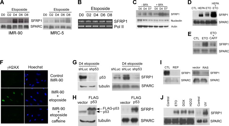
Stress-induced SFRP1 secretion. (A) Secretion of SFRP1 from etoposide-treated IMR-90 and MRC-5 cells. IMR-90 and MRC-5 cells were treated with 20 μM etoposide for 48 h. On the indicated days after initiating the etoposide treatment, conditioned medium was collected and concentrated. Conditioned medium from untreated cells (day 0 [D0]) was also included as a control. Ten micrograms of each conditioned medium was analyzed for SFRP1 levels by immunoblotting. SPARC (secreted protein, acidic, cysteine rich) served as a loading control. (B) RNA levels of SFRP1 after etoposide treatment of IMR-90 cells. IMR-90 cells were treated with 20 μM etoposide for 48 h. At the indicated time points after initiating the etoposide treatment, total RNA was isolated. Total RNA from untreated cells (D0) was also included as control. RT-PCR analysis was performed for mRNA levels of SFRP1 and RNA polymerase II (Pol II, loading control). (C) Increased SFRP1 entry to the secretory pathway upon etoposide treatment. IMR-90 cells were left untreated (D0) or treated with 20 μM etoposide for 48 h. At the indicated time points after initiating the etoposide treatment, whole-cell lysates were prepared with or without brefeldin A (BFA) treatment (5 μg/ml for 2 h) and were analyzed for SFRP1 levels by immunoblotting. Nucleolin and actin served as loading controls. (D) Etoposide (ETO) increases extracellular SFRP1 levels in the presence of heparin (HEPA). IMR-90 cells were treated with 50 μg/ml heparin or 20 μM etoposide or both for 48 h. Four days after each treatment was initiated, the SFRP1 protein present in the conditioned medium was determined by anti-SFRP1 immunoblotting. (E and F) Inhibition of SFRP1 secretion by caffeine. IMR-90 cells were left untreated, treated with 20 μM etoposide for 48 h, or treated with 5 mM caffeine (CAFF) overnight prior to treatment with 20 μM etoposide for 48 h. Four days after etoposide treatment, the secretion of SFRP1 and SPARC was analyzed by immunoblotting (E), and the DNA damage response was assessed by γH2AX staining (F). (G) Etoposide-induced SFRP1 secretion is p53 dependent. IMR-90 cells were infected with lentiviruses expressing shRNA against luciferase (shLuc, control) or shRNA against p53 (shp53) and were selected with 2 μg/ml puromycin. The selected cells were treated with 20 μM etoposide for 48 h. Five days after the etoposide treatment was initiated, conditioned medium was collected and was analyzed for SFRP1 and SPARC levels by immunoblotting (right). p53 knockdown was verified by anti-p53 immunoblotting of the whole-cell lysates (left). (H) p53 induces SFRP1 secretion. IMR-90 cells were infected with empty vector or FLAG-p53-expressing lentivirus. Four days later, the expression of p53 and tubulin was analyzed by immunoblotting of the whole-cell lysates (left), and the secretion of SFRP1 and SPARC was analyzed by immunoblotting of the conditioned medium (right). (I) Secretion of SFRP1 from IMR-90 cells undergoing replicative senescence (REP) or Ras-induced senescence (RAS) was analyzed by anti-SFRP1 immunoblotting. CTL, control. (J) Different stresses induce SFRP1 secretion. IMR-90 cells were treated with 20 μM etoposide (ETO) for 48 h, 1 μM doxorubicin (DOX) for 2 h, 500 μM H2O2 for 2 h, or 2 J/m2 UV light. Conditioned medium was collected at 4 days posttreatment and was analyzed for SFRP1 and SPARC levels by immunoblotting.
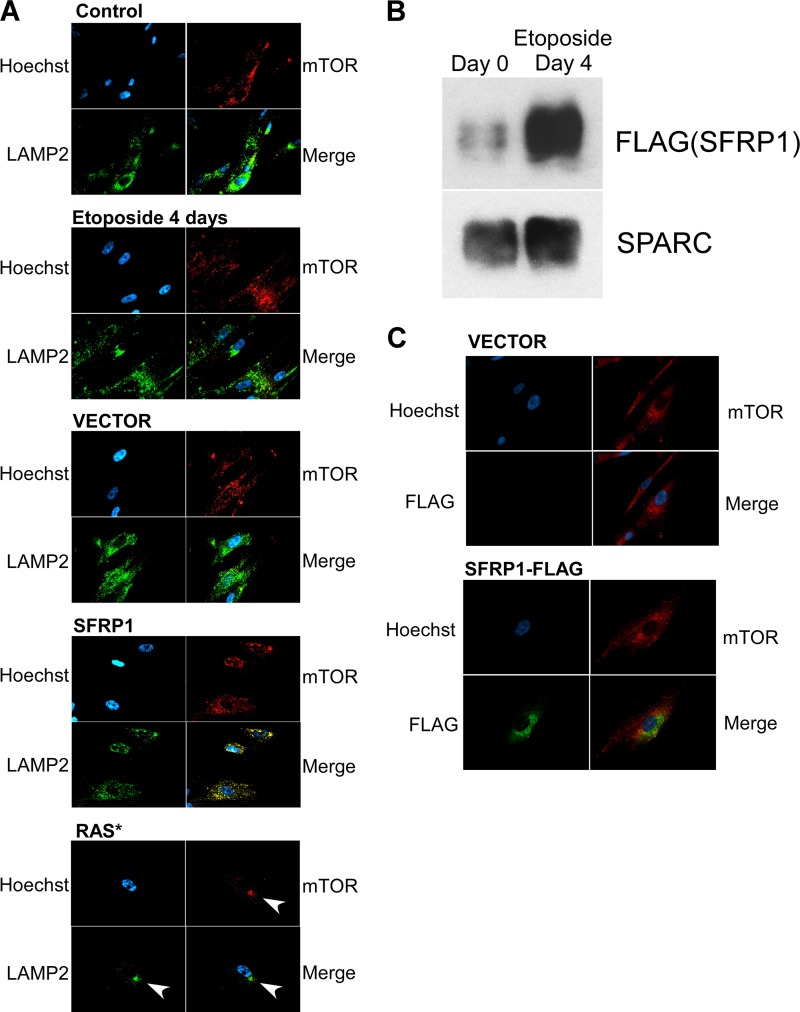
Lack of TASCC formation upon etoposide treatment or SFRP1 expression. (A) Subcellular location of mTOR and LAMP2 upon senescence induced by etoposide, SFRP1, or oncogenic Ras. The formation of TASCC (TOR-autophagy spatial coupling compartment) was assessed by staining for mTOR and LAMP2. Whereas TASCC was readily detectable upon Ras-induced senescence (arrows), TASCC formation was not observed upon etoposide-induced or SFRP1-induced senescence. (B) Etoposide increases the secretion of FLAG-tagged SFRP1. IMR-90 cells were infected with lentiviruses expressing C-terminally FLAG-tagged SFRP1 and were treated with etoposide. The levels of secreted FLAG-tagged SFRP1 were determined by anti-FLAG immunoblotting. (C) Lack of colocalization of FLAG-tagged SFRP1 and mTOR. IMR-90 cells were infected with lentiviruses expressing C-terminally FLAG-tagged SFRP1 or empty vector and were treated with etoposide. Four days later, the cells were stained for mTOR and FLAG.
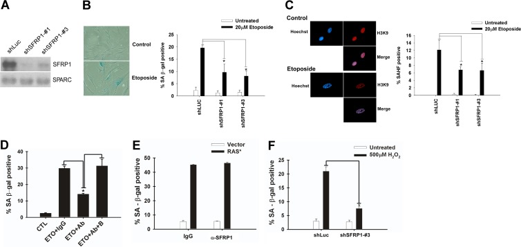
SFRP1 downregulation alleviates etoposide-induced senescence. (A) shRNA knockdown of SFRP1. IMR-90 cells were infected with lentiviruses expressing shRNA against luciferase (shLuc; control) or shRNAs against SFRP1 (shSFRP1-1 and shSFRP1-3) and were selected with 2 μg/ml puromycin. Five days after infection, conditioned medium was collected and was analyzed for SFRP1 and SPARC levels by immunoblotting. (B and C) SFRP1 knockdown attenuates etoposide-induced senescence. The effect of SFRP1 knockdown on etoposide-induced senescence of IMR-90 cells was analyzed 6 days after etoposide treatment. (B) SA-β-Gal staining. (C) Senescence-associated heterochromatic foci (SAHF). A minimum of 100 cells were counted. *, P < 0.05 compared to shLuc plus 20 μM etoposide. (D) SFRP1 antibody inhibits etoposide-induced senescence. IMR-90 cells were treated for 48 h with etoposide. Beginning 24 h after treatment, the cells were incubated with either 1 μg/ml of control rabbit IgG, anti-SFRP1 antibody (rabbit polyclonal H-90; Santa Cruz Biotechnology), or anti-SFRP1 antibody (Ab) plus immunogen (block [B]). Cells were assayed for SA-β-Gal staining 5 days after etoposide treatment. *, P < 0.05 compared to etoposide plus the IgG control or to etoposide plus anti-SFRP1 and block. (E) SFRP1 antibody does not affect Ras-induced senescence. IMR-90 cells were infected with c-H-RasV12-expressing lentivirus (empty vector lentivirus as a control). Beginning 4 days after infection, the cells were incubated with either 1 μg/ml of control rabbit IgG or anti-SFRP1 antibody. Eight days after infection, the cells were stained for SA-β-Gal. (F) SFRP1 knockdown abolishes oxidative stress-induced senescence. IMR-90 cells were treated with 500 μM H2O2 for 2 h. The following day, the cells were infected with lentiviruses expressing shRNA against luciferase or SFRP1. On day 6 after H2O2 treatment, the cells were analyzed for SA-β-Gal. *, P < 0.05.
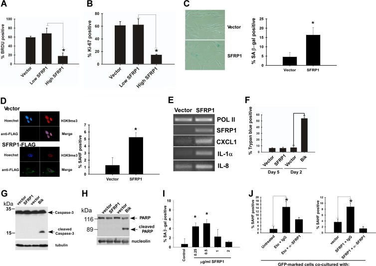
SFRP1 induces senescence. (A and B) SFRP1 inhibits cell proliferation. IMR-90 cells were infected with empty lentiviral vector or C-terminally FLAG-tagged SFRP1 lentivirus and were selected with 2 μg/ml puromycin. (A) BrdU incorporation. Four days after infection, the cells were labeled with BrdU for 24 h and stained with antibodies against BrdU and SFRP1. Immunofluorescence microscopy was used to detect the cells displaying no SFRP1 or low SFRP1 expression (Low SFRP1) and those displaying high SFRP1 expression (High SFRP1), and the percentage of BrdU-positive cells was scored. (B) Ki-67 staining. Five days after infection, the cells were stained for Ki-67 and FLAG (SFRP1). *, P < 0.05. (C and D) SFRP1 lentivirus induces senescence phenotypes. IMR-90 cells were infected with empty lentiviral vector or SFRP1 lentivirus and were selected with 2 μg/ml puromycin. Five days after infection, the cells were stained for SA-β-Gal (C) or SAHF (D). *, P < 0.05 compared to vector control. (E) SFRP1 induces senescence-associated secretory phenotype genes. The expression of CXCL1, IL-1α, and IL-8 was examined by RT-PCR 5 days after SFRP1 viral expression of IMR-90 cells. RNA polymerase II (Pol II) served as a loading control. (F) Trypan blue staining. IMR-90 cells expressing SFRP1 (5 days after infection) or Bik (2 days after infection) were assessed for cell death by dye exclusion assays. (G) Caspase 3 cleavage. IMR-90 cells expressing SFRP1 or Bik were assessed for caspase 3 cleavage by immunoblotting with tubulin as a loading control. (H) PARP cleavage. IMR-90 cells expressing SFRP1 or Bik were assessed for PARP cleavage by immunoblotting with nucleolin as loading control. (I) Recombinant SFRP1 induces senescence in IMR-90 cells. IMR-90 cells were treated with the indicated concentration of recombinant SFRP1 for 4 days and were stained for SA-β-Gal. *, P < 0.05 compared to untreated control. (J) Nonsenescent IMR-90 cells acquire a senescence phenotype upon coculture with senescent cells. Nonsenescent IMR-90 cells were infected with GFP-expressing lentivirus. These GFP-labeled IMR-90 cells were cocultured with either senescent IMR-90 cells (induced to senesce by etoposide treatment [left] or by SFRP1 lentiviral expression [right]), or nonsenescent IMR-90 cells (untreated or vector infected) for 4 days. Where indicated, the cells were treated with 1 μg/ml of control IgG or anti-SFRP1 antibody beginning 24 h after initiation of coculture. The GFP-positive cells were scored for the development of senescence-associated heterochromatic foci (SAHF). *, P < 0.05. Note that the GFP-positive cells in coculture were not positive for the DNA damage marker γH2AX, indicating that the spreading of senescence is not due to residual etoposide or SFRP1-induced DNA damage.
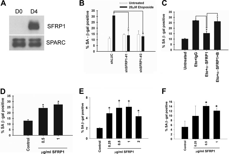
SFRP1 mediates senescence in epithelial cells. (A) Secretion of SFRP1 from etoposide-treated RPE-28 cells. RPE-28 retinal pigment epithelial cells were treated with 20 μM etoposide for 48 h. Four days after the etoposide treatment was initiated, conditioned medium was collected and concentrated. Conditioned medium from untreated cells (D0) was also included as a control. Ten micrograms of each conditioned medium was analyzed for SFRP1 levels by immunoblotting. SPARC served as a loading control. (B) SFRP1 shRNAs attenuates etoposide-induced senescence in RPE-28 cells. RPE-28 cells were infected with lentiviruses expressing shRNA against luciferase or shRNAs against SFRP1 and were selected with 2 μg/ml puromycin. The effect of SFRP1 shRNAs on etoposide-induced senescence of RPE-28 cells was analyzed 6 days after etoposide treatment. *, P < 0.05 compared to shLuc plus 20 μM etoposide. (C) SFRP1 antibody inhibits etoposide-induced senescence in RPE-28 cells. RPE-28 cells were treated for 48 h with etoposide. Beginning 24 h after treatment, the cells were incubated with 1 μg/ml of control rabbit IgG, anti-SFRP1 antibody, or anti-SFRP1 antibody plus immunogen (block [B]). Cells were assayed for SA-β-Gal staining at 5 days after etoposide treatment. *, P < 0.05 compared to etoposide plus the IgG control or to etoposide plus anti-SFRP1 plus block. (D) Recombinant SFRP1 induces senescence in RPE-28 cells. RPE-28 cells were treated with the indicated concentration of recombinant SFRP1 for 4 days and were stained for SA-β-Gal. *, P < 0.05 compared to untreated control. Human primary mammary epithelial cells (E) or MCF-7 breast cancer cells (F) were treated with the indicated concentration of recombinant SFRP1 for 4 days and were stained for SA-β-Gal. *, P < 0.05 compared to untreated control.
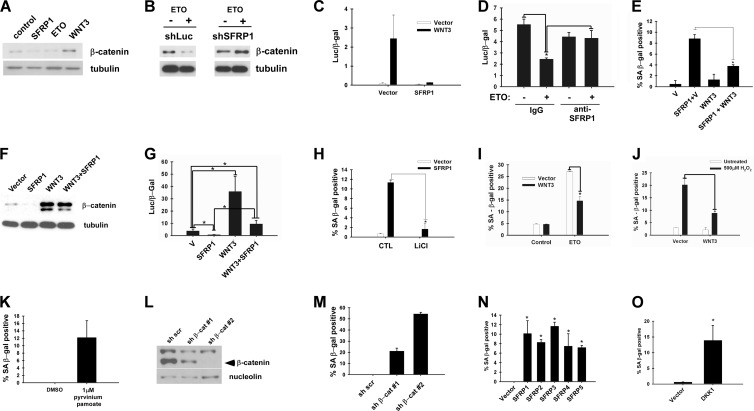
Wnt inhibition results in senescence. (A) SFRP1 reduces soluble β-catenin levels in IMR-90 cells. IMR-90 cells were infected with vector, SFRP1, or Wnt3 lentivirus or treated with 20 μM etoposide. Five days later, soluble β-catenin levels were analyzed by immunoblotting using 5 μg of each soluble fraction. (B) SFRP1 knockdown abrogates etoposide-induced reduction of soluble β-catenin levels. IMR-90 cells were infected with lentiviruses expressing shRNA against luciferase (shLuc) or shRNA against SFRP1 (shSFRP1-3). The cells were treated with 20 μM etoposide, and 5 days later, soluble β-catenin levels were analyzed by immunoblotting using 5 μg of each soluble fraction. (C) SFRP1 silences Wnt-dependent transcription in IMR-90 cells. IMR-90 cells were cotransfected with Super TopFlash reporter, cytomegalovirus (CMV)-β-Gal, and empty vector, SFRP1, or Wnt3 where indicated, and the luciferase activity was determined 48 h after transfection. Transfection efficiencies were normalized using CMV-β-Gal activity. (D) Etoposide treatment represses TopFlash reporter activity. IMR-90 cells were treated with 20 μM etoposide or left untreated for 24 h prior to transfection. The cells were transfected with Super TopFlash reporter and CMV-β-Gal for 4 h, and 1 μg/ml anti-SFRP1 antibody (H-90) or IgG was added after removal of the transfection mixture. The luciferase activity was determined 48 h after transfection. Transfection efficiencies were normalized using CMV-β-Gal activity. *, P < 0.05. (E) Wnt3 counteracts SFRP1-induced senescence. IMR-90 cells were infected with the indicated lentiviruses, and 5 days after infection, the cells were stained for SA-β-Gal. *, P < 0.05. (F) Wnt3 counteracts SFRP1-induced reduction of soluble β-catenin levels. Soluble β-catenin levels were analyzed by immunoblotting. (G) Wnt3 counteracts SFRP1-induced repression of TopFlash reporter activity. IMR-90 cells were transfected with Super TopFlash reporter and CMV-β-Gal in conjunction with SFRP1 or Wnt3 as indicated. *, P < 0.05. (H) LiCl treatment suppresses SFRP1-induced senescence. IMR-90 cells were infected with vector or SFRP1 lentivirus, and 3 days after infection, the cells were treated with 20 mM LiCl for 48 h and stained for SA-β-Gal. *, P < 0.05. (I) Wnt3 counteracts etoposide-induced senescence. IMR-90 cells were infected with Wnt3 or empty vector lentivirus. The cells were treated with 20 μM etoposide or left untreated, and the SA-β-Gal activity was analyzed 6 days after etoposide treatment. *, P < 0.05. (J) Wnt3 counteracts oxidative stress-induced senescence. IMR-90 cells were treated with 500 μM H2O2 for 2 h. The subsequent day, the cells were infected with lentiviruses expressing Wnt3 or empty vector. On day 6 after H2O2 treatment, the cells were analyzed for SA-β-Gal. *, P < 0.05. (K) Pharmacological inhibition of Wnt signaling results in senescence. IMR-90 cells were treated with dimethyl sulfoxide (DMSO) control or 1 μM pyrvinium pamoate for 4 days and were stained for SA-β-Gal. (L) shRNA knockdown of β-catenin. IMR-90 cells were infected with lentiviruses expressing scrambled shRNA (sh scr), shRNA against β-catenin-1 (sh β-cat#1), or shRNA against β-catenin-2 (sh β-cat#2) (sh β-cat 2) and 5 days after infection, β-catenin protein levels were analyzed by immunoblotting. Nucleolin served as a loading control. (M) β-Catenin knockdown results in senescence. IMR-90 cells were infected with the indicated lentiviruses and at 5 days postinfection stained for SA-β-Gal. (N) All five SFRP family members induce senescence. IMR-90 cells were infected with lentiviruses expressing the indicated SFRP family members. At 5 days postinfection, the cells were stained for SA-β-Gal. *, P < 0.05 compared to vector control. (O) DKK1 induces senescence. IMR-90 cells were infected with vector or DKK1 lentivirus. At 5 days postinfection, the cells were stained for SA-β-Gal. *, P < 0.05 compared to vector control.
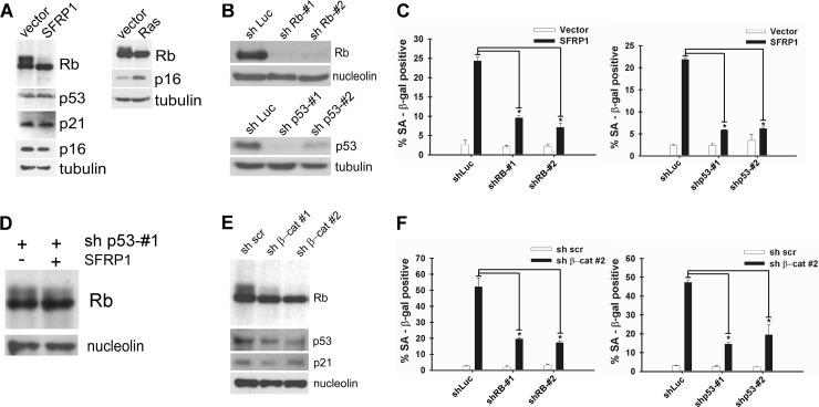
Role of Rb and p53 pathways in senescence induction by Wnt inhibition. (A) Rb dephosphorylation upon SFRP1 expression. IMR-90 cells were infected with vector or SFRP1 lentivirus, and 5 days after infection, Rb, p53, p21, p16, and tubulin expression was analyzed by immunoblotting. SFRP1 induced Rb dephosphorylation but did not affect p53, p21, or p16 expression. For comparison, Rb, p16, and tubulin expression upon Ras-induced senescence was also analyzed by immunoblotting. (B) Knockdown of Rb and p53 in IMR-90 cells. IMR-90 cells were infected with the indicated lentiviruses. Five days after infection, Rb, p53, nucleolin, and tubulin expression was analyzed by immunoblotting. sh, shRNA. (C) Knockdown of Rb or p53 abolishes SFRP1-induced senescence. IMR-90 cells were coinfected with the indicated lentiviruses. Five days postinfection, the cells were stained for SA-β-Gal. *, P < 0.05 compared to SFRP1 plus shLuc. (D) p53 knockdown attenuates Rb dephosphorylation by SFRP1. IMR-90 cells were coinfected with the indicated lentiviruses, and 5 days after infection, Rb expression was analyzed by immunoblotting. (E) β-Catenin knockdown results in Rb dephosphorylation. IMR-90 cells were infected with lentiviruses expressing scrambled shRNA (sh scr), shRNA against β-catenin-1 (sh β-cat 1), or shRNA against β-catenin-2 (sh β-cat#2), and 5 days after infection, Rb, p53, and p21 expression was analyzed by immunoblotting. Nucleolin served as a loading control. (F) Knockdown of Rb or p53 abolishes β-catenin RNAi-induced senescence. IMR-90 cells were coinfected with the indicated lentiviruses and 5 days after infection stained for SA-β-Gal. *, P < 0.05 compared to sh β-catenin 2 plus shLuc.
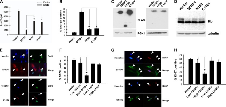
Cancer-associated SFRP1 mutants are defective for senescence induction. (A) Cancer-associated SFRP1 mutants display compromised Wnt-inhibitory activity. 293 cells were cotransfected with Super TopFlash reporter, CMV-β-Gal, and plasmids encoding Wnt3, SFRP1, SFRP1 mutants found in human cancers (N150 or C140Y), or empty vector as indicated. The luciferase activity was determined 48 h after transfection. Transfection efficiencies were normalized using CMV-β-Gal activity. *, P < 0.05. (B) Cancer-associated SFRP1 mutants are defective for senescence induction. IMR-90 cells were infected with the indicated lentiviruses and at 5 days postinfection stained for SA-β-Gal. *, P < 0.05. (C) Expression levels of wild-type and mutant SFRP1. IMR-90 cells were infected with lentiviruses encoding SFRP1 or the N150 or C140Y mutant (all C-terminally FLAG tagged). Five days postinfection, the expression levels of wild-type and mutant SFRP1 were analyzed by anti-FLAG immunoblotting of the brefeldin A-treated cell lysate (left) and the conditioned medium (right). (D) SFRP1 mutants display impaired ability to induce dephosphorylation of Rb. IMR-90 cells were infected with vector lentivirus or lentivirus encoding SFRP1, N150, or C140Y, and 5 days after infection, Rb expression was analyzed by immunoblotting. Tubulin served as a loading control. (E to H) The SFRP1 C140Y mutant is unable to inhibit cell proliferation. IMR-90 cells were infected with empty lentiviral vector or lentiviruses expressing wild-type or C140Y mutant SFRP1. Cell proliferation was assessed by BrdU incorporation (E and F) and by Ki-67 staining (G and H) as described in the legend of Fig. 4A and B. The cells expressing wild-type or C140Y SFRP1 are indicated by arrowheads in panels E and G. *, P < 0.05.
References
-
- Acosta JC, et al. 2008. Chemokine signaling via the CXCR2 receptor reinforces senescence. Cell 133: 1006–1018 - PubMed
-
- Bartkova J, et al. 2006. Oncogene-induced senescence is part of the tumorigenesis barrier imposed by DNA damage checkpoints. Nature 444: 633–637 - PubMed
-
- Bovolenta P, Esteve P, Ruiz JM, Cisneros E, Lopez-Rios J. 2008. Beyond Wnt inhibition: new functions of secreted Frizzled-related proteins in development and disease. J. Cell Sci. 121: 737–746 - PubMed
-
- Caldwell GM, et al. 2004. The Wnt antagonist sFRP1 in colorectal tumorigenesis. Cancer Res. 64: 883–888 - PubMed
Publication types
MeSH terms
Substances
Grants and funding
LinkOut - more resources
Full Text Sources
Other Literature Sources
Research Materials