

Role of calcium and mitochondria in MeHg-mediated cytotoxicity
- PMID: 22927718
- PMCID: PMC3425894
- DOI: 10.1155/2012/248764
Role of calcium and mitochondria in MeHg-mediated cytotoxicity
Abstract
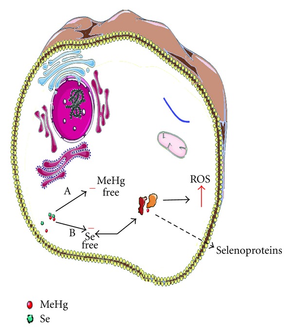
Methylmercury (MeHg) mediated cytotoxicity is associated with loss of intracellular calcium (Ca²⁺) homeostasis. The imbalance in Ca²⁺ physiology is believed to be associated with dysregulation of Ca²⁺ intracellular stores and/or increased permeability of the biomembranes to this ion. In this paper we summarize the contribution of glutamate dyshomeostasis in intracellular Ca²⁺ overload and highlight the mitochondrial dysfunctions induced by MeHg via Ca²⁺ overload. Mitochondrial disturbances elicited by Ca²⁺ may involve several molecular events (i.e., alterations in the activity of the mitochondrial electron transport chain complexes, mitochondrial proton gradient dissipation, mitochondrial permeability transition pore (MPTP) opening, thiol depletion, failure of energy metabolism, reactive oxygen species overproduction) that could culminate in cell death. Here we will focus on the role of oxidative stress in these phenomena. Additionally, possible antioxidant therapies that could be effective in the treatment of MeHg intoxication are briefly discussed.
Figures


Similar articles
-
Harmonization of Mangiferin on methylmercury engendered mitochondrial dysfunction.Environ Toxicol. 2017 Feb;32(2):630-644. doi: 10.1002/tox.22265. Epub 2016 Mar 28. Environ Toxicol. 2017. PMID: 28071871
-
Evidence for interactions between intracellular calcium stores during methylmercury-induced intracellular calcium dysregulation in rat cerebellar granule neurons.J Pharmacol Exp Ther. 2003 Mar;304(3):949-58. doi: 10.1124/jpet.102.042457. J Pharmacol Exp Ther. 2003. PMID: 12604669
-
Unbalanced ER-mitochondrial calcium homeostasis promotes mitochondrial dysfunction and associated apoptotic pathways activation in methylmercury exposed rat cortical neurons.J Biochem Mol Toxicol. 2022 Sep;36(9):e23136. doi: 10.1002/jbt.23136. Epub 2022 Jun 9. J Biochem Mol Toxicol. 2022. PMID: 35678294
-
Mitochondria and Ca(2+)in cell physiology and pathophysiology.Cell Calcium. 2000 Nov-Dec;28(5-6):339-48. doi: 10.1054/ceca.2000.0170. Cell Calcium. 2000. PMID: 11115373 Review.
-
Methylmercury and brain development: A review of recent literature.J Trace Elem Med Biol. 2016 Dec;38:99-107. doi: 10.1016/j.jtemb.2016.03.001. Epub 2016 Mar 4. J Trace Elem Med Biol. 2016. PMID: 26987277 Free PMC article. Review.
Cited by
-
Low-Dose Methylmercury-Induced Apoptosis and Mitochondrial DNA Mutation in Human Embryonic Neural Progenitor Cells.Oxid Med Cell Longev. 2016;2016:5137042. doi: 10.1155/2016/5137042. Epub 2016 Jul 21. Oxid Med Cell Longev. 2016. PMID: 27525052 Free PMC article.
-
L-Ascorbic Acid 2-Phosphate Attenuates Methylmercury-Induced Apoptosis by Inhibiting Reactive Oxygen Species Accumulation and DNA Damage in Human SH-SY5Y Cells.Toxics. 2023 Feb 2;11(2):144. doi: 10.3390/toxics11020144. Toxics. 2023. PMID: 36851019 Free PMC article.
-
The catecholaminergic neurotransmitter system in methylmercury-induced neurotoxicity.Adv Neurotoxicol. 2017;1:47-81. doi: 10.1016/bs.ant.2017.07.002. Epub 2017 Sep 1. Adv Neurotoxicol. 2017. PMID: 32346666 Free PMC article. No abstract available.
-
Methylmercury alters proliferation, migration, and antioxidant capacity in human HTR8/SV-neo trophoblast cells.Reprod Toxicol. 2018 Jun;78:60-68. doi: 10.1016/j.reprotox.2018.03.008. Epub 2018 Mar 23. Reprod Toxicol. 2018. PMID: 29581082 Free PMC article.
-
The Putative Role of Environmental Mercury in the Pathogenesis and Pathophysiology of Autism Spectrum Disorders and Subtypes.Mol Neurobiol. 2018 Jun;55(6):4834-4856. doi: 10.1007/s12035-017-0692-2. Epub 2017 Jul 22. Mol Neurobiol. 2018. PMID: 28733900 Review.
References
-
- Bakir F, Damluji SF, Amin Zaki I. Methylmercury poisoning in Iraq: an interuniversity report. Science. 1973;181(4096):230–241. - PubMed
Publication types
MeSH terms
Substances
LinkOut - more resources
Full Text Sources
Miscellaneous
