

Histopathology of optic nerve pit-associated maculopathy
- PMID: 22927726
- PMCID: PMC3422135
- DOI: 10.2147/OPTH.S34706
Histopathology of optic nerve pit-associated maculopathy
Abstract
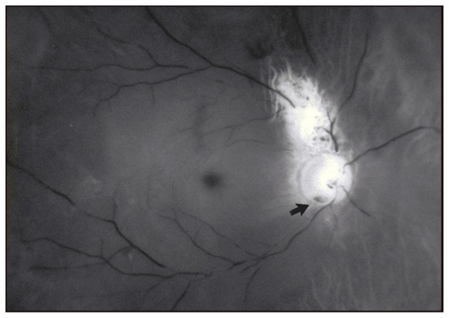
Purpose: To describe the histopathologic findings of an eye bank specimen containing an optic nerve pit with associated serous elevation of the macula and cavernous atrophy of the optic nerve.
Methods: An eye bank specimen found to have an optic nerve pit with serous elevation of the macula was grossly examined and photographed. The globe was processed for both light and scanning electron microscopy.
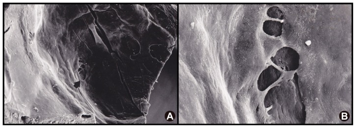
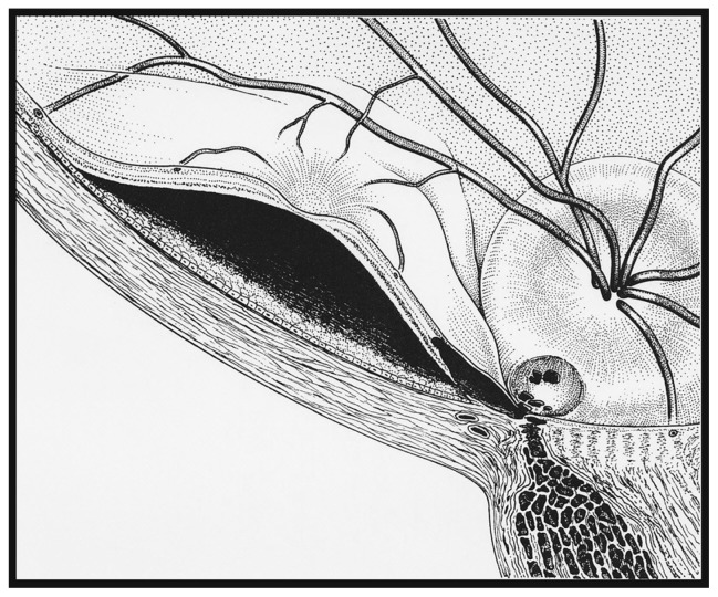
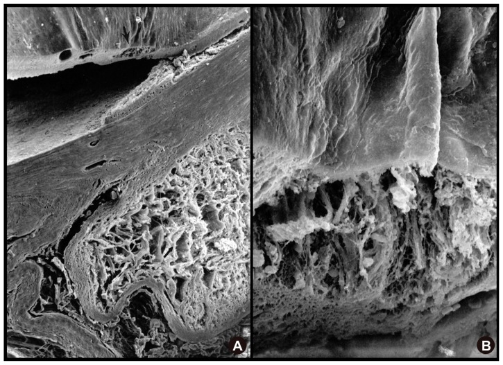
Results: The scanning electron microscopic study of this eye with an optic nerve pit revealed holes in the diaphanous membrane overlying the nerve at the edge of the optic pit. Serial histopathology sections revealed a connection between the holes overlying the optic pit and the subretinal space via a schisis-like cavity in the retina.
Conclusion: The discovery of an optic nerve pit with coexisting serous detachment of the macula in an eye bank eye and subsequent pathological evaluation provides support for current theories into the mechanism of the visual loss in this condition. Our finding supports syneretic vitreous to be the source of the subretinal fluid.
Keywords: electron microscopy; light microscopy; optic nerve pit; retinal detachment.
Figures

References
-
- Wiethe T. Ein FaIl von Angeborener Difformitat der Schnervenpapille. Arch F Augenheilkd. 1882;11:14–19.
-
- Brown GC, Shields JA, Goldberg RE. Congenital pits of the optic nerve head: II. Clinical studies in humans. Ophthalmology. 1980;87(1):51–65. - PubMed
-
- Reed D. Congenital pits of the optic nerve 1999. Clin Eye Vis Care. 11(2):75–80.
-
- Reis W. Ein wenig bekannte typische Missbildung am Sehnerveneintritt, Z. Augenheilk. 1908;19:505–528.
-
- Brown GC, Tasman WS. Congenital Anomalies of the Optic Disc. New York, NY: Grune and Stratton; 1983. pp. 97–126.
LinkOut - more resources
Full Text Sources
