. 2012;10(8):e1001379.
doi: 10.1371/journal.pbio.1001379. Epub 2012 Aug 21.

Evolutionarily repurposed networks reveal the well-known antifungal drug thiabendazole to be a novel vascular disrupting agent

Affiliations

Evolutionarily repurposed networks reveal the well-known antifungal drug thiabendazole to be a novel vascular disrupting agent

Hye Ji Cha et al. PLoS Biol. 2012.

Abstract

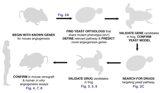
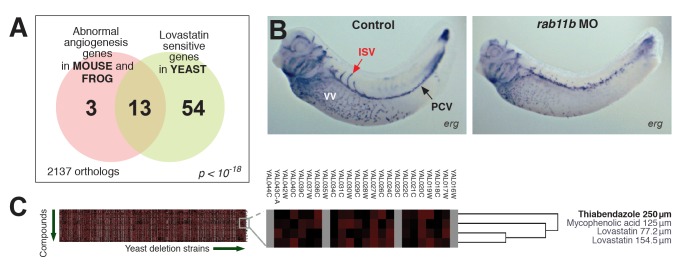
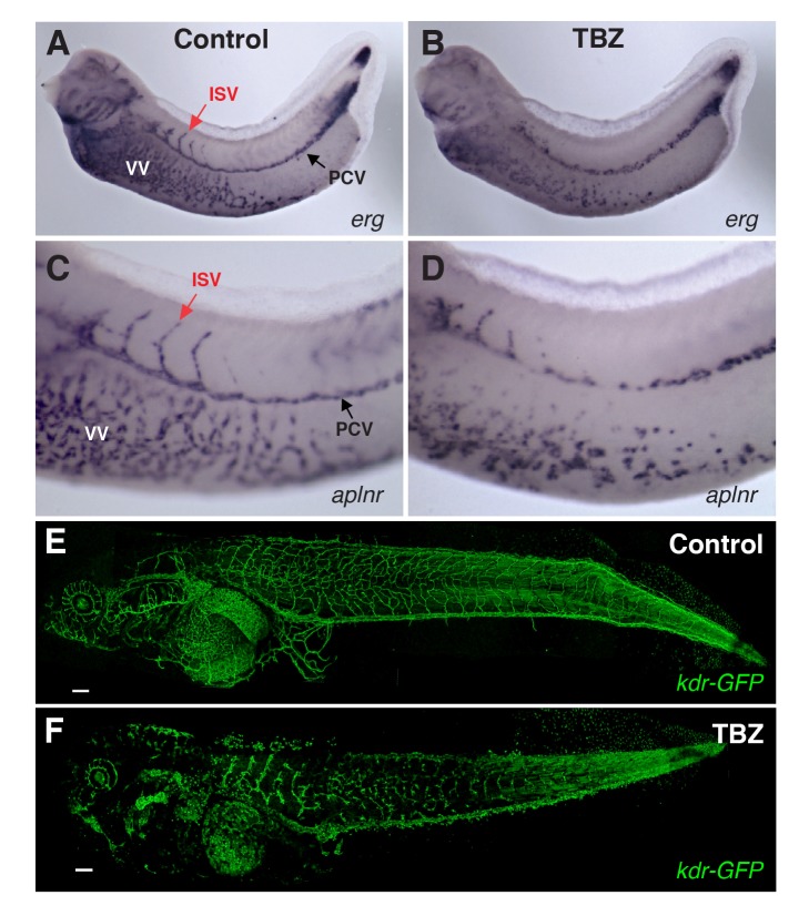
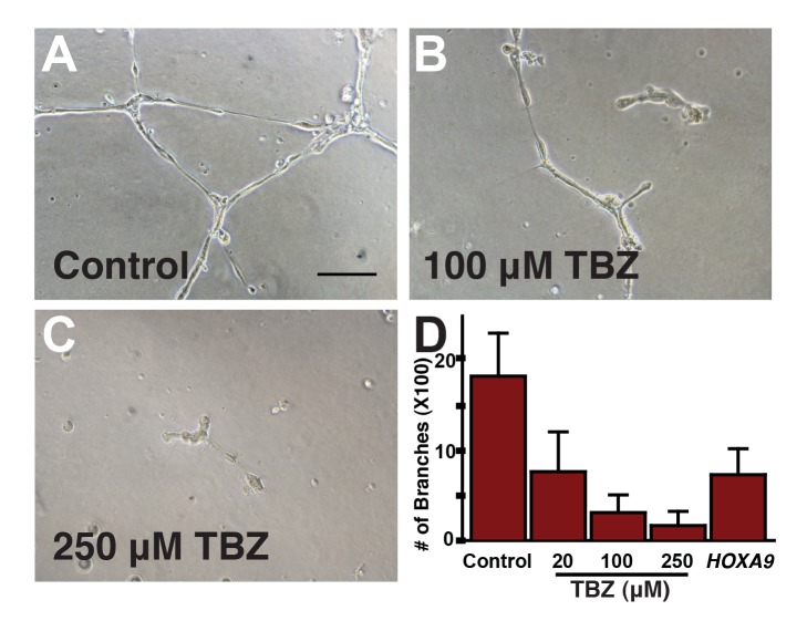
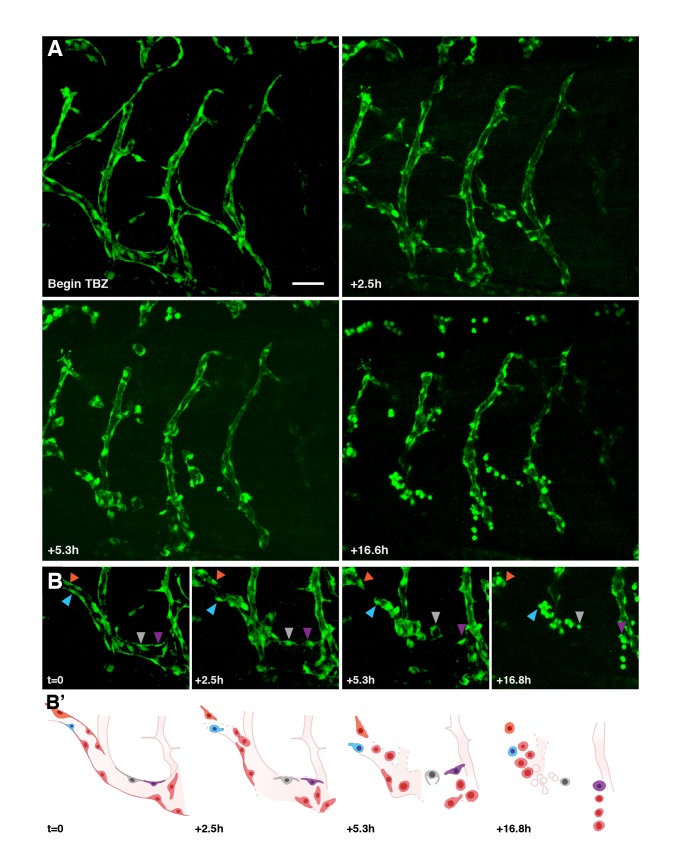
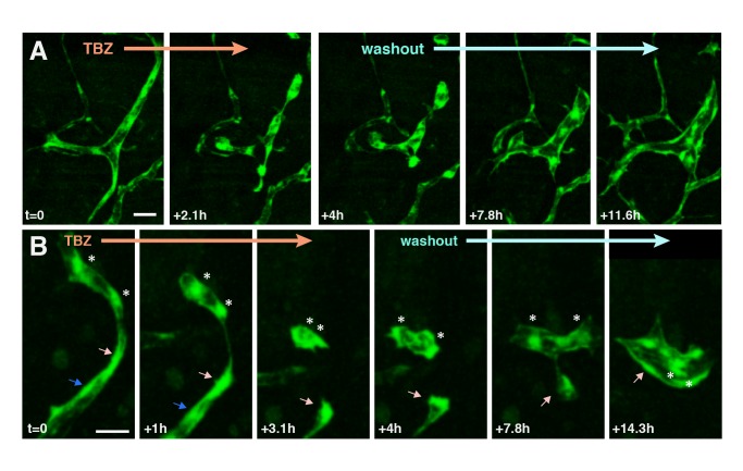
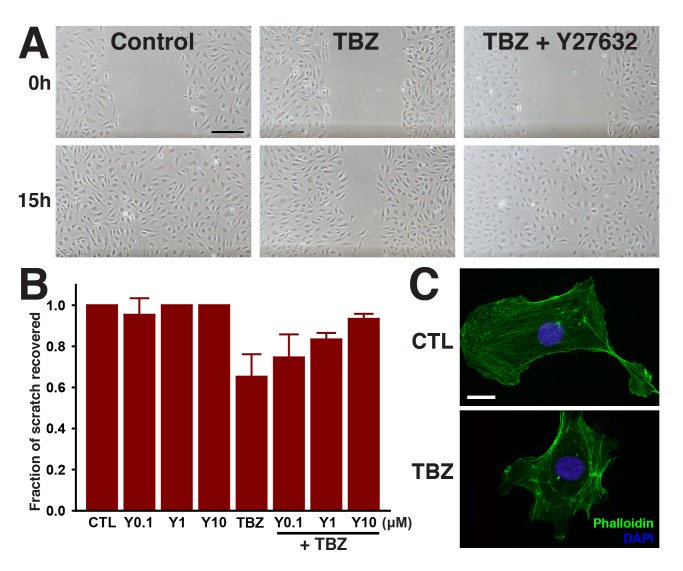
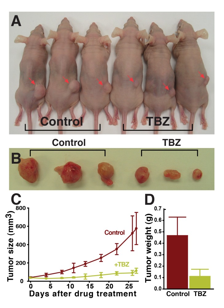
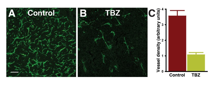
Studies in diverse organisms have revealed a surprising depth to the evolutionary conservation of genetic modules. For example, a systematic analysis of such conserved modules has recently shown that genes in yeast that maintain cell walls have been repurposed in vertebrates to regulate vein and artery growth. We reasoned that by analyzing this particular module, we might identify small molecules targeting the yeast pathway that also act as angiogenesis inhibitors suitable for chemotherapy. This insight led to the finding that thiabendazole, an orally available antifungal drug in clinical use for 40 years, also potently inhibits angiogenesis in animal models and in human cells. Moreover, in vivo time-lapse imaging revealed that thiabendazole reversibly disassembles newly established blood vessels, marking it as vascular disrupting agent (VDA) and thus as a potential complementary therapeutic for use in combination with current anti-angiogenic therapies. Importantly, we also show that thiabendazole slows tumor growth and decreases vascular density in preclinical fibrosarcoma xenografts. Thus, an exploration of the evolutionary repurposing of gene networks has led directly to the identification of a potential new therapeutic application for an inexpensive drug that is already approved for clinical use in humans.


Conflict of interest statement

A patent application based on this work has been filed. The authors have declared that no other competing interests exist.

Figures

Figure 1
Figure 1. Overview of the evolutionary method used to discover a novel vascular disrupting agent.
Strict reliance on the evolutionary conservation of the relevant gene module allowed for an experimental design exploiting the unique experimental advantages of each model organism, thus speeding the search for novel angiogenesis inhibitors. (Vector female silhouette under Creative Commons Attribution 2.0 from ‘Keep Fit’ Vector Pack, Blog.SpoonGraphics.)
Figure 2
Figure 2. Identification of candidate angiogenesis inhibitors based upon genetic interactions with a yeast gene module.
(A) Summary of the gene module (modified from [1]). Tests of genes associated with the yeast phenotype (lovastatin sensitivity) correctly identified novel angiogenesis genes, as in and additionally shown in (B) for the gene rab11b. Morpholino (MO) knockdown of rab11B induces vascular defects in developing Xenopus laevis (frog) embryos, measured by in situ hybridization versus marker gene erg. ISV, intersomitic vein; PCV, posterior cardinal vein; VV, vitellin vein. (C) In an unbiased hierarchical clustering of compounds by their synthetic genetic interaction profiles with yeast genes (analyzing data from [13]), the action of TBZ is among those interacting with this gene module and also most similar to lovastatin, the signature compound affiliated with the angiogenesis gene module; hence, TBZ is a likely candidate angiogenesis inhibitor. Here, complete linkage clustering employing uncentered correlation coefficients is shown; additional clustering methods are illustrated in Figure S2.
Figure 3
Figure 3. TBZ inhibits angiogenesis in vivo in Xenopus embryos.
Formation of Xenopus embryo veins is disrupted, marked by expression of vascular reporter genes (A, B) erg and (C, D) aplnr, contrasting treatment with 1% DMSO control only (A, C) with 1% DMSO, 250 µM TBZ, treated at stage 31 and imaged at stages 35–36 (B, D). PCV, posterior cardinal vein; ISV, intersomitic vein; VV, vitellin veins. Similarly, TBZ disrupts vasculature imaged within a living Xenopus embryo and visualized by vascular specific GFP in kdr:GFP frogs from , contrasting the vasculature of stage 46 animals treated from stage 41 with the 1% DMSO control (E) or 1% DMSO, 250 µM TBZ (F). Scale bar, 200 µm.
Figure 4
Figure 4. TBZ significantly disrupts tube formation in cultured human umbilical vein endothelial cells (HUVECs), an in vitro capillary model.
Here, we show effects of 1% DMSO-treated control (A) versus 1% DMSO, 100 µM TBZ (B) and 1% DMSO, 250 µM TBZ (C). Scale bar, 100 µm. (D) Tube disruption is dose-dependent and comparable to that from silencing known pro-angiogenic gene HOXA9.
Figure 5
Figure 5. TBZ disrupts newly established vasculature, as visualized in vivo using time-lapse fluorescence microscopy within kdr:GFP frogs.
Retraction and rounding of vascular endothelial cells (arrowheads) is apparent in TBZ-treated embryos (A, time lapse of frogs treated as in Figure 3) as compared with continued vascular growth in control animals (Figure S7). Scale bar, 80 µm. (B) A series with intermediate time points is shown for a sub-region of that shown in (A). (B′) Schematics of the images in (B) indicate positions of specific cells.
Figure 6
Figure 6. After disassembly of the vasculature by TBZ, washout of the drug leads to partial recovery of the vascular network.
Two time series are shown in (A) and (B), imaged as in Figure 5. Following washout, cells dissociated by TBZ treatment recommence cell elongation and connection. Individual cells are indicated by arrows/asterisks. Scale bar in (A), 50 µm; in (B), 40 µm.
Figure 7
Figure 7. TBZ impedes migration of HUVECs in a wound scratch assay, but treatment with the Rho Kinase inhibitor Y27632 reverses TBZ's effects.
(A) The effects of 1% DMSO-treated control versus 1% DMSO, 250 µM TBZ, and 1% DMSO, 250 µM TBZ, 10 µM Y27632. Scale bar, 200 µm. (B) quantifies the dose-dependent suppression of TBZ inhibition by Y27632. Error bars represent the mean ± 1 s.d. across 3 wells (1 of 3 trials). TBZ results in disorganization of actin stress fibers, as shown in (C) for 1% DMSO-treated control versus 1% DMSO, 250 µM TBZ-treated cells. Scale bar, 20 µm.
Figure 8
Figure 8. TBZ slows the growth of human HT1080 fibrosarcoma xenograft tumors in athymic Cre nu/nu mice.
Tumors are significantly reduced in size in TBZ-treated animals (A), shown in (B) biopsied from mice after 27 d of 50 mg/kg (corresponding to 250 µM) TBZ treatment, and quantified in (C) and (D) (1 of 2 trials).
Figure 9
Figure 9. Blood vessel density is significantly reduced within TBZ-treated tumors.
(A) and (B) show tumor vasculature visualized by immunohistochemistry of microdissected tumor sections using an anti-CD31 (PECAM-1) antibody staining for vasculature in the region of highest vessel density (“hot spots”; scale bar, 100 µm), and the total area of PECAM-1 staining above a fluorescence intensity threshold (arbitrary units) is quantified in (C).

Comment in

References

    1. McGary KL, Park TJ, Woods JO, Cha HJ, Wallingford JB, et al. (2010) Systematic discovery of nonobvious human disease models through orthologous phenotypes. Proc Natl Acad Sci U S A 107: 6544–6549. - PMC - PubMed
    1. Carmeliet P, Jain RK (2011) Molecular mechanisms and clinical applications of angiogenesis. Nature 473: 298–307. - PMC - PubMed
    1. Kerbel RS (2008) Tumor angiogenesis. N Engl J Med 358: 2039–2049. - PMC - PubMed
    1. Heath VL, Bicknell R (2009) Anticancer strategies involving the vasculature. Nat Rev Clin Oncol 6: 395–404. - PubMed
    1. Folkman J (2007) Angiogenesis: an organizing principle for drug discovery? Nat Rev Drug Discov 6: 273–286. - PubMed

Publication types

MeSH terms