. 2012;8(8):e1002854.
doi: 10.1371/journal.ppat.1002854. Epub 2012 Aug 23.

A novel virulence strategy for Pseudomonas aeruginosa mediated by an autotransporter with arginine-specific aminopeptidase activity

Affiliations

A novel virulence strategy for Pseudomonas aeruginosa mediated by an autotransporter with arginine-specific aminopeptidase activity

Jeni C A Luckett et al. PLoS Pathog. 2012.

Abstract

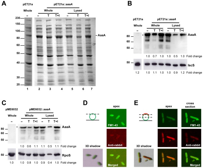
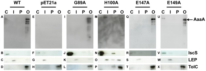
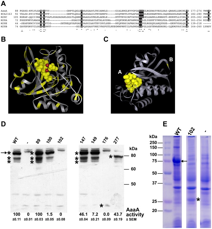
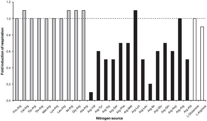
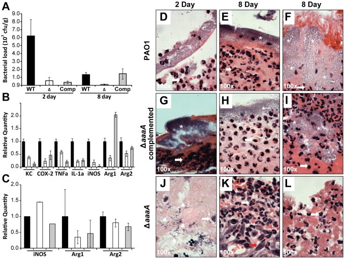
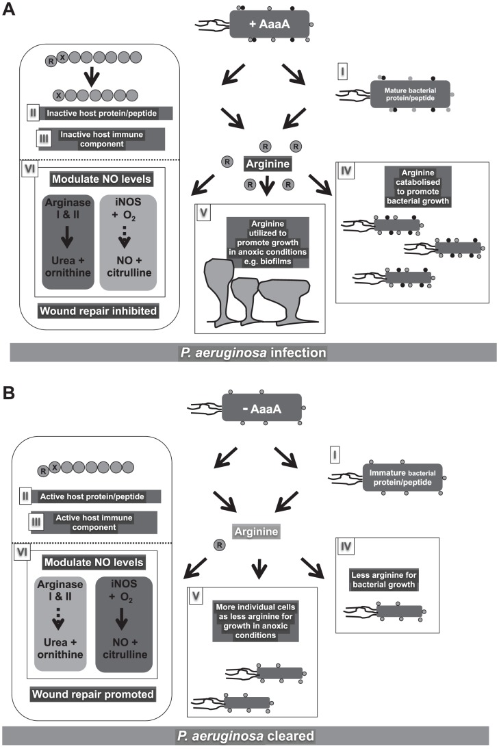
The opportunistic human pathogen, Pseudomonas aeruginosa, is a major cause of infections in chronic wounds, burns and the lungs of cystic fibrosis patients. The P. aeruginosa genome encodes at least three proteins exhibiting the characteristic three domain structure of autotransporters, but much remains to be understood about the functions of these three proteins and their role in pathogenicity. Autotransporters are the largest family of secreted proteins in Gram-negative bacteria, and those characterised are virulence factors. Here, we demonstrate that the PA0328 autotransporter is a cell-surface tethered, arginine-specific aminopeptidase, and have defined its active site by site directed mutagenesis. Hence, we have assigned PA0328 with the name AaaA, for arginine-specific autotransporter of P. aeruginosa. We show that AaaA provides a fitness advantage in environments where the sole source of nitrogen is peptides with an aminoterminal arginine, and that this could be important for establishing an infection, as the lack of AaaA led to attenuation in a mouse chronic wound infection which correlated with lower levels of the cytokines TNFα, IL-1α, KC and COX-2. Consequently AaaA is an important virulence factor playing a significant role in the successful establishment of P. aeruginosa infections.


Conflict of interest statement

The authors have declared that no competing interests exist.

Figures

Figure 1
Figure 1. The passenger and β-barrel domains of AaaA remain connected and are tethered to the cell surface.
E. coli LEMO21 bearing the empty vector pET21a or pET21a::aaaA was grown to mid exponential phase in LB, and induced with 1 mM IPTG for 1 h. Following harvesting, washing and resuspension in PBS-Hepes, half of the cells were lysed by sonication. The whole and lysed cells were split into three aliquots and incubated with (T) or without (−) trypsin according to the Materials and Methods. Trypsin inhibitor was added at the same time as trypsin to one of the aliquots (T+I). Proteins were separated through a 9% SDS PAGE and stained with Coomassie Blue (Panel A) or subjected to immunoblotting with either α-AaaA (Panel B, top), or α-IscS (Panel B, bottom) antisera. A parallel experiment was performed with P. aeruginosa ΔaaaA bearing either pME6032 or pME6032::aaaA. LB overnight cultures were diluted 1∶100 in fresh LB, grown for 3 h at 37°C, and induced with 1 mM IPTG for 1 h. The immunoblot of the P. aeruginosa proteins is shown in Panel C, with the cytoplasmic control protein being detected with α-RpoS in the bottom panel. The sizes of molecular weight markers are shown in kDa on the left, and the position of AaaA is indicated. In Panels B and C, densitometry was used to estimate the quantity of the cytoplasmic protein and the full length AaaA (indicated with the asterisk) detected in the immunoblots using imageJ software. The fold change of AaaA, IscS and RpoS are shown below the images of the respective immunoblots. The images in Panels D and E were captured by confocal fluorescent microscopy. P. aeruginosa ΔaaaA(pME6032::aaaA) was grown and induced as described for Panel C, probed with FM1-43 and either α-AaaA (Panel E) or pre-immune serum (Panel D). Incubation with donkey α-rabbit alexa fluor 680-conjugated secondary antibody (red) was performed before images were captured at either the apex or cross section of individual cells (as indicated in the dotted lines of the cartoon). Green fluorescence from FM1-43 (top Panel, green circle in cartoon), red fluorescence from alexa fluor 680 (middle Panel, red stars in cartoon), merged 2D and merged 3D shadowed images are shown.
Figure 2
Figure 2. AaaA localises to the outer membrane of E. coli.
E. coli LEMO21 bearing the empty vector (pET21a: Panels E–H), pET21a::aaaA (WT: Panels A–D) or similar plasmids producing one of four site directed mutants (pET21a::aaaA G89A (G89A: Panels I–L), pET21a::aaaA H100A (H100A: Panels M–P), pET21a::aaaA E147A (E147A: Panels Q–T), pET21a::aaaA E149A (E149A: Panels U–X)) were grown until mid-exponential phase in LB, and induced with 1 mM IPTG for 1 h. Each strain was divided into aliquots to obtain the different fractions of the cell according to the materials and methods (C: cytoplasm; I: inner membrane; P: periplasm; O: outer membrane). Proteins were separated through a 9% SDS PAGE and immunoblotted with α-AaaA (Panels A,E,I,M,Q,U), α-IscS (Panels B,F,J,N,R,V), α-LEP (Panels C,G,K,O,S,W) and α-TolC (Panels D,H,L,P,T,X) antisera. The positions of AaaA, IscS, LEP and TolC protein are indicated on the left.
Figure 3
Figure 3. AaaA is a member of the M28 family of aminopeptidases and site directed mutagenesis confirms that predicted active site residues of AaaA contribute to arginine aminopeptidase activity.
(Panel A) ClustalW2 multiple sequence alignment of the predicted active sites of the holotype enzymes for the four M28 subfamilies plus the two M28C ATs AaaA and ECA2163 (from Pectobacterium caratovora subspecies carotovorum). Identical residues are indicated by an asterisk, and similar residues by a colon or full stop. The residues highlighted in the black box are those shown to be functional within the active site. Underlining indicates the position of the conserved residues chosen for site directed mutagenesis. The holotype enzymes shown are: Streptomyces griseus aminopeptidase S (SGAP) M28.003/MER002161 (M28A), glutamate carboxypeptidase II M28.010/MER002104 (M28B), E. coli IAP aminopeptidase M28.05/MER001290 (M28C), and aminopeptidase AP1 M28.002/MER001284 (M28E). All the sequences were taken from UniProt database software (http://www.uniprot.org/). (Panel B) Crystal structure of the M28.003 founding aminopeptidase (SGAP) with the residues that are conserved in an alignment with PA0328 highlighted in yellow. The Red balls indicate the two intercalated metal ions. Panel C indicates the positions of the residues in AaaA that were selected for mutagenesis. The structure shown was predicted for AaaA using an alignment with and crystal structure of SGAP as the guide. All residues mutated were predicted to be in the active site (A) except G89 which is predicted to lie on an outward facing loop of the protein (B). All mutations were substitutions to Alanine. E. coli LEMO21 containing a pET21a vector alone (−) or with WT AaaA or one of the mutated versions (indicated by the mutation) were grown in LB until OD600 of 0.5, and induced with IPTG for 3 h. Whole cell extracts were separated through a 9% SDS PAGE and stained with Coomassie Blue (Panel E), or subjected to immunoblotting using α-AaaA antibody (Panel D). The asterisks indicate products of aaaA, and the arrow indicates full length AaaA. The relative activities of each mutant AaaA in the arginine-p-nitroanilide assay determined as described in Figure 4 are listed below their respective lane on the immunoblot in Panel D. The activity following incubation of cells with the substrate for 6.5 h is shown as this was the point when wild type AaaA reached maximal absorbance at 405 nm. The absorbance at 405 nm was adjusted to the level of AaaA made in each particular case by dividing by the amount of AaaA quantified from the immunoblot using densitometry performed with the ImagJ software. The standard error of the mean (SEM) for each is also shown.
Figure 4
Figure 4. AaaA can remove arginine from p-nitroanilide.
Panel A. The P. aeruginosa ΔaaaA mutant alone (open triangles) or bearing either the empty plasmid pME6032 (open circles) or its derivative carrying aaaA (pME6032::aaaA: closed circles) were treated as described in Figure S2B except arginine-p-nitroanilide was used as a substrate. WT PAO1 cells were treated similarly (closed triangles), and activities (measured as changes in A405 nm) are compared against a growth media blank (crosses). Panel B. E. coli DH5α bearing either the empty plasmid pME6032 (open circles) or its derivative carrying aaaA (pME6032::aaaA: closed circles) were grown in LB until exponential phase, induced with 1 mM IPTG, and then incubated with arginine-p-nitroanilide as described in Figure S2B. Activities are compared against a growth media blank (crosses). Error bars are+/−1 S.D. (n = 15). All measurements have been corrected for differential growth of bacteria by normalising to an initial OD600 nm of 0.1.
Figure 5
Figure 5. The activity of AaaA enables P. aeruginosa to grow using the tripeptide arg-gly-asp as the sole source of carbon and nitrogen.
P. aeruginosa PAO1 (closed circles) and its derived aaaA deficient mutant (ΔaaaA, open circles) alone or bearing pME6032 (vector, open triangles) or pME6032::aaaA (complemented, closed triangles) were grown to mid-exponential phase before the induction of AaaA production by 1 mM IPTG. Cells were resuspended in MMP to OD600 of 1, and subsequently 20 µl of this solution diluted into 200 µl of MMP containing arginine at 10 mM (Panel A), or 10 mM of the tripeptide arg-gly-asp (Panel B). The graph shows the subsequent growth in the Tecan monitored by observing the increase in OD492 over time. The data is representative of 3 independent repetitions of this experiment.
Figure 6
Figure 6. AaaA promotes the ability of P. aeruginosa to respire dipeptides with N-terminal arginine except when adjacent to Arginine or Lysine.
P. aeruginosa PAO1 and its derived aaaA deficient mutant were inoculated into nitrogen minimal media (NMM) alone or NMM containing the indicated nitrogen source. Cellular respiration/metabolic activity is reported via reduction of tetrazolium dye and plotted against time. The area under the curve (AUC) for a selection of nitrogen sources following 24 h incubation in each condition is plotted here. The values have been normalised by subtraction of the AUC of the control (no nitrogen source added) on the respective Biolog plate. Relative respiration is calculated by the difference between the normalised AUC of wild type and mutant divided by their sum and multiplied by 100. The fold induction was calculated by dividing the normalised AUC of the mutant by that of the wild type, so a value of 1.0 is no change. Biolog Phenotype microarray plates PM03B and PM06-08 were used as indicated, and each condition performed in duplicate (results from one are shown).
Figure 7
Figure 7. The AaaA deficient mutant is less virulent in the chronic mouse wound model.
Either the P. aeruginosa wild type PAO1 (black bars), the ΔaaaA mutant (white bars), or the complemented ΔaaaA mutant PAJL2 (grey bars) was inoculated (104 CFU) into a chronic wound in each of 9 mice. After 2 (3 mice per group) or 8 (7 mice per group) days, wound tissue was removed and the bacterial load was estimated by calculating the colony forming units (Panel A). Chronically-wounded mice were euthanized at post infection day 2 (3 mice per group) or day 8 (7 mice per group), and wound tissue was harvested for qRT-PCR to detect the mRNA of the indicated cytokines and other host enzymes in the infected wound tissue as described in the materials and methods (Panel B and C). Tissue from the wounds of the 2 day infected mice (Panels D,G,J) or 8 day infected mice (Panels E,F,H,I,K,L) was stained with H&E and is shown at 100× magnification. Images of the P. aeruginosa wild type PAO1 (Panels D,E,F), ΔaaaA mutant (Panels G,H,I), and the complemented ΔaaaA mutant PAJL2 (Panels J,K,L) are shown with infiltrating neutrophils indicated by white arrows, elongated fibroblasts with a red arrow, single bacterial cells with white arrow heads and clumps of bacteria with a white asterisk. Panels D–E,G–H,J–K are representative of the wound site and Panels F,I,L are representative of the site of infection below the wound.
Figure 8
Figure 8. Cartoon Model illustrating a selection of the potential roles AaaA may have within a chronic wound.
In Panel A, the P. aeruginosa WT scenario is depicted, where AaaA (black dots) is present on the surface of P. aeruginosa cells colonising a host. Panel B shows infection with an AaaA deficient mutant that only has non-AaaA proteins on its surface (grey dots). It is possible that AaaA may: Panel I degrade a protein on the surface of P. aeruginosa, causing activation that aids infection (represented by removal of the black outline around the grey dots in Panel A, but not in Panel B), Panel II degrade a host protein/peptide, that may be a component of the host immune system (Panel III) by removing an aminoterminal arginine (R in circle). These activities may be sufficient to aid pathogenicity, however they may serve to liberate arginine that can be catabolised by the bacteria (Panel IV) resulting in growth promotion in Panel A that is not evident in the absence of AaaA (Panel B). This may provide a fitness advantage to the bacteria that improves virulence. In conditions where oxygen is limited, the arginine may provide a particular advantage (Panel V), potentially enabling formation of biofilms that could both serve to promote colonisation and provide resistance against the immune system. If only some of the released arginine is utilized by the bacteria, local arginine levels may rise in the host (Panel VI). This could induce arginase production in host cells (depicted by dark grey box and solid black arrows in Panel A:VI). The arginase enzymes will degrade the arginine, reducing its availability as a substrate for iNOS (indicated by pale grey box and dashed grey arrows in Panel A:VI). Consequently, there will be lower levels of nitric oxide (NO) and P. aeruginosa will be able to successfully establish an infection. Alternatively, in Panel B:VI, AaaA is absent from the invading P. aeruginosa, so there is no degradation of proteins and peptides. This maintains the limited arginine concentration and avoids induction of arginase in host cells. Consequently, arginine would be available to serve as a substrate for iNOS, and the nitric oxide generated could disable the bacterial cells and promote wound healing.

References

    1. Kerr KG, Snelling AM (2009) Pseudomonas aeuginosa: a formidable and ever-present adversary. J Hosp Infect 73: 338–344. - PubMed
    1. George AM, Jones PM, Middleton PG (2009) Cystic fibrosis infections: treatment strategies and prospects. FEMS Microbiol Lett 300: 153–164. - PubMed
    1. Doring G, Conway S, Heijerman H, Hodson M, Hoiby N, et al. (2000) Antibiotic therapy against Pseudomonas aeruginosa in cystic fibrosis: a European consensus. Eur Respir J 16: 749–467. - PubMed
    1. El Solh AA, Alhajhusain A (2009) Update on the treatment of Pseudomonas aeruginosa pneumonia. J Antimicrob Chemother 64: 229–238. - PubMed
    1. Lister PD, Wolter DJ, Hanson ND (2009) Antibacterial-resistant Pseudomonas aeruginosa: clinical impact and complex regulation of chromosomally encoded resistance mechanisms. Clin Microbiol Rev 22: 582–610. - PMC - PubMed

Publication types

MeSH terms