. 2012;7(8):e42559.
doi: 10.1371/journal.pone.0042559. Epub 2012 Aug 21.

Pre-clinical development of BCG.HIVA(CAT), an antibiotic-free selection strain, for HIV-TB pediatric vaccine vectored by lysine auxotroph of BCG

Affiliations

Pre-clinical development of BCG.HIVA(CAT), an antibiotic-free selection strain, for HIV-TB pediatric vaccine vectored by lysine auxotroph of BCG

Narcís Saubi et al. PLoS One. 2012.

Abstract

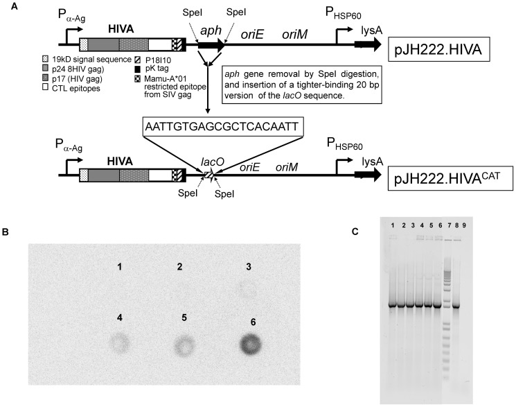
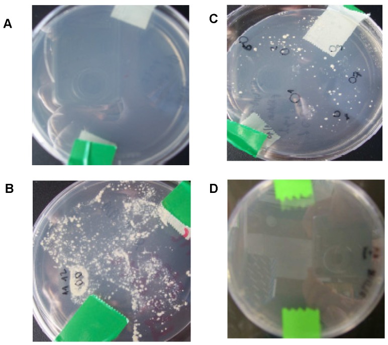
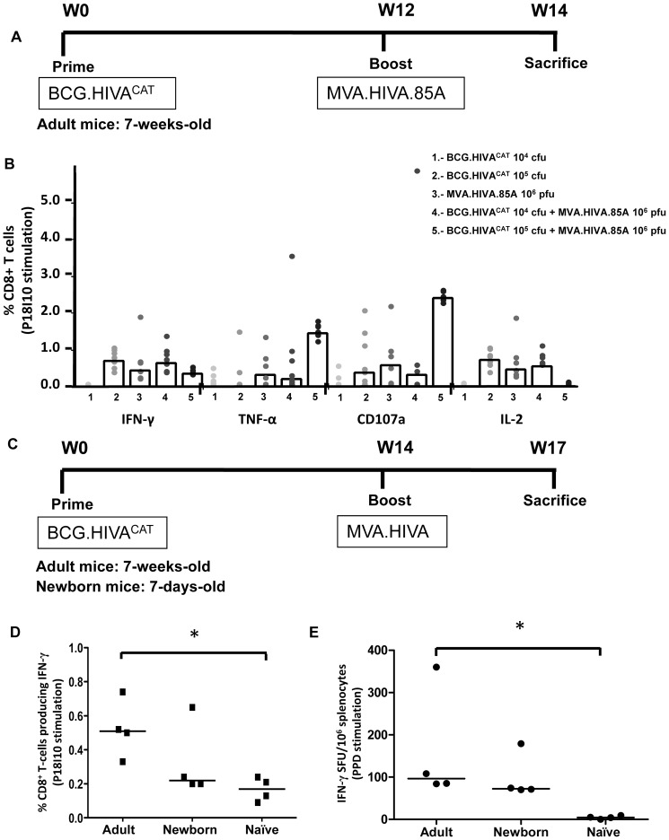
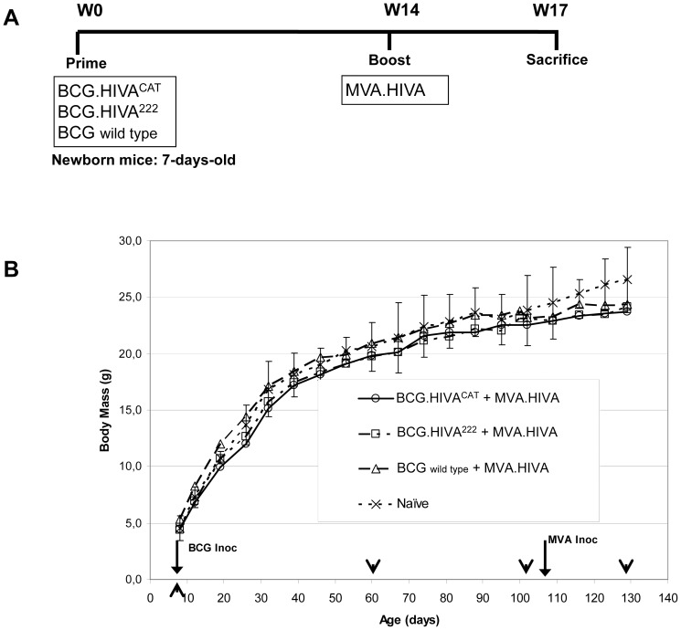
In the past, we proposed to develop a heterologous recombinant BCG prime-recombinant modified vaccinia virus Ankara (MVA) boost dual pediatric vaccine platform against transmission of breast milk HIV-1 and Mycobacterium tuberculosis (Mtb). In this study, we assembled an E. coli-mycobacterial shuttle plasmid pJH222.HIVA(CAT) expressing HIV-1 clade A immunogen HIVA. This shuttle vector employs an antibiotic resistance-free mechanism based on Operator-Repressor Titration (ORT) system for plasmid selection and maintenance in E. coli and lysine complementation in mycobacteria. This shuttle plasmid was electroporated into parental lysine auxotroph (safer) strain of BCG to generate vaccine BCG.HIVA(CAT). All procedures complied with Good Laboratory Practices (GLPs). We demonstrated that the episomal plasmid pJH222.HIVA(CAT) was stable in vivo over a 20-week period, and genetically and phenotypically characterized the BCG.HIVA(CAT) vaccine strain. The BCG.HIVA(CAT) vaccine in combination with MVA.HIVA induced HIV-1- and Mtb-specific interferon γ-producing T-cell responses in newborn and adult BALB/c mice. On the other hand, when adult mice were primed with BCG.HIVA(CAT) and boosted with MVA.HIVA.85A, HIV-1-specific CD8(+) T-cells producing IFN-γ, TNF-α, IL-2 and CD107a were induced. To assess the biosafety profile of BCG.HIVA(CAT)-MVA.HIVA regimen, body mass loss of newborn mice was monitored regularly throughout the vaccination experiment and no difference was observed between the vaccinated and naïve groups of animals. Thus, we demonstrated T-cell immunogenicity of a novel, safer, GLP-compatible BCG-vectored vaccine using prototype immunogen HIVA. Second generation immunogens derived from HIV-1 as well as other major pediatric pathogens can be constructed in a similar fashion to prime protective responses soon after birth.


Conflict of interest statement

Competing Interests: The authors have declared that no competing interests exist.

Figures

Figure 1
Figure 1. Construction of the BCG.HIVACAT vaccine strain.
(A) A synthetic GC-rich HIVA gene was fused to the region encoding the 19-kDa lipoprotein signal sequence and inserted into the episomal pJH222 E. coli-mycobacterium shuttle plasmid. This plasmid contains kanamycin resistance (aph) and complementing lysA genes and an E. coli origin of replication (oriE). In addition, pJH222 contained the mycobacterial origin of replication (oriM). The BALB/c mouse T-cell and MAb Pk epitopes used in this work are depicted. P α-Ag, M. tuberculosis α-antigen promoter; PHSP60, heat shock protein 60 gene promoter. The aph gene was removed by SpeI digestion and the lacO sequence was inserted and transformed into E. coli DH1lacdapD strain. (B) Immunodot of BCG.HIVACAT lysates. Dot 1: BCG wild type (negative control); Dot 2, 3, 4 and 5: clone 3, clone 7, clone 9 and clone 10 of BCG.HIVACAT; Dot 6: BCG.HIVA222 (positive control). HIVA peptide was detected using the anti-Pk MAb followed by horseradish peroxidase-Goat-anti-Mouse and enhanced chemiluminescence (ECL) detection. (C) In vivo plasmid stability of BCG.HIVACAT harboring pJH222.HIVACAT. Mice were injected s.c. with 105 cfu of BCG.HIVACAT and boosted i.m. with 106 pfu of MVA.HIVA, spleens were homogenized 20 weeks after BCG inoculation and the recovered rBCG colonies were tested for the presence of the HIVA DNA coding sequence by PCR. Lanes 1 to 6: Six rBCG colonies were recovered in the non-lysine supplemented plate; lane 7: Molecular weight marker; lane 8: Plasmid DNA positive control; lane 9: Distilled water (negative control).
Figure 2
Figure 2. Genetic characterization of the BCG.HIVACAT.
GenoType MTBC assay and Multiplex PCR assay. (A) The BCG.HIVACAT strain identification results representative of all of the patterns obtained with the GenoType MTBC assay. The positions of the oligonucleotides, the marker line and the BCG hybridization pattern are shown on the right. The specificity and targeted genes of the lines are as follows: 1, conjugate control; 2, amplification control (23S rRNA); 3, MTBC specific (23S rRNA); 4 to 12, discriminative for the MTBC species (gyrB); 13, M. bovis BCG (RD1). The samples analyzed were: Strip 1: BCG Connaught; Strip 2: BCG.HIVACAT; Strip 3: BCG.HIVA222; Strip 4: BCG wild type. All four strains presented the same hybridization pattern corresponding to M. bovis BCG. (B) The BCG.HIVACAT Pasteur substrain identification by multiplex PCR assay. Lane 1 and 12: molecular weight marker; lane 2: negative control; lane 3, 6–11: BCG.HIVACAT(clone10); lane 4: BCG Danish strain (using 1 µl of template); lane 5: BCG Danish strain (using 4 µl template); The samples were analyzed by multiplex primer assay or single primer pair assay. Lane 2–5: multiplex primers; lane 6: ET1-3 primers; lane 7: RD2 primers; lane 8: RD8 primers; lane 9: RD14 primers; lane 10: RD16 primers; lane 11: C3–C5 primers. (C) Enzymatic restriction analysis of pJH222.HIVACAT plasmid DNA extracted from both the Master Seed (MS, lanes 1–5) and the Working Stock (WS, lanes 7–11) of BCG.HIVACAT cultures. Lane 1 and lane 7: uncut plasmid; lane 2 and lane 8: HpaI digestion; lane 3 and 9: KpnI digestion; lane 4 and 10: digestion with SpeI; lane 5 and 11: digestion with HindIII; lane 6 and 12: Molecular Weight Marker (1 kb Plus, Invitrogen). (D) PCR analysis of HIVA DNA coding sequence using as template the cultures of BCG.HIVACAT Master Seed (lane 1), and Working Stock (lane 2), Molecular Weight Marker (lane 3), positive control plasmid DNA pJH222.HIVA (lane 4).
Figure 3
Figure 3. Phenotypic characterization of the BCG.HIVACAT.
We assessed the phenotype of lysine auxotrophy, lysine complementation and kanamycin resistance of BCG.HIVACAT strain. (A) BCG lysine auxotroph strain plated on non-lysine supplemented 7H10; (B) BCG lysine auxotroph strain plated on lysine supplemented 7H10; (C) BCG.HIVACAT plated on 7H10 without lysine and kanamycin supplementation; (D) BCG.HIVACAT plated on 7H10 without lysine supplementation and with kanamycin.
Figure 4
Figure 4. Induction of HIV-1- and Mtb-specific T-cells responses by the BCG.HIVACAT prime - MVA.HIVA boost regimen.
(A) Adult mice (7-weeks-old) immunized with either 104 or 105 cfu of BCG.HIVACAT alone (subcutaneously), 106 pfu of MVA.HIVA.85A alone (intramuscularly), or 104 or 105 cfu of BCG.HIVACAT as a prime and boosted with 106 pfu of MVA.HIVA.85A (left to right). Mice were sacrificed 2 weeks later for T-cell analysis. (B) Analysis of IFN-γ, TNF-α, CD107a and IL-2 vaccine elicited HIV-1-specific CD8+ T-cell responses. The frequencies of cells producing cytokine are shown. Data are presented as group medians as well as individual animal responses (n = 5). (C) Adult and newborn mice (7-days-old) were either left unimmunized or immunized with 2×10⁶ cfu of BCG.HIVACAT (intradermal and subcutaneous route respectively) and subsequently given a booster dose of 106 pfu of MVA.HIVA (intramuscularly) at 14 weeks post BCG immunization, and sacrificed 3 weeks later. (D) Analysis of IFN-γ vaccine elicited HIV-1-specific CD8+ T-cell responses. The frequencies of cells producing cytokine are shown. Data are presented as group medians as well as individual animal responses (n = 4). (E) PPD-specific T-cell responses elicited by BCG.HIVACAT. Immune responses to BCG were assessed in an ex vivo IFN-γ ELISPOT assay using PPD as the antigen. The median spot-forming units (SFU) per 106 splenocytes for each group of mice (n = 4) as well as individual animal responses is shown. * = p<0.05.
Figure 5
Figure 5. BCG.HIVACAT prime and MVA.HIVA boost safety in newborn mice.
(A) Newborn mice were either left unimmunized or immunized with 2×10⁶ cfu of BCG wild type, BCG:HIVA222 or BCG.HIVACAT by subcutaneous route and subsequently given a booster dose of 106 pfu of MVA.HIVA at week 14. (B) The body weight was weekly recorded, and the mean for each group of mice is shown (n = 10). Data from naive mice are presented as mean ± 2 SEM (n = 6); At specific time points the weight differences between vaccinated and naïve mice group were analyzed by ANOVA test (arrowheads).

References

    1. UNAIDS World AIDS Day Report 2011 (2011) Joint United Nations Programme on HIV/AIDS (UNAIDS). Available: http://www.unaids.org/en/media/unaids/contentassets/documents/unaidspubl.... Accessed 2012 Aug 1.
    1. Cavarelli M, Scarlatti G (2011) Human immunodeficiency virus type 1 mother-to-child transmission and prevention: successes and controversies. J Intern Med 270: 561–579 doi: 10.1111/j.1365-2796.2011.02458.x. - PubMed
    1. Borkowsky W, Wara D, Fenton T, McNamara J, Kang M, et al. (2000) Lymphoproliferative responses to recombinant HIV-1 envelope antigens in neonates and infants receiving gp120 vaccines. J Infect Dis 181: 890–896 doi: 10.1086/315298. - PubMed
    1. Johnson DC, McFarland EJ, Muresan P, Fenton T, McNamara J, et al. (2005) Safety and immunogenicity of an HIV-1 recombinant canarypox vaccine in newborns and infants of HIV-1-infected women. J Infect Dis 192: 2129–2133 doi: 10.1086/498163. - PubMed
    1. McFarland EJ, Johnson DC, Muresan P, Fenton T, Tomaras GD, et al. (2006) HIV-1 vaccine induced immune responses in newborns of HIV-1 infected mothers. AIDS 20: 1481–1489 doi:10.1097/01.aids.0000237363.33994.45. - DOI - PubMed

Publication types