. 2012;6(8):e1763.
doi: 10.1371/journal.pntd.0001763. Epub 2012 Aug 21.

Transcriptomic signature of Leishmania infected mice macrophages: a metabolic point of view

Collaborators, Affiliations

Transcriptomic signature of Leishmania infected mice macrophages: a metabolic point of view

Imen Rabhi et al. PLoS Negl Trop Dis. 2012.

Abstract

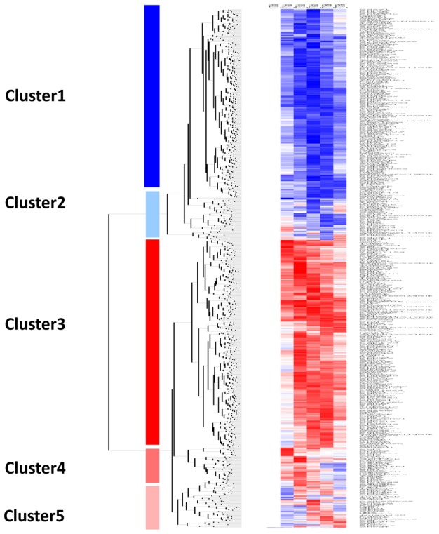
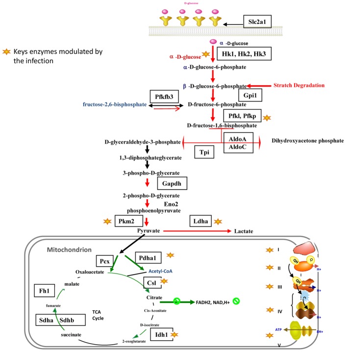
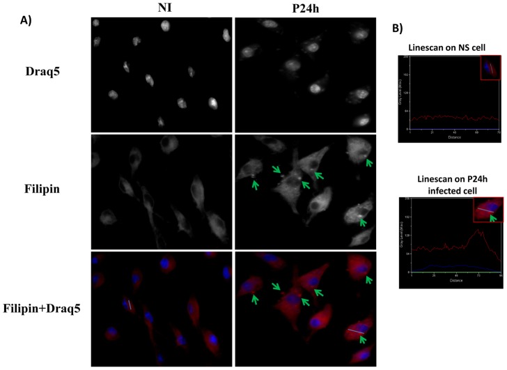
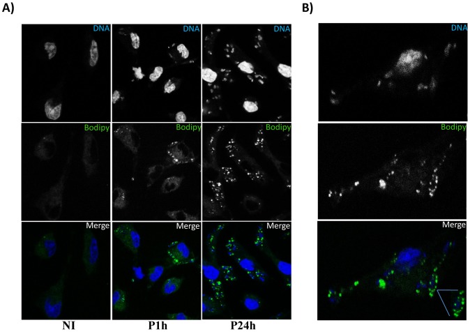
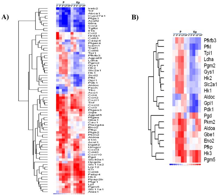
We analyzed the transcriptional signatures of mouse bone marrow-derived macrophages at different times after infection with promastigotes of the protozoan parasite Leishmania major. Ingenuity Pathway Analysis revealed that the macrophage metabolic pathways including carbohydrate and lipid metabolisms were among the most altered pathways at later time points of infection. Indeed, L. major promastiogtes induced increased mRNA levels of the glucose transporter and almost all of the genes associated with glycolysis and lactate dehydrogenase, suggesting a shift to anaerobic glycolysis. On the other hand, L. major promastigotes enhanced the expression of scavenger receptors involved in the uptake of Low-Density Lipoprotein (LDL), inhibited the expression of genes coding for proteins regulating cholesterol efflux, and induced the synthesis of triacylglycerides. These data suggested that Leishmania infection disturbs cholesterol and triglycerides homeostasis and may lead to cholesterol accumulation and foam cell formation. Using Filipin and Bodipy staining, we showed cholesterol and triglycerides accumulation in infected macrophages. Moreover, Bodipy-positive lipid droplets accumulated in close proximity to parasitophorous vacuoles, suggesting that intracellular L. major may take advantage of these organelles as high-energy substrate sources. While the effect of infection on cholesterol accumulation and lipid droplet formation was independent on parasite development, our data indicate that anaerobic glycolysis is actively induced by L. major during the establishment of infection.


Conflict of interest statement

The authors have declared that no competing interests exist.

Figures

Figure 1
Figure 1. Hierarchical clustering of genes differentially expressed in Leishmania major infected BALB/c macrophages.
Five distinct clusters were identified by hierarchical clustering analysis using dChip software. Two clusters represent genes that were significantly down-regulated (cluster 1) and up-regulated (cluster 3) over the time course. The second (cluster 2), contains genes that start to be up-regulated from 6 to 24 hours after infection Two other clusters contain genes up-regulated during the early hours (1,3 and 6 hrs) of infection (cluster 4) and at 6, 12 and 24 hrs (cluster 5). Each row represents a spot on the microarray and each column a separate microarray. BMdM response was studied at five different time points (from the left to the right: P-T1h, P-T3h, P-T6h, P-T12h and P-T24h). The left-hand column show non-infected cells (NI) that were used as internal control.
Figure 2
Figure 2. Canonical Metabolic pathways identified by Inguenuity Pathway Analysis software as significantly altered (p<0.05) in L. major infected BALB/c macrophages for each time point (3 h (orange), 6 h (violet), 12 h (red) and 24 hours post-infection (green)).
The negative log10(p-value) are plotted on the Y-axis.
Figure 3
Figure 3. L. major-infected macrophages display a modulation of several genes involved in glycolysis, TCA cycle and mitochondrial respiration.
Figure 4
Figure 4. Free cholesterol accumulation in Leishmania major infected macrophages.
BMdM were infected by Leishmania major promastigotes at different time points. Cells were fixed by formaldehyde, stained by Filipin for free cholesterol accumulation and visualized by Epifluorescence microscopy. The results are representative of two independent experiments. Only the results obtained 24 h post-infection is shown here. Similar results were observed for the other infection time point. B) Representative linescans (Metamorph software) of infected and non-infected cells that allow to quantify intensity differences in Filipin staining.
Figure 5
Figure 5. Leishmania major infection induces lipid droplet accumulation.
BALB/c BMdM cells were infected by Leishmania major promastigotes at different time points. Cells were fixed by formaldehyde, stained by Bodipy493/503 for lipid droplet accumulation and visualized by confocal microscopy. A) Bodipy accumulation in Leishmania infected Macrophages. Only the results obtained one hour and twenty four hours after infection are shown here. Similar results were observed for the other infection time points. B) Lipid accumulation in close association with the parasitophorus vacuole. The results are representative of four independent experiments.
Figure 6
Figure 6. Leishmania major infection does not induce COX-2 protein production.
Raw264.7 cells were either stimulated by LPS or infected by L. major promastigotes. Cells were then lysed at the indicated times, collected and finally subjected to Western blotting with antibodies specific for COX-2. Total ERK immunoblot was shown as loading control.
Figure 7
Figure 7. Hierarchic clustering of genes differentially regulated in macrophages following lived and heat-inactivated Leishmania promastigotes.
The Heat Map show a partial list of genes classified through Ingenuity Pathways Analysis as (A) related to Cell Metabolic Pathways and significantly altered (p<0.05) in macrophages infected with live parasite (P) or heat-killed parasite (KP) versus non-infected macrophages across the time course of infection. (B) Implicated in carbohydrates between live and heat killed promastigotes. Red indicates gene induction; blue, gene repression.

References

    1. Chaussabel D, Semnani RT, McDowell MA, Sacks D, Sher A, et al. (2003) Unique gene expression profiles of human macrophages and dendritic cells to phylogenetically distinct parasites. Blood 102: 672–681. - PubMed
    1. Buates S, Matlashewski G (2001) General suppression of macrophage gene expression during Leishmania donovani infection. J Immunol 166: 3416–3422. - PubMed
    1. Rodriguez NE, Chang HK, Wilson ME (2004) Novel program of macrophage gene expression induced by phagocytosis of Leishmania chagasi . Infect Immun 72: 2111–2122. - PMC - PubMed
    1. Osorio y Fortea J, de La Llave E, Regnault B, Coppee JY, Milon G, et al. (2009) Transcriptional signatures of BALB/c mouse macrophages housing multiplying Leishmania amazonensis amastigotes. BMC Genomics 10: 119. - PMC - PubMed
    1. Gentleman RC, Carey VJ, Bates DM, Bolstad B, Dettling M, et al. (2004) Bioconductor: open software development for computational biology and bioinformatics. Genome Biol 5: R80. - PMC - PubMed

Publication types