Review
. 2012:53:69-82.
doi: 10.1042/bse0530069.

Cell wars: regulation of cell survival and proliferation by cell competition

Affiliations
Review

Cell wars: regulation of cell survival and proliferation by cell competition

Silvia Vivarelli et al. Essays Biochem. 2012.

Abstract

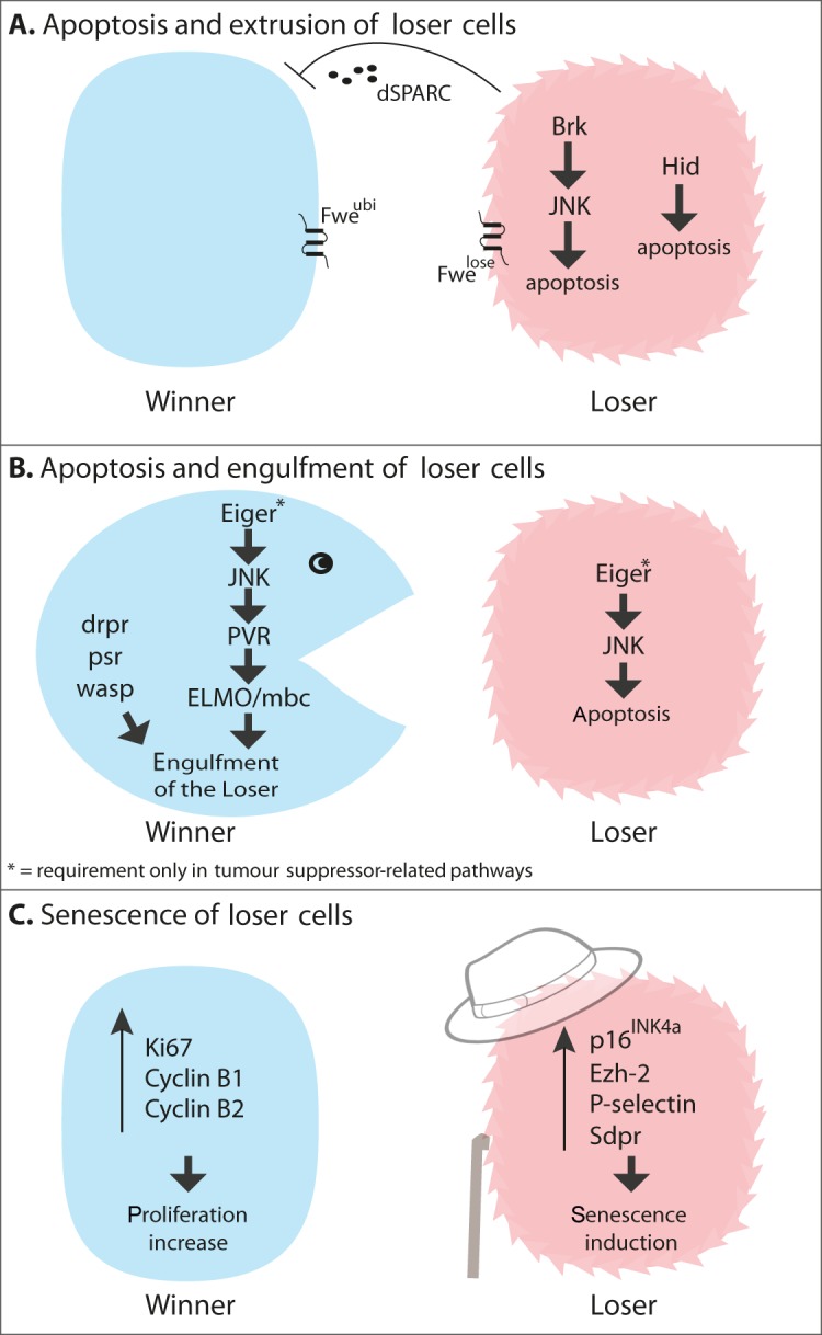
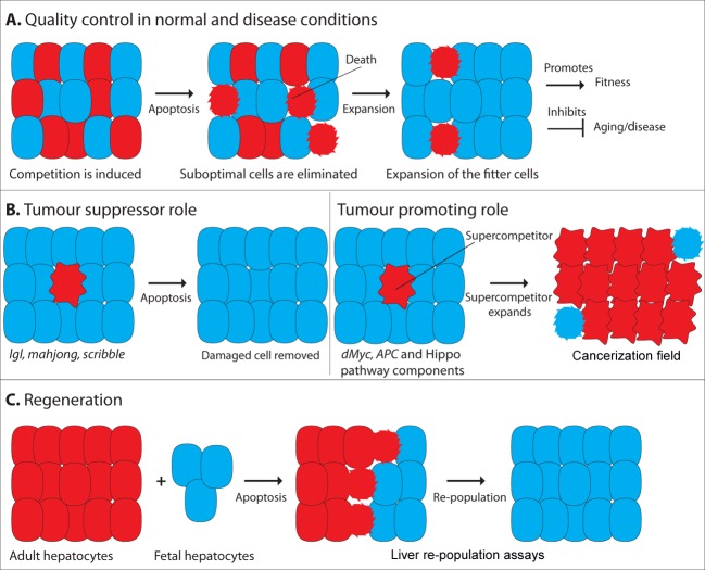
During cell competition fitter cells take over the tissue at the expense of viable, but less fit, cells, which are eliminated by induction of apoptosis or senescence. This probably acts as a quality-control mechanism to eliminate suboptimal cells and safeguard organ function. Several experimental conditions have been shown to trigger cell competition, including differential levels in ribosomal activity or in signalling pathway activation between cells, although it is unclear how those differences are sensed and translated into fitness levels. Many of the pathways implicated in cell competition have been previously linked with cancer, and this has led to the hypothesis that cell competition could play a role in tumour formation. Cell competition could be co-opted by cancer cells to kill surrounding normal cells and boost their own tissue colonization. However, in some cases, cell competition could have a tumour suppressor role, as cells harbouring mutations in a subset of tumour suppressor genes are killed by wild-type cells. Originally described in developing epithelia, competitive interactions have also been observed in some stem cell niches, where they play a role in regulating stem cell selection, maintenance and tissue repopulation. Thus competitive interactions could be relevant to the maintenance of tissue fitness and have a protective role against aging.


Figures

Figure 1.
Figure 1.. Mechanisms involved in the elimination of loser cells
(A) Apoptosis and extrusion of loser cells. Two mechanisms of apoptosis induction have been identified. In the first, JNK is activated via Brk (Brinker), whereas in the second activation of the pro-apototic gene Hid leads to death. Different Fwe (Flower) isoforms label winner and loser cells. dSPARC is secreted from loser cells and acts as a temporary shield to protect them from apoptosis. (B) Apoptosis and engulfment of loser cells. In some contexts, winner cells engulf neighbouring loser cells, via activation of genes involved in phagocytosis (listed in the Figure). This is regulated by Eiger/JNK activation, which promotes apoptosis in the losers and engulfment in the winners. (C) Senescence of loser cells. Competition among HSPCs results in the activation of senescence-related genes in the loser cells (listed in the Figure). Conversely, winner cells activate proliferation markers (listed in the Figure).
Figure 2.
Figure 2.. Potential links between cell competition and disease
(A) Quality control in normal and disease conditions. Suboptimal cells are eliminated from epithelial tissues through apoptosis, whereas the fitter cells show a corresponding increase in proliferation. (B) Tumour suppressor role. Cell competition could act as a protective mechanism to remove potentially dangerous cells from a tissue, thus preventing tumour formation. Tumour promoting role. By contrast, cell competition could be exploited by mutant cells to promote tumour formation. Mutant supercompetitor cells with precancerous lesions could outcompete and cull surrounding wild-type cells, leading to the generation of cancerization fields. (C) Regeneration. On the basis of experiments in liver repopulation assays, it has been proposed that cell competition could be exploited in regenerative medicine.

References

    1. Morata G., Ripoll P. Minutes: mutants of Drosophila autonomously affecting cell division rate. Dev. Biol. 1975;42:211–221. - PubMed
    1. Simpson P. Parameters of cell competition in the compartments of the wing disc of Drosophila. Dev. Biol. 1979;69:182–193. - PubMed
    1. Simpson P., Morata G. Differential mitotic rates and patterns of growth in compartments in the Drosophila wing. Dev. Biol. 1981;85:299–308. - PubMed
    1. Oliver E.R., Saunders T.L., Tarle S.A., Glaser T. Ribosomal protein L24 defect in belly spot and tail (Bst), a mouse Minute. Development. 2004;131:3907–3920. - PMC - PubMed
    1. Johnston L.A., Prober D.A., Edgar B.A., Eisenman R.N., Gallant P. Drosophila myc regulates cellular growth during development. Cell. 1999;98:779–790. - PMC - PubMed