Review
. 2012 Oct;14 Suppl 3(0 3):109-19.
doi: 10.1111/j.1463-1326.2012.01652.x.

Repair of diverse diabetic defects of β-cells in man and mouse by pharmacological glucokinase activation

Affiliations
Review

Repair of diverse diabetic defects of β-cells in man and mouse by pharmacological glucokinase activation

Nicolai M Doliba et al. Diabetes Obes Metab. 2012 Oct.

Abstract

Glucokinase activators (GKAs) are being developed and clinically tested for potential antidiabetic therapy. The potential benefits and limitations of this approach continue to be intensively debated. To contribute to the understanding of experimental pharmacology and therapeutics of GKAs, we have tested the efficacy of one of these agents (Piragliatin) in isolated islets from humans with type 2 diabetes mellitus (T2DM), from mice with glucokinase (GK) mutations induced by ethyl-nitroso-urea (ENU) as models of Maturity Onset Diabetes of the Young linked to GK and Permanent Neonatal Diabetes Mellitus linked to GK (PNDM-GK) and finally of islets rendered glucose insensitive by treatment with the sulphonyl urea compound glyburide in organ culture. We found that the GKA repaired the defect in all three instances as manifest in increased glucose-induced insulin release and elevated intracellular calcium responses. The results show the remarkable fact that acute pharmacological activation of GK reverses secretion defects of β-cells caused by molecular mechanism that differ vastly in nature, including the little understood multifactorial lesion of β-cells in T2DM of man, the complex GK mutations in mice resembling GK disease and acute sulphonylurea failure of mouse β-cells in tissue culture. The implications of these results are to be discussed on the theoretical basis underpinning the strategy of developing these drugs and in light of recent results of clinical trials with GKAs that failed for little understood reasons.


Conflict of interest statement

Conflict of Interests

The authors declare no conflict of interest.

Figures

Figure 1
Figure 1
The network of glucokinase (GK)-containing tissues throughout the body and their connections. Expression of GK in the pancreatic islet α and β-cells, other endocrine cells (including pituitary and gut) and cells in the portal vascular tree and central nervous system (including the hypothalamus and brain stem) makes up about 0.1% of the body’s total GK complement. The liver contains the rest (about 99.9%). The pancreatic islet cells and the liver constitute the basic GK-dependent regulatory system maintaining glucose homeostasis. ANS, autonomic nervous system; GKA, glucokinase activator. With permission from reference [1].
Figure 2
Figure 2
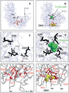
Effects of glucokinase activator (GKA) binding on glucokinase (GK) structure. Panels A and B: Synergistic binding of glucose and GKA to GK induces a large conformational change of the protein structure which allows the full visualization of the glucose-binding site in the crystal structure and causes a major rearrangement of the allosteric drug receptor site. Panels C and D provide a detailed view of the glucose-binding site in the open and closed conformation. Panels E and F illustrate how glucose binding provides access of the GKA to the allosteric site, which is occluded in the open conformation. The orientation of the cutouts in panels C and D is the same as in panels A and B but it is slightly changed in panels E and F to better visualize the critical opening of the V62–G72 loop when ligands bind. Figure design based on Kamata et al. [22].
Figure 3
Figure 3
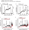
Blood glucose levels of mice glucokinase (GK)P417R/P417R and GKP417R/+ mice. Panel A shows the blood sugars in the fed state of the GK-P417R line measured before shipping the animals from Chicago to Philadelphia; Panel B: blood glucose levels before sacrificing the animals for islet isolation at the University of Pennsylvania. The small difference in blood sugar levels of the fed animals is more likely due to reduced food intake as a result of shipping the animals which were used 1 day after arrival.
Figure 4
Figure 4
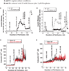
Effects of glucose and Piragliatin on insulin secretion and intracellular calcium concentrations of islets isolated from control mice and from mice with a GKP417R/P417R mutation induced by ethyl-nitroso-urea (ENU). Panel A: insulin release profile. Islets were cultured for 3–4 days in RPMI 1640 medium with 10 mM glucose present before they were used for the perifusion experiment. A physiological amino acid mixture was present throughout the entire run at 4.0 mM. Initially, islets were perfused in the absence of glucose for 30 min and then a glucose ramp from 0 to 20 mM was applied during the next 25 min. The highest 20 mM concentration was maintained for another 15 min and then 3 µM of Piragliatin was added to the perfusate for the next 25 min. Afterwards, all stimuli were removed for 20 min and islets were then depolarized with 30 mM KCl. Panel B: islets were cultured for 3–4 days with 25 mM glucose and 3 µM Piragliatin. The same experimental protocol as describe in panel A was employed to study insulin secretion. Panels C and D show Ca2+ profiles for the same islets preparations and conditions comparable to those presented in panels A and B.
Figure 5
Figure 5
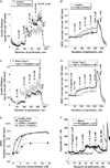
Amino acid- and glyburide-stimulated insulin secretion and intracellular Ca2+ in islets isolated from control and glucokinase (GK)P417R/P417R mice. Panels A and C: insulin release and Ca2+ profiles of islets cultured for 3–4 days in regular RPMI 1640 culture medium with 10 mM glucose. Panels B and D: insulin release and Ca2+ profiles of islets cultured for 3–4 days with 25 mM glucose and 3 µM Piragliatin. Note that in contrast to experiments presented in figure 4, the amino acid stimulus was introduced later during the course of the perifusion.
Figure 6
Figure 6
Piragliatin restores glucose sensitivity and enhances amino acid induced insulin release in glyburide-treated mouse islets. Three experimental conditions were used during islet culturing (for 3 days) and perifusion: (i) control islets were cultured in RPMI 1640 media containing 10 mM glucose without further additions; (ii) 0.1 µM glyburide was added to the culture medium described in condition 1; (iii) 0.1 µM glyburide plus 3 µM Piragliatin were added to the culture medium described under 1. During perifusion, the drugs were present at the same concentrations as during culture. Panel A: insulin secretion was stimulated by a glucose ramp from 0 to 25 mM extending over a period of 50 min. Panel B: insulin release was stimulated by a physiological amino acid mixture ramp from 0 to 12 mM extending over a period of 50 min. Results are presented as mean ± s.e. of three to four experiments.
Figure 7
Figure 7
Impaired insulin release, oxygen consumption and intracellular Ca2+ concentration of isolated islets from type 2 diabetic organ donors and the restoration of insulin secretion and respiration by a glucokinase activator (GKA). Panel A shows the insulin release patterns of healthy and type diabetic islets with glucose stimulation using stepwise increases of glucose from 0 to 3, 6, 12 and 24 mM. Panel B shows the oxygen consumption profiles for the same experiments during stepwise increase of glucose concentration followed by treatment with 5 µM of the uncoupler of Ox/Phos FCCP and 1 mM Na-azide. O2 consumption was determined with a method based on phosphorescence quenching of metalloporphyrins by oxygen [63]. Panels C and D show the insulin release and oxygen consumption patterns of type 2 diabetic islets in the absence and presence of Piragliatin (3 µM). Panel E: comparison of OCR glucose dependencies of healthy islets and those of diabetics and of the effect of Piragliatin on these two sets of islets. Panel F: GKA effect on intracellular calcium concentrations in islets from type 2 diabetic organ donors. HbA1c levels for the pancreas donors with T2DM were 9.3, 11.0 and 7.4% as compared to an average of 5.6 % for controls.
Figure 8
Figure 8
Hypothetical involvement of glucokinase (GK) and therefore glucokinase activators (GKAs) in the preservation and growth promotion of pancreatic β-cells. The interaction between GK and protein factors governing apoptosis [(BCL-2, BCL-XL, BAD, BAK, BAX) the latter two proapoptotic factors situated furthest downstream in the pathway but not shown] is highlighted. The P-BAD/GK complex and its association with mitochondria is shown [53,58]. For more details on P-BAD formation and the targeting of the proapoptotic factor to mitochondria and the ER, see [57,59]. The glucose and thus GK dependency of glucagon-like peptide-1 (GLP-1), insulin and acetylcholine initiated signalling processes is sketched in. It is hypothesized that activation of these pathways and their enhancement by GKAs protects β-cells from proapoptotic diabetogenic factors and enhances β-cell replication or neogenesis. It is further speculated that persistent deviations of free intracellular calcium from a basal set point may favour apoptosis but that transient, perhaps oscillatory changes of calcium may be beneficial. Note that for simplicity’s sake, the well established PKA, EPAC and PKC signalling in insulin release is not shown. Selected abbreviations: Akt or PKB, protein kinase B; Ach, acetylcholine; BAD, P-BAD, BCL-2 and BCL-x, mediators of apoptosis and antiapoptosis; CAC, citric acid cycle; DAG, diacylglycerol; ER, endoplasmic reticulum; EPAC, Rap GTPase guanine nucleotide exchange factor; ET, electron transport and oxidative phosphorylation; FoxO, mediator of insulin and insulin like growth factor signalling; GLP-1, glucagon like peptide 1; IRS-2, insulin receptor substrate 2; IP3, trisphosphoinositol; NRs, G-protein-coupled nutrient receptors; Pdx-1 or IPF-1, insulin promoter factor 1; PIP2, phosphotidyl inositol biphosphate; PKA, protein kinase A; PKC, protein kinase C; Pyr, pyruvate. Modified from [24].

Similar articles

Cited by

References

    1. Matschinsky FM, Magnuson MA, Zelent D, et al. The network of glucokinase-expressing cells in glucose homeostasis and the potential of glucokinase activators for diabetes therapy. Diabetes. 2006;55:1–12. - PubMed
    1. Matschinsky FM, Porte D. Glucokinase activators (GKAs) promise a new pharmacotherapy for diabetics. F1000 Med Rep. 2010;2:43. - PMC - PubMed
    1. Cuesta-Munoz AL, Huopio H, Otonkoski T, et al. Severe persistent hyperinsulinemic hypoglycemia due to a de novo glucokinase mutation. Diabetes. 2004;53:2164–2168. - PubMed
    1. Njolstad PR, Sovik O, Cuesta-Munoz A, Bjorkhaug L, et al. Neonatal diabetes mellitus due to complete glucokinase deficiency. N Engl J Med. 2001;344:1588–1592. - PubMed
    1. Njolstad PR, Sagen JV, Bjorkhaug L, et al. Permanent neonatal diabetes caused by glucokinase deficiency: inborn error of the glucose-insulin signaling pathway. Diabetes. 2003;52:2854–60. - PubMed

Publication types

MeSH terms