Coassembled cytotoxic and pegylated peptide amphiphiles form filamentous nanostructures with potent antitumor activity in models of breast cancer
- PMID: 22928955
- PMCID: PMC3458188
- DOI: 10.1021/nn302503s
Coassembled cytotoxic and pegylated peptide amphiphiles form filamentous nanostructures with potent antitumor activity in models of breast cancer
Abstract
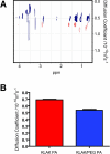
Self-assembled peptide amphiphiles (PAs) consisting of hydrophobic, hydvrogen-bonding, and charged hydrophilic domains form cylindrical nanofibers in physiological conditions and allow for the presentation of a high density of bioactive epitopes on the nanofiber surface. We report here on the use of PAs to form multifunctional nanostructures with tumoricidal activity. The combination of a cationic, membrane-lytic PA coassembled with a serum-protective, pegylated PA was shown to self-assemble into nanofibers. Addition of the pegylated PA to the nanostructure substantially limited degradation of the cytolytic PA by the protease trypsin, with an 8-fold increase in the amount of intact PA observed after digestion. At the same time, addition of up to 50% pegylated PA to the nanofibers did not decrease the in vitro cytotoxicity of the cytolytic PA. Using a fluorescent tag covalently attached to PA nanofibers we were able to track the biodistribution in plasma and tissues of tumor-bearing mice over time after intraperitoneal administration of the nanoscale filaments. Using an orthotopic mouse xenograft model of breast cancer, systemic administration of the cytotoxic pegylated nanostructures significantly reduced tumor cell proliferation and overall tumor growth, demonstrating the potential of multifunctional PA nanostructures as versatile cancer therapeutics.
Figures


Similar articles
-
Supramolecular Assembly of Peptide Amphiphiles.Acc Chem Res. 2017 Oct 17;50(10):2440-2448. doi: 10.1021/acs.accounts.7b00297. Epub 2017 Sep 6. Acc Chem Res. 2017. PMID: 28876055 Free PMC article.
-
Self-assembly of cytotoxic peptide amphiphiles into supramolecular membranes for cancer therapy.Adv Healthc Mater. 2013 Jan;2(1):126-33. doi: 10.1002/adhm.201200118. Epub 2012 Jul 31. Adv Healthc Mater. 2013. PMID: 23184589 Free PMC article.
-
Dip-pen patterning and surface assembly of peptide amphiphiles.Langmuir. 2005 Jun 7;21(12):5242-6. doi: 10.1021/la0501785. Langmuir. 2005. PMID: 15924443
-
Self-assembly of peptide amphiphiles: from molecules to nanostructures to biomaterials.Biopolymers. 2010;94(1):1-18. doi: 10.1002/bip.21328. Biopolymers. 2010. PMID: 20091874 Free PMC article. Review.
-
Urea-Modified Self-Assembling Peptide Amphiphiles That Form Well-Defined Nanostructures and Hydrogels for Biomedical Applications.ACS Appl Bio Mater. 2022 Oct 17;5(10):4599-4610. doi: 10.1021/acsabm.2c00158. Epub 2022 Jun 2. ACS Appl Bio Mater. 2022. PMID: 35653507 Review.
Cited by
-
Stab2-Mediated Clearance of Supramolecular Polymer Nanoparticles in Zebrafish Embryos.Biomacromolecules. 2020 Mar 9;21(3):1060-1068. doi: 10.1021/acs.biomac.9b01318. Epub 2020 Feb 21. Biomacromolecules. 2020. PMID: 32083854 Free PMC article.
-
Novel Peptide Therapeutic Approaches for Cancer Treatment.Cells. 2021 Oct 27;10(11):2908. doi: 10.3390/cells10112908. Cells. 2021. PMID: 34831131 Free PMC article. Review.
-
Proapoptotic Peptide Brush Polymer Nanoparticles via Photoinitiated Polymerization-Induced Self-Assembly.Angew Chem Int Ed Engl. 2020 Oct 19;59(43):19136-19142. doi: 10.1002/anie.202006385. Epub 2020 Aug 26. Angew Chem Int Ed Engl. 2020. PMID: 32659039 Free PMC article.
-
D-amino acids modulate the cellular response of enzymatic-instructed supramolecular nanofibers of small peptides.Biomacromolecules. 2014 Oct 13;15(10):3559-68. doi: 10.1021/bm5010355. Epub 2014 Sep 17. Biomacromolecules. 2014. PMID: 25230147 Free PMC article.
-
Supramolecular Assembly of Peptide Amphiphiles.Acc Chem Res. 2017 Oct 17;50(10):2440-2448. doi: 10.1021/acs.accounts.7b00297. Epub 2017 Sep 6. Acc Chem Res. 2017. PMID: 28876055 Free PMC article.
References
-
- Petros RA, DeSimone JM. Strategies in the Design of Nanoparticles for Therapeutic Applications. Nat. Rev. Drug Discovery. 2010;9:615–627. - PubMed
-
- Peer D, Karp JM, Hong S, Farokhzad OC, Margalit R, Langer R. Nanocarriers as an Emerging Platform for Cancer Therapy. Nat. Nanotechnol. 2007;2:751–760. - PubMed
-
- Matsumura Y, Maeda H. A New Concept for Macromolecular Therapeutics in Cancer Chemotherapy: Mechanism of Tumoritropic Accumulation of Proteins and the Antitumor Agent Smancs. Cancer Res. 1986;46:6387–6392. - PubMed
Publication types
MeSH terms
Substances
Grants and funding
LinkOut - more resources
Full Text Sources
Other Literature Sources
Medical

