. 2012 Oct 2;126(14):1728-38.
doi: 10.1161/CIRCULATIONAHA.112.115089. Epub 2012 Aug 28.

Therapeutic impact of follistatin-like 1 on myocardial ischemic injury in preclinical models

Affiliations

Therapeutic impact of follistatin-like 1 on myocardial ischemic injury in preclinical models

Yasuhiro Ogura et al. Circulation. .

Abstract

Background: Acute coronary syndrome is a leading cause of death in developed countries. Follistatin-like 1 (FSTL1) is a myocyte-derived secreted protein that is upregulated in the heart in response to ischemic insult. Here, we investigated the therapeutic impact of FSTL1 on acute cardiac injury in small and large preclinical animal models of ischemia/reperfusion and dissected its molecular mechanism.

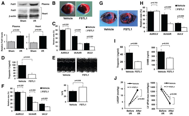
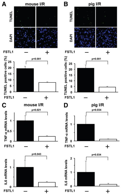
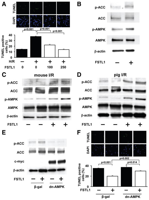
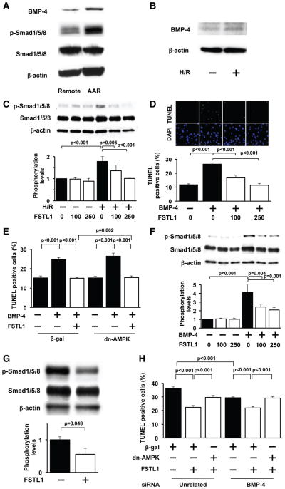
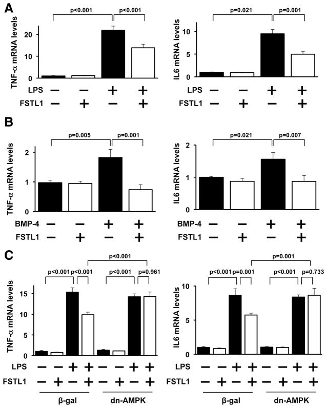
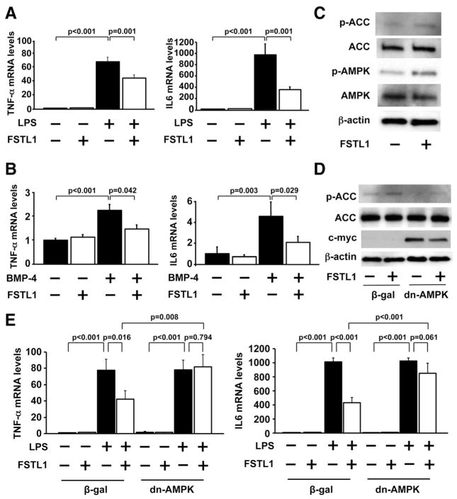
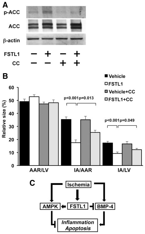
Methods and results: Administration of human FSTL1 protein significantly attenuated myocardial infarct size in a mouse or pig model of ischemia/reperfusion, which was associated with a reduction of apoptosis and inflammatory responses in the ischemic heart. Administration of FSTL1 enhanced the phosphorylation of AMP-activated protein kinase in the ischemia/reperfusion-injured heart. In cultured cardiac myocytes, FSTL1 suppressed apoptosis in response to hypoxia/reoxygenation and lipopolysaccharide-stimulated expression of proinflammatory genes through its ability to activate AMP-activated protein kinase. Ischemia/reperfusion led to enhancement of bone morphogenetic protein-4 expression and Smad1/5/8 phosphorylation in the heart, and FSTL1 suppressed the increased phosphorylation of Smad1/5/8 in ischemic myocardium. Treating cardiac myocytes with FSTL1 abolished the bone morphogenetic protein-4-stimulated increase in apoptosis, Smad1/5/8 phosphorylation, and proinflammatory gene expression. In cultured macrophages, FSTL1 diminished lipopolysaccharide-stimulated expression of proinflammatory genes via activation of AMP-activated protein kinase and abolished bone morphogenetic protein-4-dependent induction of proinflammatory mediators.

Conclusions: Our data indicate that FSTL1 can prevent myocardial ischemia/reperfusion injury by inhibiting apoptosis and inflammatory response through modulation of AMP-activated protein kinase- and bone morphogenetic protein-4-dependent mechanisms, suggesting that FSTL1 could represent a novel therapeutic target for post-myocardial infarction, acute coronary syndrome.


Figures

Figure 1
Figure 1
Administration of follistatin-like 1 (FSTL1) protein decreases myocardial infarct size in animal models of ischemia/reperfusion (I/R). A, Fstl1 protein levels in the heart and plasma in mice at 24 hours after myocardial I/R injury or sham operation. Fstl1 protein expression was analyzed by Western blotting and expressed relative to β-actin levels (n=4). B and C, Systemic delivery of human FSTL1 protein reduces myocardial infarct size in mouse I/R models. After intravenous injection of human FSTL1 protein (100 ng/g mouse) or vehicle, wild-type mice were subjected to 60 minutes of ischemia followed by 24 hours of reperfusion. Representative photographs of the mouse heart sections stained with Evans blue and subsequent 2,3,5-triphenyl tetrazolium chloride (TTC) are shown (B). Left ventricular (LV) area, the area at risk (AAR), and the infarct area (IA) were measured, and quantitative analysis of infarct size is shown (C; n=7– 8). D, FSTL1 reduces plasma troponin I levels of mice at 3 hours after I/R (n=4). E, Administration of FSTL1 protein improves LV function as assessed by echocardiography. Representative M-mode echocardiograms for vehicle- or FSTL1-treated mice at 24 hours after I/R are shown (top); quantitative analysis of LV fractional shortening (%FS) in mice treated with vehicle or FSTL1 protein is also shown (bottom; n=5). F, FSTL1 administration during reperfusion reduces infarct size in mice. Recombinant human FSTL1 protein (100 ng/g mouse) or vehicle was administered intravenously to mice at 5 minutes after reperfusion. Quantitative analysis of infarct size is shown (n=7). G and H, Intracoronary administration of human FSTL1 protein suppresses myocardial infarct size after I/R in pigs. Pigs were subjected to 45 minutes of ischemia followed by 24 hours of reperfusion, and recombinant human FSTL1 protein (3 μg/kg pig) or vehicle was injected through the wire lumen of the catheter during the first 10 minutes of ischemia. Representative photographs of the heart sections stained with Evans blue and TTC are shown (G). Quantitative analysis of the AAR/LV, IA/AAR, and IA/LV ratios is shown (H; n=5). I, FSTL1 treatment inhibits circulating levels of troponin I and creatine phosphokinase-MB (CKMB) in pigs at 24 hours after I/R (n=5). J, FSTL1 decreases LV end-diastolic pressure (LVEDP) and increases LV dP/dtmax in pigs at 24 hours after I/R (n=3–5).
Figure 2
Figure 2
Administration of follistatin-like 1 (FSTL1) protein attenuates apoptosis and inflammatory responses in the ischemic myocardium of mice and pigs. A, Systemic delivery of FSTL1 attenuates apoptosis in the ischemic heart of mice. Upper panels show representative photographs of mouse heart sections stained with terminal deoxynucleotidyl transferase-mediated dUTP nick-end labeling (TUNEL; green) and DAPI (blue). Lower graph shows quantitative analysis of TUNEL-positive nuclei (n=5). B, Intracoronary administration of FSTL1 to pigs inhibits apoptosis in the ischemic myocardium. Upper panels show representative pictures of heart sections stained with TUNEL (green) and DAPI (blue). Lower graph shows quantitative analysis of TUNEL-positive nuclei (n=4). C and D, Delivery of human FSTL1 suppresses expression of proinflammatory cytokines in the ischemic hearts of mice (C) and pigs (D). The mRNA expression of tumor necrosis factor-α (TNF-α) and interleukin-6 (IL-6) was measured by reverse transcription–polymerase chain reaction method and expressed relative to β-actin levels (n=5). I/R indicates ischemia/reperfusion.
Figure 3
Figure 3
Follistatin-like 1 (FSTL1) inhibits apoptosis via activation of AMP-regulated protein kinase (AMPK). A, FSTL1 suppresses hypoxia/reoxygenation (H/R)–induced apoptosis of neonatal rat ventricular myocytes (NRVMs). NRVMs were treated with FSTL1 protein (100 or 250 ng/mL) or vehicle under conditions of normoxia or H/R. NRVMs were stained with terminal deoxynucleotidyl transferase-mediated dUTP nick-end labeling (TUNEL; green) and DAPI (blue), and quantitative analysis of TUNEL-positive myocytes was performed (n=4). B, FSTL1 promotes AMPK phosphorylation in NRVMs. NRVMs were treated with FSTL1 protein (250 ng/mL) or vehicle for 30 minutes. The phosphorylation status of acetyl-coenzyme A carboxylase (p-ACC) and AMPK (p-AMPK) was analyzed by Western blotting. C and D, FSTL1 promotes AMPK signaling pathways in I/R-injured hearts of mice (C) and pigs (D). The phosphorylation of ACC and AMPK was assessed by Western blot analysis. Representative blots are shown from 4 independent experiments. E, AMPK inhibition abolishes FSTL1-induced ACC phosphorylation as assessed by Western blot analysis. After transduction with a dominant-negative form of AMPK tagged with c-myc (Ad-dn-AMPK) or β-galactosidase (Ad-β-gal) at a multiplicity of infection of 10 for 24 hours, NRVMs were treated with FSTL1 (250 ng/mL) or vehicle for 30 minutes. Representative blots are shown from 3 independent experiments. F, AMPK is involved in the suppressive action of FSTL1 on H/R-induced apoptosis of NRVMs. NRVMs were transduced with Ad-dn-AMPK or Ad-β-gal and cultured in the presence or absence of FSTL1 (250 ng/mL) under conditions of H/R. Apoptotic nuclei were identified by TUNEL staining (n=4).
Figure 4
Figure 4
Follistatin-like 1 (FSTL1) antagonizes bone morphogenetic protein-4 (BMP-4) signaling in the ischemic heart and cardiac myocytes. A, BMP-4 is upregulated in the heart in response to ischemia/reperfusion (I/R). Representative blots of BMP-4 and phosphorylated Smad1/5/8 (p-Smad1/5/8) at remote nonischemic area (remote) and ischemic area at risk (AAR) of pigs are shown from 4 independent experiments. B, Hypoxia/reoxygenation (H/R) increases the expression of BMP-4 in NRVMs. Representative blots of BMP-4 are shown from 4 independent experiments. C, FSTL1 attenuates the phosphorylation of Smad1/5/8 in neonatal rat ventricular myocytes (NRVMs) under conditions of H/R. NRVMs were treated with FSTL1 (100 or 250 ng/mL) or vehicle under conditions of normoxia or H/R. The phosphorylation levels of Smad1/5/8 (p-Smad1/5/8) were analyzed by Western blotting and expressed relative to β-actin levels (n=3). D, FSTL1 abolishes BMP-4 –induced apoptosis of NRVMs. NRVMs were treated with BMP-4 protein (100 ng/mL) or vehicle along with FSTL1 protein (100 or 250 ng/mL) or vehicle for 18 hours under normoxic conditions. Top, Representative pictures of NRVMs stained with terminal deoxynucleotidyl transferase-mediated dUTP nick-end labeling (TUNEL; green) and DAPI (blue). Bottom, Quantitative analysis of TUNEL-positive NRVMs (n=4). E, Effect of AMP-regulated protein kinase (AMPK) inactivation on FSTL1-mediated inhibition of BMP4-stimulated apoptosis of NRVMs. NRVMs were transduced with dominant-negative form of AMPK tagged with c-myc (Ad-dn-AMPK) or β-galactosidase (Ad-β-gal) at a multiplicity of infection of 10 for 24 hours and treated with BMP-4 protein (100 ng/mL) or vehicle along with FSTL1 protein (250 ng/mL) or vehicle for 18 hours under normoxic conditions (n=4). F, FSTL1 suppresses BMP-4 –stimulated phosphorylation of Smad1/5/8 in NRVMs. NRVMs were treated with BMP-4 protein (100 ng/mL) or vehicle along with FSTL1 protein (100 or 250 ng/mL) or vehicle for 18 hours under normoxic conditions. The phosphorylation levels of Smad1/5/8 (p-Smad1/5/8) were determined by Western blot analysis and expressed relative to β-actin levels (n=4). G, Intracoronary administration of FSTL1 attenuates the phosphorylation of Smad1/5/8 in the ischemic myocardium in pigs. Top, Representative blots of p-Smad1/5/8, Smad1/5/8, and β-actin. Bottom, Quantitative analysis of relative phosphorylation levels of Smad1/5/8 (n=4). H, Involvement of BMP-4 antagonization and AMPK activation in the inhibitory action of FSTL1 on H/R-induced myocyte apoptosis. NRVMs are transfected with siRNAs targeting BMP-4 or unrelated siRNAs (40 nmol/L) and transduced with an Ad-dn-AMPK or Ad-β-gal followed by treatment with FSTL1 (250 ng/mL) or vehicle (n=4).
Figure 5
Figure 5
Follistatin-like 1 (FSTL1) suppresses inflammatory responses in cultured cardiac myocytes. A, FSTL1 diminishes lipopolysaccharide (LPS)-stimulated expression of proinflammatory genes in neonatal rat ventricular myocytes (NRVMs). NRVMs were pretreated with FSTL1 (250 ng/mL) or vehicle for 30 minutes, followed by stimulation with LPS (100 ng/mL) or vehicle for 6 hours. The mRNA expression of tumor necrosis factor-α (TNF-α) and interleukin-6 (IL-6) was measured by reverse transcription–polymerase chain reaction (RT-PCR) and expressed relative to β-actin levels (n=4). B, Effect of FSTL1 on bone morphogenetic protein-4 (BMP-4)–stimulated expression of proinflammatory cytokines. NRVMs were treated with BMP-4 protein (100 ng/mL) or vehicle along with FSTL1 protein (100 or 250 ng/mL) or vehicle for 18 hours. The transcript levels of TNF-α and IL-6 were determined by RT-PCR and expressed relative to β -actin levels (n=4). C, AMPK participates in the effect of FSTL1 on the LPS-induced increase in proinflammatory gene expression. After transduction with a dominant-negative form of AMPK tagged with c-myc (Ad-dn-AMPK) or β -galactosidase (Ad-β -gal) at a multiplicity of infection of 10 for 24 hours, NRVMs were pre-treated with FSTL1 (250 ng/mL) or vehicle for 30 minutes, followed by treatment with LPS (100 ng/mL) or vehicle for 6 hours. The mRNA levels were analyzed by RT-PCR and expressed relative to β -actin levels (n=4).
Figure 6
Figure 6
Follistatin-like 1 (FSTL1) protein diminishes inflammatory responses in cultured macrophages. A, FSTL1 inhibits lipopolysaccharide (LPS)-stimulated expression of proinflammatory genes in macrophages. Macrophage RAW264.7 cells were cultured in the presence of FSTL1 (250 ng/mL) or vehicle for 30 minutes, followed by treatment with LPS (100 ng/mL) or vehicle for 6 hours. The mRNA expression of tumor necrosis factor-α (TNF-α) and interleukin-6 (IL-6) was quantified by reverse transcription–polymerase chain reaction (RT-PCR) analysis and expressed relative to β-actin levels (n=4). B, FSTL1 abolishes bone morphogenetic protein-4 (BMP-4)–stimulated expression of proinflammatory mediators in macrophages. Macrophages were treated with BMP-4 protein (100 ng/mL) or vehicle along with FSTL1 protein (250 ng/mL) or vehicle for 18 hours. The transcript levels of TNF-α and IL-6 were analyzed by RT-PCR and expressed relative to β-actin levels (n=4). C, FSTL1 promotes the AMP-regulated protein kinase (AMPK) signaling pathway in macrophages. Macrophages were treated with FSTL1 (250 ng/mL) or vehicle for 15 minutes. The phosphorylation levels of ACC (p-ACC) and AMPK (p-AMPK) were determined by Western blot analysis. D, AMPK inactivation cancels FSTL1-stimulated ACC phosphorylation in macrophages as determined by Western blot analysis. Macrophages were transduced with Ad- dominant-negative form of AMPK tagged with c-myc (Ad-dn-AMPK) or β-galactosidase (Ad-β-gal), and treated with FSTL1 (250 ng/mL) or vehicle for 15 minutes. E, AMPK signaling is involved in FSTL1-mediated inhibition of LPS-stimulated expression of proinflammatory cytokines. After transduction with Ad-dn-AMPK or Ad-β-gal, macrophages were treated with FSTL1 (250 ng/mL) or vehicle for 30 minutes followed by stimulation with LPS (100 ng/mL) or vehicle for 6 hours. Transcript levels were determined by RT-PCR analysis and expressed relative to β-actin levels (n=4).
Figure 7
Figure 7
AMP-regulated protein kinase (AMPK) is involved in the myocardial infarct-sparing effect of follistatin-like 1 (FSTL1). A, Effect of AMPK inactivation on acetyl-coenzyme A carboxylase (ACC) phosphorylation in the ischemic heart. AMPK inhibitor compound C (CC; 20 mg/kg) dissolved in dimethyl sulfoxide (DMSO) or DMSO was injected intraperitoneally into mice. After intravenous injection of human FSTL1 protein (100 ng/g mouse) or vehicle, wild-type mice were subjected to ischemia/reperfusion (I/R). The phosphorylation of ACC (p-ACC) in the ischemic heart was assessed by Western blot analysis. Representative blots are shown from 4 independent experiments. B, Role of AMPK in FSTL1-mediated inhibition of infarct size. Quantitative analysis of the ratios of area at risk (AAR) to left ventricle (LV), infarct area (IA) to AAR, and the IA to LV is shown (n=6). C, Proposed scheme for the mechanism by which FSTL1 protects the heart from ischemic injury. FSTL1 is upregulated in the heart and plasma in response to ischemic insult. FSTL1 promotes AMPK signaling pathways in the ischemic heart, thereby leading to a reduction of inflammation and apoptosis. FSTL1 also antagonizes bone morphogenetic protein-4 (BMP-4)– dependent proinflammatory and proapoptotic signals in the myocardium. Therefore, FSTL1 confers beneficial actions on the ischemic hearts by reducing inflammatory response and apoptosis through modulation of AMPK- and BMP-4 – dependent mechanisms.

Similar articles

Cited by

References

    1. Roger VL, Go AS, Lloyd-Jones DM, Adams RJ, Berry JD, Brown TM, Carnethon MR, Dai S, de Simone G, Ford ES, Fox CS, Fullerton HJ, Gillespie C, Greenlund KJ, Hailpern SM, Heit JA, Ho PM, Howard VJ, Kissela BM, Kittner SJ, Lackland DT, Lichtman JH, Lisabeth LD, Makuc DM, Marcus GM, Marelli A, Matchar DB, McDermott MM, Meigs JB, Moy CS, Mozaffarian D, Mussolino ME, Nichol G, Paynter NP, Rosamond WD, Sorlie PD, Stafford RS, Turan TN, Turner MB, Wong ND, Wylie-Rosett J. Heart disease and stroke statistics—2011 update: a report from the American Heart Association. Circulation. 2011;123:e18– e209. - PMC - PubMed
    1. Lange RA, Hillis LD. Immediate angioplasty for acute myocardial infarction. N Engl J Med. 1993;328:726–728. - PubMed
    1. Fox KA, Steg PG, Eagle KA, Goodman SG, Anderson FA, Jr, Granger CB, Flather MD, Budaj A, Quill A, Gore JM. Decline in rates of death and heart failure in acute coronary syndromes, 1999 –2006. JAMA. 2007;297:1892–1900. - PubMed
    1. Simoons ML, Windecker S. Controversies in cardiovascular medicine: chronic stable coronary artery disease: drugs vs. revascularization. Eur Heart J. 2010;31:530–541. - PubMed
    1. Verma S, Fedak PW, Weisel RD, Butany J, Rao V, Maitland A, Li RK, Dhillon B, Yau TM. Fundamentals of reperfusion injury for the clinical cardiologist. Circulation. 2002;105:2332–2336. - PubMed

Publication types

Substances