

The peptide derived from the Ig-like domain of human herpesvirus 8 K1 protein induces death in hematological cancer cells
- PMID: 22929310
- PMCID: PMC3517441
- DOI: 10.1186/1756-9966-31-69
The peptide derived from the Ig-like domain of human herpesvirus 8 K1 protein induces death in hematological cancer cells
Abstract
Background: Although significant progress has been made in the treatment of lymphomas, many lymphomas exhibit resistance to cell death, suggesting a defective Fas signaling, which remains poorly understood. We previously reported that cells expressing the K1 protein of human herpesvirus 8 (HHV-8) resist death through the complex formation of the Ig-like domain of K1 with Fas. Recently, we investigated whether peptides derived from the Ig-like domain of the K1 protein may affect cell death.
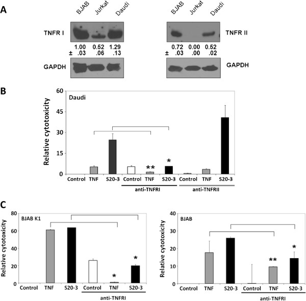
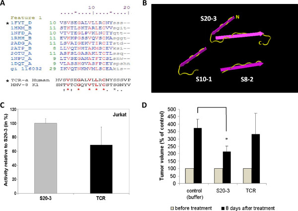
Methods: K1 positive and negative cell lines were incubated with the K1-derived peptides, and cell death (apoptotic and necrotic) was assessed by flow cytometry and LDH assay. Activation of caspases was assessed by fluorometric assay and flow cytometry. Fas receptor-independent, peptide-mediated cell killing was tested in the Fas-resistant Daudi cell line and Jurkat cell clones deficient in caspase-8 and FADD functionality. Activation of TNF receptors I and II was blocked by pre-incubation with corresponding blocking antibodies. The effect of the K1 peptide in vivo was tested in a mouse xenograft model.
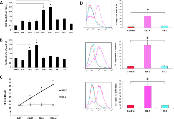
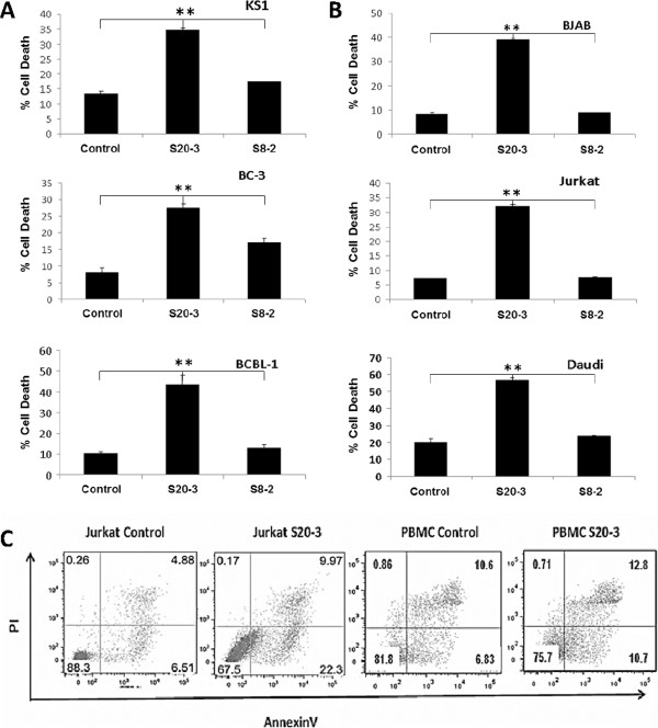
Results: We observed that the peptide S20-3 enhanced cell death in K1-positive BJAB cells and HHV-8 positive primary effusion lymphoma (PEL) cell lines. Similar effects of this peptide were observed in B-cell lymphoma and T-lymphoblastic leukemia cells without K1 expression but not in normal human peripheral blood mononuclear cells. A single intratumoral injection of the S20-3 peptide decreased the growth of Jurkat xenografts in SCID mice. The mechanism of tumor cell death induced by the S20-3 peptide was associated with activation of caspases, but this activity was only partially inhibited by the pan-caspase inhibitor z-VAD. Furthermore, the K1 peptide also killed Fas-resistant Daudi cells, and this killing effect was inhibited by pre-incubation of cells with antibodies blocking TNFRI.
Conclusion: Taken together, these findings indicate that the S20-3 peptide can selectively induce the death of malignant hematological cell lines by Fas- and/or TNFRI-dependent mechanisms, suggesting the K1-derived peptide or peptidomimetic may have promising therapeutic potential for the treatment of hematological cancers.
Figures

Similar articles
-
Mechanism of Fas signaling regulation by human herpesvirus 8 K1 oncoprotein.J Natl Cancer Inst. 2009 Mar 18;101(6):399-411. doi: 10.1093/jnci/djn516. Epub 2009 Mar 10. J Natl Cancer Inst. 2009. PMID: 19276446 Free PMC article.
-
Fas-associated death domain protein and caspase-8 are not recruited to the tumor necrosis factor receptor 1 signaling complex during tumor necrosis factor-induced apoptosis.J Biol Chem. 2003 Jul 11;278(28):25534-41. doi: 10.1074/jbc.M303399200. Epub 2003 Apr 29. J Biol Chem. 2003. PMID: 12721308
-
Necrotic death pathway in Fas receptor signaling.J Cell Biol. 2000 Dec 11;151(6):1247-56. doi: 10.1083/jcb.151.6.1247. J Cell Biol. 2000. PMID: 11121439 Free PMC article.
-
Cellular Dynamics of Fas-Associated Death Domain in the Regulation of Cancer and Inflammation.Int J Mol Sci. 2024 Mar 12;25(6):3228. doi: 10.3390/ijms25063228. Int J Mol Sci. 2024. PMID: 38542202 Free PMC article. Review.
-
T cell intrinsic roles of autophagy in promoting adaptive immunity.Curr Opin Immunol. 2010 Jun;22(3):321-5. doi: 10.1016/j.coi.2010.03.005. Epub 2010 Apr 12. Curr Opin Immunol. 2010. PMID: 20392618 Free PMC article. Review.
Cited by
-
CD74 interferes with the expression of fas receptor on the surface of lymphoma cells.J Exp Clin Cancer Res. 2014 Oct 10;33(1):80. doi: 10.1186/s13046-014-0080-y. J Exp Clin Cancer Res. 2014. PMID: 25304249 Free PMC article.
-
Nucleolin inhibits Fas ligand binding and suppresses Fas-mediated apoptosis in vivo via a surface nucleolin-Fas complex.Blood. 2013 Jun 6;121(23):4729-39. doi: 10.1182/blood-2012-12-471094. Epub 2013 Apr 18. Blood. 2013. PMID: 23599269 Free PMC article.
-
Obesity, Bioactive Lipids, and Adipose Tissue Inflammation in Insulin Resistance.Nutrients. 2020 May 3;12(5):1305. doi: 10.3390/nu12051305. Nutrients. 2020. PMID: 32375231 Free PMC article. Review.
-
Endoplasmic reticulum stress in the adipose tissue and monocyte chemoattractant protein-1 are involved in tacrolimus-induced diabetes mellitus.Pharmacol Res Perspect. 2023 Jun;11(3):e01081. doi: 10.1002/prp2.1081. Pharmacol Res Perspect. 2023. PMID: 37195045 Free PMC article.
References
-
- Muller M, Strand S, Hug H, Heinemann EM, Walczak H, Hofmann WJ, Stremmel W, Krammer PH, Galle PR. Drug-induced apoptosis in hepatoma cells is mediated by the CD95 (APO-1/Fas) receptor/ligand system and involves activation of wild-type p53. J Clin Invest. 1997;99:403–413. doi: 10.1172/JCI119174. - DOI - PMC - PubMed
-
- Vega MI, Huerta-Yepez S, Jazirehi AR, Garban H, Bonavida B. Rituximab (chimeric anti-CD20) sensitizes B-NHL cell lines to Fas-induced apoptosis. Oncogene. 2005;24:8114–8127. - PubMed
Publication types
MeSH terms
Substances
Grants and funding
LinkOut - more resources
Full Text Sources
Research Materials
Miscellaneous
