Review
. 2012 Nov;32(11):1959-72.
doi: 10.1038/jcbfm.2012.126. Epub 2012 Aug 29.

Drug transport across the blood-brain barrier

Affiliations
Review

Drug transport across the blood-brain barrier

William M Pardridge. J Cereb Blood Flow Metab. 2012 Nov.

Abstract

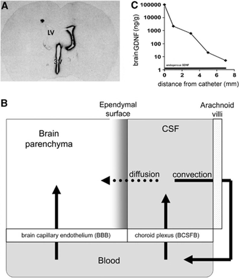
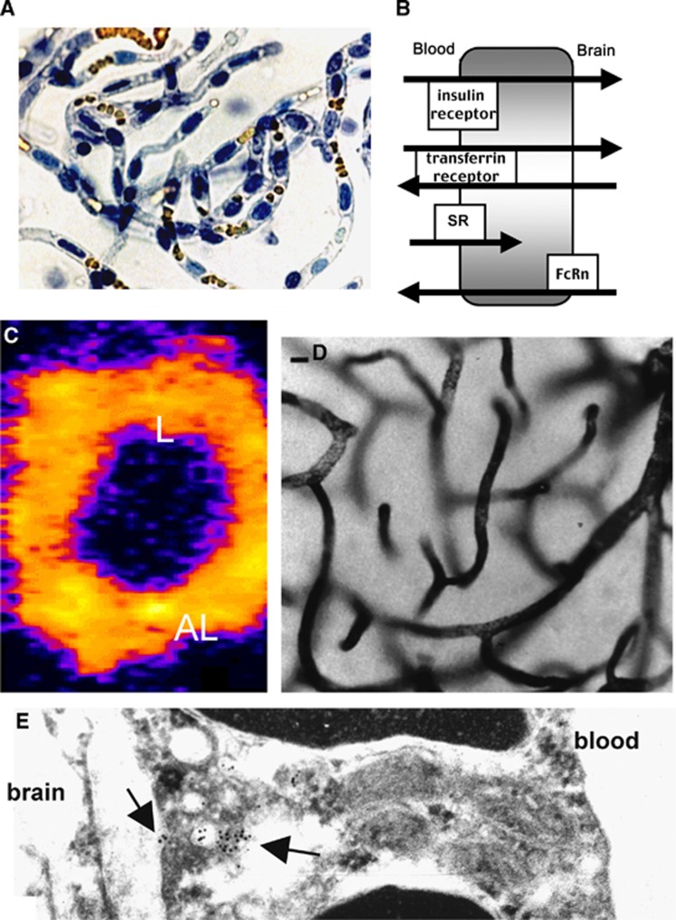
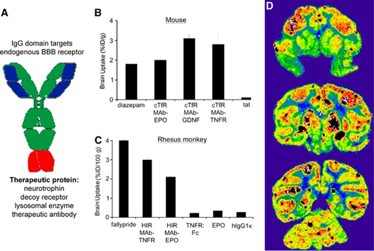
The blood-brain barrier (BBB) prevents the brain uptake of most pharmaceuticals. This property arises from the epithelial-like tight junctions within the brain capillary endothelium. The BBB is anatomically and functionally distinct from the blood-cerebrospinal fluid barrier at the choroid plexus. Certain small molecule drugs may cross the BBB via lipid-mediated free diffusion, providing the drug has a molecular weight <400 Da and forms <8 hydrogen bonds. These chemical properties are lacking in the majority of small molecule drugs, and all large molecule drugs. Nevertheless, drugs can be reengineered for BBB transport, based on the knowledge of the endogenous transport systems within the BBB. Small molecule drugs can be synthesized that access carrier-mediated transport (CMT) systems within the BBB. Large molecule drugs can be reengineered with molecular Trojan horse delivery systems to access receptor-mediated transport (RMT) systems within the BBB. Peptide and antisense radiopharmaceuticals are made brain-penetrating with the combined use of RMT-based delivery systems and avidin-biotin technology. Knowledge on the endogenous CMT and RMT systems expressed at the BBB enable new solutions to the problem of BBB drug transport.


Figures

Figure 1
Figure 1
(A) Whole body autoradiogram of a mouse euthanized 5 minutes after the intravenous injection of [14C]-histamine. (B) India ink visualization of complexity of the microvasculature of the cortex of rat brain. (C) Vascular cast of the microvasculature of the human cerebellar cortex. The distance between capillaries in brain is about 40 μm.
Figure 2
Figure 2
(A) Film autoradiogram of rat brain section 20 hours after the injection of [125I]-BDNF (brain-derived neurotrophic factor) into the lateral ventricle (LV). (B) Solute distribution into brain parenchyma from the cerebrospinal fluid (CSF) compartment is limited by the slow rate of solute diffusion from the ependymal surface, relative to the rapid rate of bulk flow (convection) of CSF from the ventricles to the systemic circulation across the arachnoid villi. (C) Logarithmic decrease in the brain concentration of glial-derived neurotrophic factor (GDNF) in the brain of the Rhesus monkey relative to the distance from the intracerebral catheter used for brain drug delivery with convection-enhanced diffusion.
Figure 3
Figure 3
(A) Predicted secondary structure of the bovine blood–brain barrier (BBB) GLUT1 glucose transporter is shown, which is formed by 12 transmembrane domains, a glycosylated extracellular loop between transmembrane domains 1 and 2, and intracellular amino and carboxyl termini. (B) Northern blot of mRNA derived from either total rabbit brain (lane 1) or capillary-depleted rabbit brain (lane 2) for either the GLUT1 glucose transporter or actin. (C) Electron microscopic immunogold study of human brain with a primary antiserum against the purified human erythrocyte glucose transporter, and a secondary antibody conjugated with 10 nm gold particles, shows abundant expression of immunoreactive GLUT1 glucose transporter on the luminal (L) and abluminal (AL) membranes of the capillary endothelium in human brain.
Figure 4
Figure 4
(A) Light micrograph of freshly isolated bovine brain capillaries stained with trypan blue, which stains nuclei blue and luminal red blood cells yellow. The capillaries are isolated free of adjoining brain tissue. (B) Schematic diagram of different brain endothelial receptors, including the insulin receptor, the transferrin (Tf) receptor, the scavenger receptor (SR), which mediates only the endocytosis from blood into the endothelial compartment for ligands such as acetylated low density lipoprotein; and the neonatal Fc receptor (FcRn), which mediates the asymmetric transcytosis of IgG molecules selectively from brain to blood, but not blood to brain. (C) Confocal microscopy of isolated rat brain capillaries showing the expression of the Tf receptor on both luminal (L) and abluminal (AL) membranes of the endothelium. (D) Light micrograph of rat brain removed after a 10-minute carotid artery infusion of a conjugate of 5 nm gold and the mouse OX26 monoclonal antibody (MAb) against the rat TfR. The staining of the capillary compartment represents TfRMAb present in the intraendothelial compartment of the brain. (E) Electron microscopy of rat brain after perfusion with the conjugate of gold and TfRMAb shows the MAb concentrated in intraendothelial vesicles (intracellular arrow), as well as MAb molecules undergoing exocytosis from the endothelial compartment to the brain interstitial space (extracellular arrow).
Figure 5
Figure 5
(A) Structure of an IgG fusion protein formed by fusion of a therapeutic protein to the carboxyl terminus of the heavy chain of a blood–brain barrier (BBB) targeting monoclonal antibody (MAb). (B) Brain uptake in the mouse, expressed as % injected dose (ID)/g brain, is shown for diazepam, a cTfRMAb–EPO fusion protein, a cTfRMAb–GDNF fusion protein, a cTfRMAb–TNFR fusion protein, and the cationic import peptide, tat. (C) Brain uptake in the Rhesus monkey is expressed as % ID/100 g brain, since the size of the primate brain is 100 g. Brain uptake is shown for fallypride, a HIRMAb–TNFR fusion protein, a HIRMAb–EPO fusion protein, the TNFR alone, EPO alone, and a human IgG1κ isotype control antibody (hIgG1κ), which is confined to the vascular compartment. (D) Film autoradiogram of Rhesus monkey brain removed 2 hours after the intravenous (IV) injection of [125I]-HIRMAb–IDUA fusion protein, showing global distribution in brain with higher uptake in gray matter relative to white matter.
Figure 6
Figure 6
(A) Conjugate of an IgG–avidin fusion protein and a monobiotinylated peptide radiopharmaceutical. The biotin group on the peptide binds to the avidin domain of the fusion protein to form a high affinity linkage between the IgG molecular Trojan horse and the peptide radiopharmaceutical. (B, C) Scans of Rhesus monkey brain at 3 hours after the intravenous (IV) injection of either [125I, N-biotinyl]-Aβ1–40 alone (B), or [125I, N-biotinyl]-Aβ1–40 conjugated to the HIRMAb (monoclonal antibody) molecular Trojan horse via a streptavidin linker (C). (D) A rat brain tumor model was produced by the intracerebral injection of human U87 glioma cells in nude rats. Immunocytochemistry (ICC) of brain removed 16 days after tumor implantation with an antibody to the human EGFR shows abundant expression of the EGFR in the brain tumor. (E) Scan of rat brain removed 2 hours after the IV injection of [111 In-DTPA, Lys-biotinyl]–EGF conjugated to the TfRMAb molecular Trojan horse via a streptavidin linker. The section used for the film autoradiography was parallel to the section used for the ICC study in (D).
Figure 7
Figure 7
(A) Structures are shown for model peptide nucleic acids (PNAs) with sequences that target either the rat glial fibrillary acidic protein (GFAP) mRNA or the rat caveolin-1α (CAV) mRNA. The methionine initiation codon sequence is underlined. (B) Confocal microscopy of brain from a rat with an intracranial RG-2 glioma is shown after double labeling with an antibody against GFAP (green channel) and an antibody against CAV (red channel). (C) Scan of brain removed from a rat with an intracranial RG-2 glioma at 1 hour after the intravenous (IV) injection of the [111In-DTPA, Lys-biotinyl]-GFAP PNA conjugated to the TfRMAb (monoclonal antibody) via a streptavidin linkage. The image over the tumor is of low intensity, as the RG-2 tumor does not express the GFAP mRNA. (D) Scan of brain removed from a rat with an intracranial RG-2 glioma at 6 hours after the IV injection of the [111In-DTPA, Lys-biotinyl]-CAV PNA conjugated to the TfRMAb via a streptavidin linkage. The image over the tumor is of high intensity, as the RG-2 tumor expresses abundant CAV mRNA.

Comment in

  • Accessing the brain: the nose may know the way.
    Djupesland PG, Mahmoud RA, Messina JC. Djupesland PG, et al. J Cereb Blood Flow Metab. 2013 May;33(5):793-4. doi: 10.1038/jcbfm.2013.41. Epub 2013 Mar 13. J Cereb Blood Flow Metab. 2013. PMID: 23486291 Free PMC article. No abstract available.

References

    1. Pardridge WM. The blood-brain barrier: bottleneck in brain drug development. NeuroRx. 2005;2:3–14. - PMC - PubMed
    1. Brightman MW, Reese TS. Junctions between intimately apposed cell membranes in the vertebrate brain. J Cell Biol. 1969;40:648–677. - PMC - PubMed
    1. Reiber H, Felgenhauer K. Protein transfer at the blood cerebrospinal fluid barrier and the quantitation of the humoral immune response within the central nervous system. Clin Chim Acta. 1987;163:319–328. - PubMed
    1. Mott FW. The late Professor Edwin Goldmann's investigations on the central nervous system by vital staining. Br Med J. 1913;2:871–873. - PMC - PubMed
    1. Friedemann U. Blood-brain barrier. Physiol Rev. 1942;22:125–145.

MeSH terms

Substances