. 2012 Oct;103(4):427-53.
doi: 10.1097/HP.0b013e318266eb4c.

The prolonged gastrointestinal syndrome in rhesus macaques: the relationship between gastrointestinal, hematopoietic, and delayed multi-organ sequelae following acute, potentially lethal, partial-body irradiation

Affiliations

The prolonged gastrointestinal syndrome in rhesus macaques: the relationship between gastrointestinal, hematopoietic, and delayed multi-organ sequelae following acute, potentially lethal, partial-body irradiation

Thomas J MacVittie et al. Health Phys. 2012 Oct.

Abstract

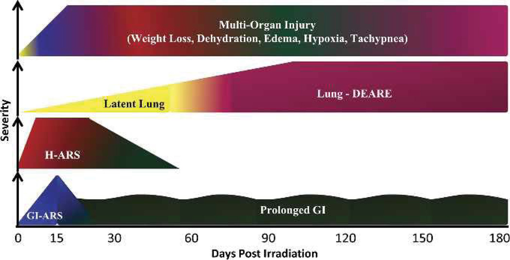
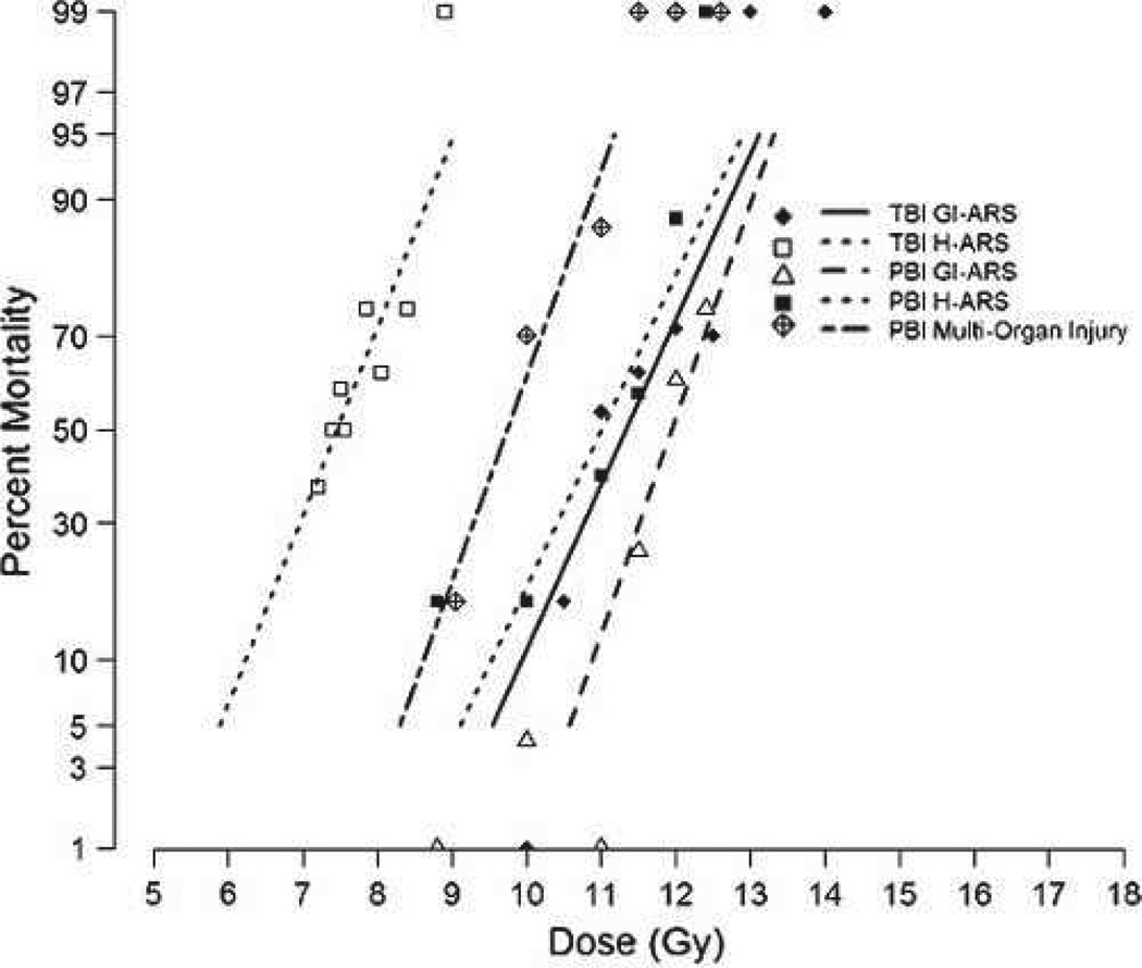
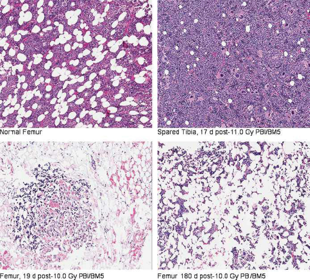
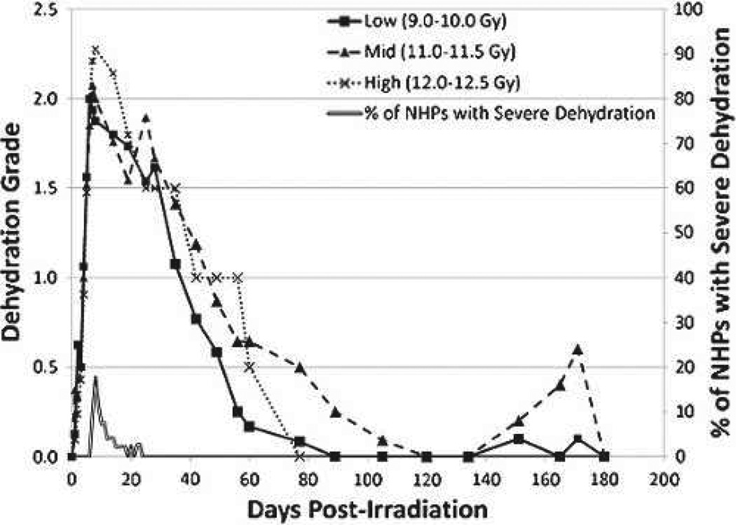
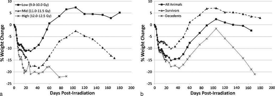
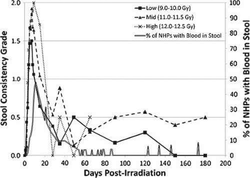
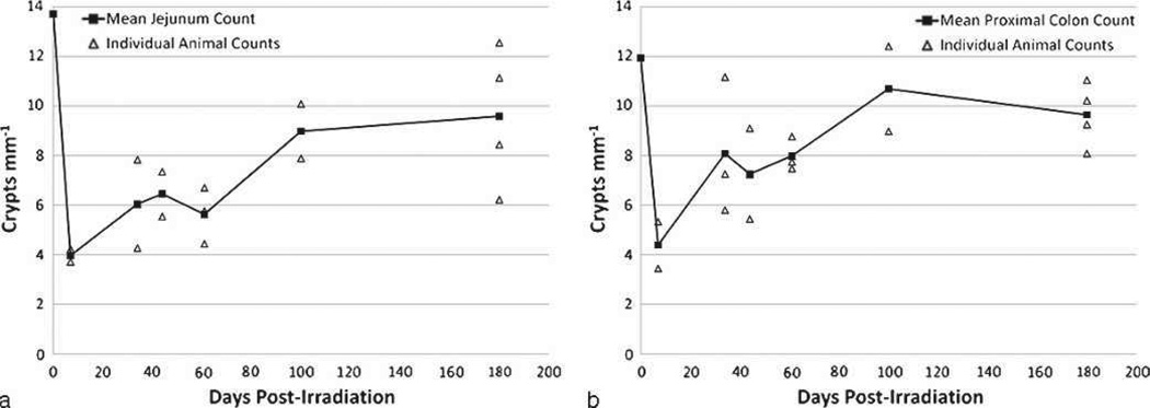
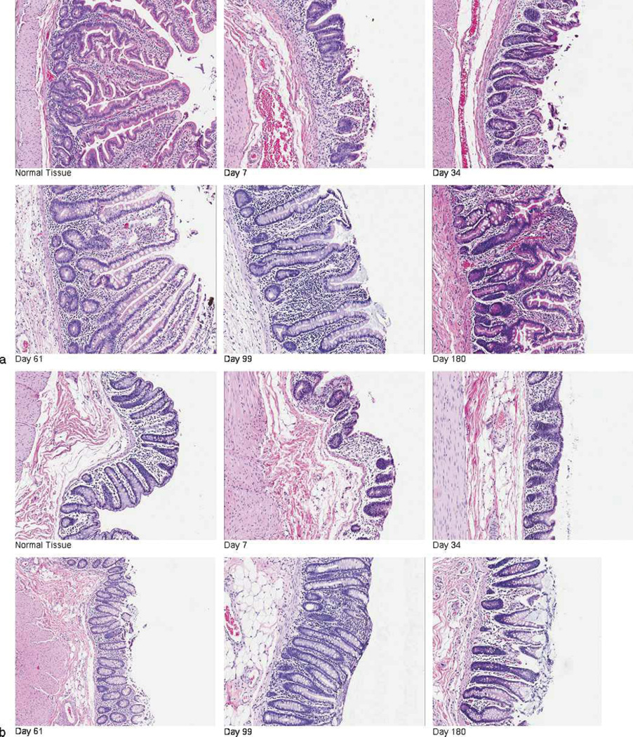
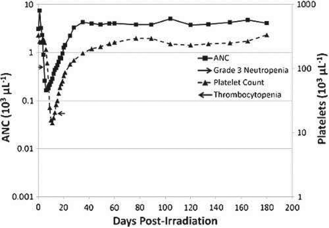
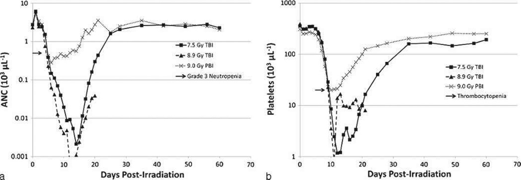
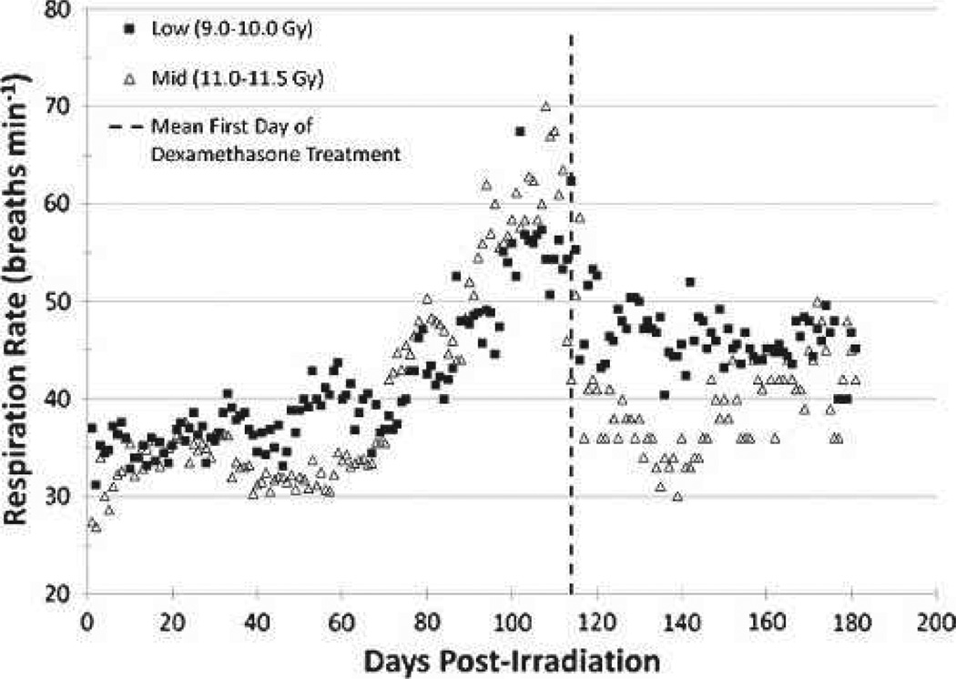
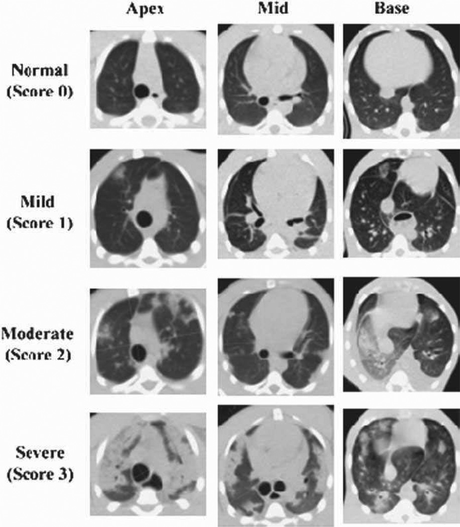
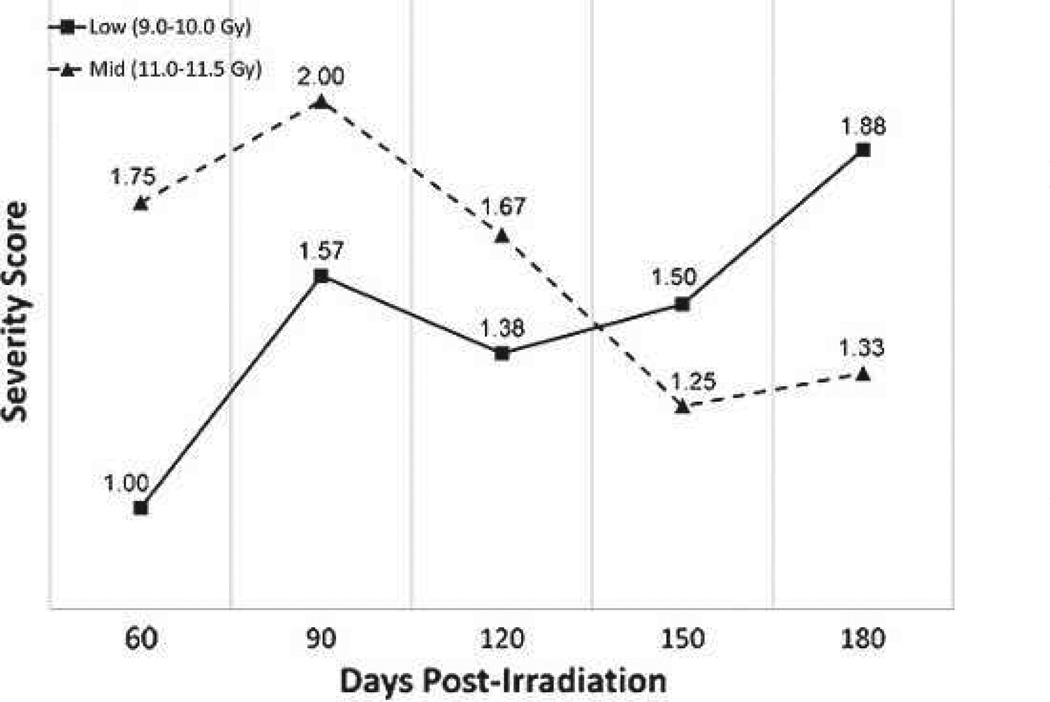
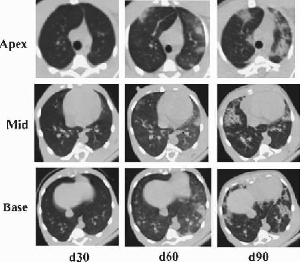
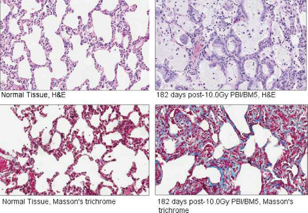
The dose response relationship for the acute gastrointestinal syndrome following total-body irradiation prevents analysis of the full recovery and damage to the gastrointestinal system, since all animals succumb to the subsequent 100% lethal hematopoietic syndrome. A partial-body irradiation model with 5% bone marrow sparing was established to investigate the prolonged effects of high-dose radiation on the gastrointestinal system, as well as the concomitant hematopoietic syndrome and other multi-organ injury including the lung. Herein, cellular and clinical parameters link acute and delayed coincident sequelae to radiation dose and time course post-exposure. Male rhesus Macaca mulatta were exposed to partial-body irradiation with 5% bone marrow (tibiae, ankles, feet) sparing using 6 MV linear accelerator photons at a dose rate of 0.80 Gy min(-1) to midline tissue (thorax) doses in the exposure range of 9.0 to 12.5 Gy. Following irradiation, all animals were monitored for multiple organ-specific parameters for 180 d. Animals were administered medical management including administration of intravenous fluids, antiemetics, prophylactic antibiotics, blood transfusions, antidiarrheals, supplemental nutrition, and analgesics. The primary endpoint was survival at 15, 60, or 180 d post-exposure. Secondary endpoints included evaluation of dehydration, diarrhea, hematologic parameters, respiratory distress, histology of small and large intestine, lung radiographs, and mean survival time of decedents. Dose- and time-dependent mortality defined several organ-specific sequelae, with LD50/15 of 11.95 Gy, LD50/60 of 11.01 Gy, and LD50/180 of 9.73 Gy for respective acute gastrointestinal, combined hematopoietic and gastrointestinal, and multi-organ delayed injury to include the lung. This model allows analysis of concomitant multi-organ sequelae, thus providing a link between acute and delayed radiation effects. Specific and multi-organ medical countermeasures can be assessed for efficacy and interaction during the concomitant evolution of acute and delayed key organ-specific subsyndromes.


Conflict of interest statement

The authors declare no conflict of interest.

Figures

Fig. 1
Fig. 1
Time course and severity of multiple organ injury following PBI/BM5. Exposure to PBI/BM5 results in the expression of key sequelae of ARS and DEARE, to include the acute GI-ARS, H-ARS with coincident prolonged GI damage, continued prolonged GI damage, lung injury, and multi-organ injury through 180 d post-exposure.
Fig. 2
Fig. 2
DRR for ARS and DEARE due to TBI and PBI/BM5 of rhesus macaques. Rhesus macaques were exposed to TBI or PBI/BM5 using 6MV LINAC-derived photons at a dose rate of 0.80 Gy min−1. Irradiations and animal care for each DRR were performed at the same radiation and veterinary facilities with the same research team (Farese et al. 2012; MacVittie et al. 2012). All exposures were measured as midline tissue dose. The DRRs were defined over time frames to assess organ-specific subsyndromes (15 and 60 d for GI-ARS and H-ARS, respectively) or multi-organ syndromes (180 d).
Fig. 3
Fig. 3
Histology of irradiated and spared bone marrow in rhesus macaques exposed to PBI/BM5. Bone marrow samples were procured from the tibia and femur. In vivo dosimetry was used to estimate the dose received to the tibiae and midline of each animal as described in Materials and Methods. The average dose to surface of tibiae was 0.53 Gy across all doses (9.0–12.5 Gy PBI) delivered to the animals. Representative photographs were taken at 10× magnification. Samples were stained with H&E.
Fig. 4
Fig. 4
Dehydration over time in rhesus macaques exposed to PBI/BM5. The grade of hydration and % of NHPs with severe dehydration versus time (d) post-PBI/BM5. Mean values are plotted for NHPs grouped into Low (9.0–10.0 Gy), Mid (11.0–11.5 Gy), and High (12.0–12.5 Gy) dose cohorts. Grades for dehydration were (0) for normal hydration, (1) for mild dehydration, (2) for moderate dehydration, and (3) for severe dehydration. Hydration status was evaluated daily.
Fig. 5
Fig. 5
Weight loss over time in rhesus macaques exposed to PBI/BM5. The % weight change relative to pre-irradiation body weight versus time (d) post-irradiation. Mean values are plotted for (a) NHPs grouped into dose cohorts, and (b) all studied NHPs, survivors, and decedents. Animals were weighed daily.
Fig. 6
Fig. 6
Diarrhea over time in rhesus macaques exposed to PBI/BM5. The severity and duration of diarrhea, assessed as grade of stool consistency and % of NHPs with bloody stool, versus time (days) post-irradiation. Mean values are plotted for NHPs grouped into dose cohorts. Each animal’s stool consistency was monitored twice a day for the duration of the in vivo phase of the study. The presence of blood in stool was evaluated concurrently. Stool grades were (0) for formed stool, (1) for soft stool, and (2) for diarrhea.
Fig. 7
Fig. 7
Radiation-induced loss and recovery of crypts in the jejunum and proximal colon of rhesus macaques exposed to PBI/BM5. Crypts were counted in the jejunum (a) and proximal colon (b) of animals exposed to 10.0 Gy PBI/BM5. Tissue was procured at days 7, 34, 44, 61, 100, and 180 post-exposure. Tissues were fixed in formalin, sectioned, and stained with H&E. A correction factor was applied to correct for error due to variation in crypt width, as detailed in Materials and Methods. The mean values are plotted with the values for each individual animal that was analyzed. The day 0 values are means from counts performed on non-irradiated, normal NHPs (n = 10).
Fig. 8
Fig. 8
Radiation-induced GI damage and recovery in rhesus macaques exposed to PBI/BM5. Animals were exposed to 10.0 Gy PBI/BM5. Tissue was procured from (a) the jejunum and (b) proximal colon at necropsy on selected study days (days 7, 44, 62, 100, and 180) across the 180 day in vivo time course. Tissue was fixed in formalin, sectioned, and stained with H&E. Representative photographs show the loss and disorganized regeneration of crypts and villi over time post-PBI/BM5. All photographs were taken at 10× magnification.
Fig. 9
Fig. 9
The time course of neutropenia and thrombocytopenia in rhesus macaques exposed to PBI/BM5. Mean values are plotted for ANC and platelet counts over time in days post-irradiation. Grade 3 neutropenia is defined as ANC ≤ 500 µL−1, and thrombocytopenia is defined as platelets <20,000 µl−1.
Fig. 10
Fig. 10
Comparison of the time course of neutropenia and thrombocytopenia in rhesus macaques exposed to PBI/BM5 or TBI. NHPs were exposed to TBI or PBI/BM5 using 6 MV LINAC-derived photons at 0.80 Gy min−1. The 9.0 Gy PBI/BM5 exposure is estimated to be an approximate LD50/60, whereas TBI at 8.9 Gy or 7.5 Gy are LD100/60 and LD50/60 doses, respectively. Mean values are plotted for (a) ANC and (b) platelet counts over time post-irradiation.
Fig. 11
Fig. 11
Respiratory rate in rhesus macaques exposed to PBI/BM5. Changes in mean NSRR are plotted as a function of time post-irradiation. This analysis was restricted to NHPs that survived >60 d post-exposure (e.g., survivors of GI-ARS and H-ARS coincident with prolonged GI; n = 28) and for whom serial daily NSRR data were available (n = 15/28). Mean values are plotted for NHPs grouped into dose cohorts. Animals in the High (12.0–12.5 Gy) dose cohort are not shown as only two of those NHPs survived >60 d, neither of which had serial NSRR data. The mean time to initiation of dexamethasone supportive care treatment was 114 d post-exposure for all NHPs on the plot.
Fig. 12
Fig. 12
Radiographic severity scoring of pulmonary injury in rhesus macaques exposed to PBI/BM5. Representative images characteristic of the qualitative scoring of radiation-induced lung injury are shown. CT images were assessed for evidence of radiation-induced lung injury (pneumonitis and fibrosis) based on characteristic changes in radiodensity. All CT scans were assessed by a radiation oncologist with expertise in thoracic radiography and scored as mild (1), moderate (2), or severe (3) based on the volume and distribution of injured lung relative to the total lung volume.
Fig. 13
Fig. 13
Time course of radiographic severity of pulmonary injury in rhesus macaques exposed to PBI/BM5. Serial, non-contrast enhanced, high-resolution CT scans were obtained in rhesus macaques approximately every 30 d post-irradiation. This analysis was restricted to NHPs that survived >60 d post-exposure (e.g., survivors of GI-ARS and H-ARS coincident with prolonged GI; n = 28) and for whom CT data were available (n = 24/28). Mean values are plotted for NHPs grouped into dose cohorts. Animals in the High (12.0–12.5 Gy) dose cohort are not shown, as only two of those NHPs survived >60 d, neither of which had serial CT data.
Fig. 14
Fig. 14
Evolution of radiographic injury in rhesus macaques exposed to PBI/BM5. Representative example of evolving radiation-induced pulmonary injury in a rhesus macaque post-11.0 Gy PBI/BM5. Images from the apex, mid, and base of the lung are shown at 30, 60, and 90 d post-exposure. This NHP was subsequently treated with dexamethasone (day 115 post-exposure) but succumbed to lung injury within 1 wk of beginning the corticosteroid treatment.
Fig. 15
Fig. 15
Histopathologic changes in the lungs of rhesus macaques exposed to PBI/BM5. Samples from normal (non-irradiated) NHPs are shown on the left, and samples from an NHP euthanized at day 182 post-10.0 Gy PBI/BM5 are shown on the right. H&E staining of the post-irradiation samples demonstrated macrophage infiltration with foamy macrophages, alveolar wall thickening, distortion of alveolar architecture, and hyaline membranes in the alveolar airspaces. Masson’s trichrome staining demonstrated collagen deposition indicative of evolving fibrosis in the post-irradiation samples.

Similar articles

Cited by

References

    1. Anno GH, Young RW, Bloom RM, Mercier JR. Dose response relationships for acute ionizing radiation lethality. Health Phys. 2003;84:565–575. - PubMed
    1. Bagdasarov AA, Raushenbakk MO, Abdullaev GM, Beliaeva BF, Lagutina NY. The treatment of acute radiation sickness with packed platelets. Probl Hematol Blood Transfus. 1959;4:1–5. - PubMed
    1. Baltschukat K, Nothdurft W. Hematological effects of unilateral and bilateral exposures of dogs to 300-kVp x rays. Radiat Res. 1990;123:7–16. - PubMed
    1. Baranov AE. Allogenic bone marrow transplantation after severe, uniform total-body irradiation: experience from recent (Nyasvizh, Belarus) and previous radiation accidents. In: MacVittie TJ, Weiss JF, Browne D, editors. Advances in the treatment of radiation injuries: advances in the biosciences. Vol. 94. Tarrytown, NY: Pergamon; Elsevier Science Ltd; 1996. pp. 281–293.
    1. Baranov AE, Guskova AK. Acute radiation disease in Chernobyl accident victims. In: Ricks RC, Fry SA, editors. The medical basis for radiation accident preparedness II; clinical experience and follow-up since 1979. New York: Elsevier; 1990. pp. 79–87.

Publication types

MeSH terms