. 2013 Mar;6(2):379-92.
doi: 10.1038/mi.2012.81. Epub 2012 Aug 29.

Molecular organization of the mucins and glycocalyx underlying mucus transport over mucosal surfaces of the airways

Affiliations

Molecular organization of the mucins and glycocalyx underlying mucus transport over mucosal surfaces of the airways

M Kesimer et al. Mucosal Immunol. 2013 Mar.

Abstract

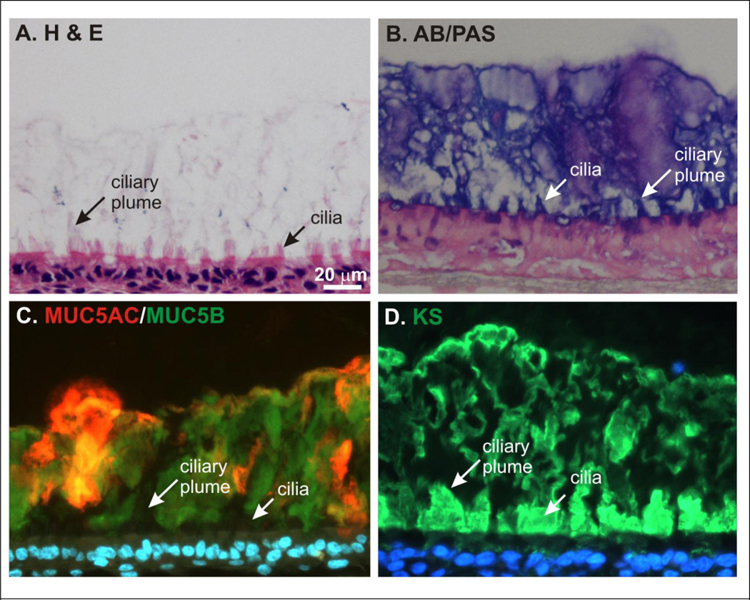
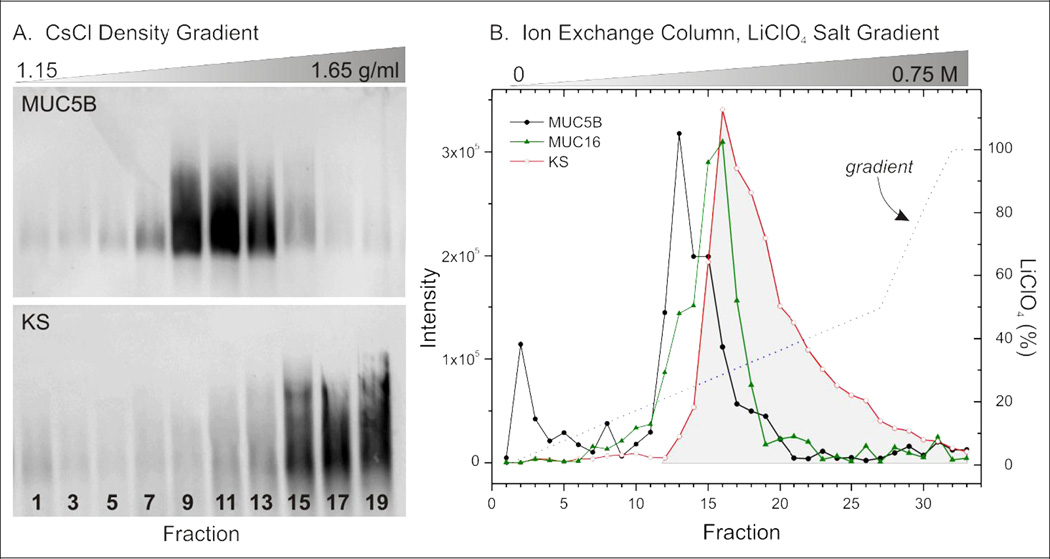
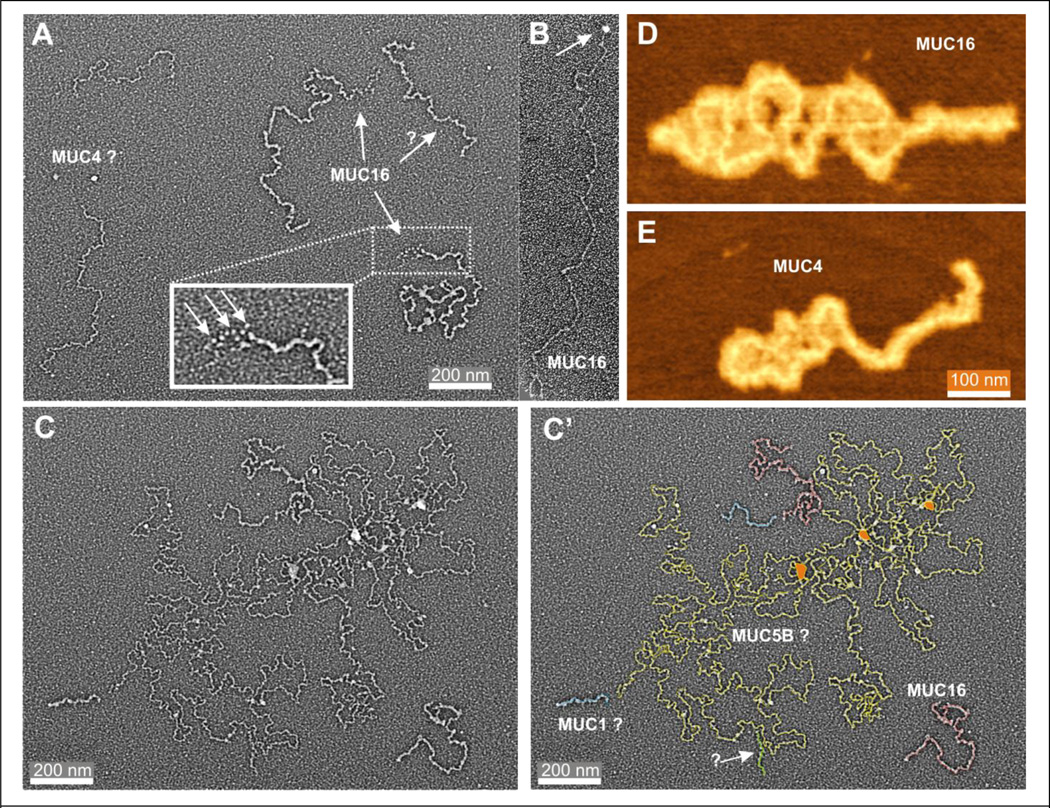
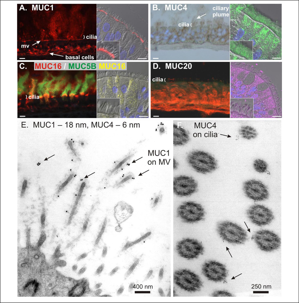
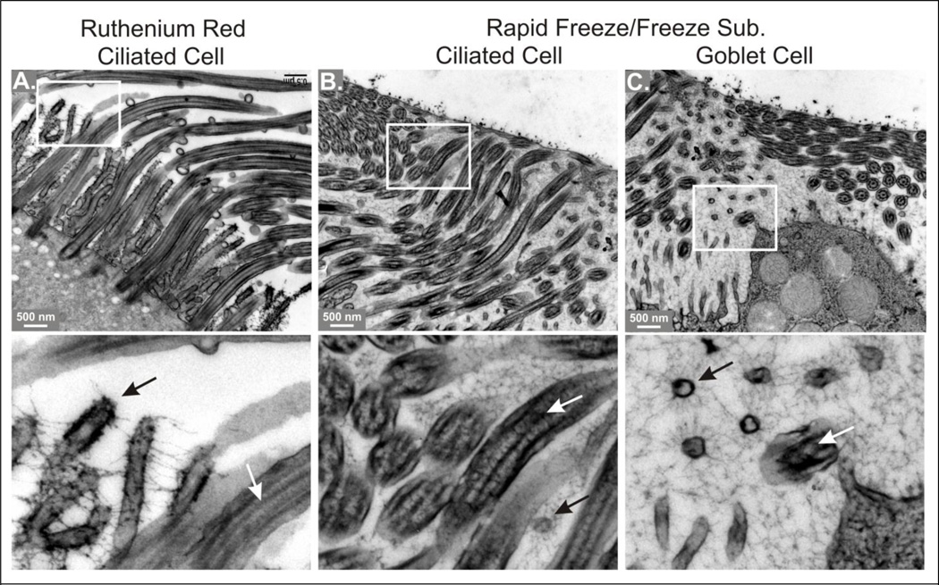
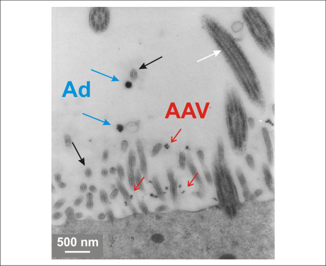
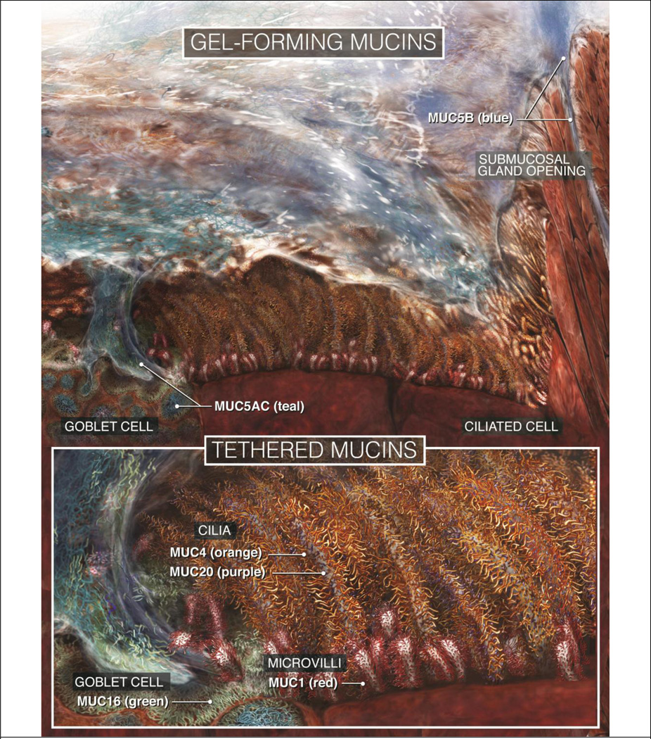
Mucus, with its burden of inspired particulates and pathogens, is cleared from mucosal surfaces of the airways by cilia beating within the periciliary layer (PCL). The PCL is held to be "watery" and free of mucus by thixotropic-like forces arising from beating cilia. With radii of gyration ~250 nm, however, polymeric mucins should reptate readily into the PCL, so we assessed the glycocalyx for barrier functions. The PCL stained negative for MUC5AC and MUC5B, but it was positive for keratan sulfate (KS), a glycosaminoglycan commonly associated with glycoconjugates. Shotgun proteomics showed KS-rich fractions from mucus containing abundant tethered mucins, MUC1, MUC4, and MUC16, but no proteoglycans. Immuno-histology by light and electron microscopy localized MUC1 to microvilli, MUC4 and MUC20 to cilia, and MUC16 to goblet cells. Electron and atomic force microscopy revealed molecular lengths of 190-1,500 nm for tethered mucins, and a finely textured glycocalyx matrix filling interciliary spaces. Adenoviral particles were excluded from glycocalyx of the microvilli, whereas the smaller adenoassociated virus penetrated, but were trapped within. Hence, tethered mucins organized as a space-filling glycocalyx function as a selective barrier for the PCL, broadening their role in innate lung defense and offering new molecular targets for conventional and gene therapies.


Figures

Figure 1
Figure 1. Density map of cilia on airway epithelial cells
Using a published electron micrograph (Figure 3 in Ref 60) of guinea-pig tracheal epithelial cells, individual cilia were mapped, manually, and are displayed as white circles. Reverse contrast was used to emphasize interciliary spaces. The micrograph represented a cross-sectional plane through the basal region of the cilia, where microvilli were apparent between the cilia, so the distribution approximates the positions of the basal bodies. Red dotted lines indicate approximate cell borders. Note the magnitude of interciliary distances, ~100 nm to >1 µm, especially between cilia on adjacent cells.
Figure 2
Figure 2. Mucus, mucins and keratan sulfate on the mucosal surface of HTBE cell cultures
Histological sections of HBTE cell cultures with accumulated mucus secretions were stained for, (A) hematoxylin and eosin, and (B) alcian blue/periodic acid-Schiffs, or immunoprobed with antibodies against (C) MUC5AC (red, monoclonal Ab, 45M1) and MUC5B (green, polyclonal, Cys-rich domains within the mucin repeat domains) or (D) keratan sulfate (green, monoclonal Ab 5D4). Panels C and D were counterstained with DAPI. Note height of the accumulated mucus layer ~ 100µm, the intense staining of KS in the periciliary layer, and in all panels, the plumes of material that appear to extend from ciliary tips and into the mucus (ciliary plumes), while excluding the polymeric mucins. In panel C, intracellular mucins are not apparent, as their fluorescence intensities were too weak to be imaged at gain settings appropriate for the highly intense extracellular signal. Scale bar (Panel A) = 20 µm.
Figure 3
Figure 3. Separation of mucins and KS-conjugated molecules collected in HTBE cell culture secretions
Mucus collected from HTBE cell cultures and solubilized in GuHCl was separated using two different methods. A. CsCl isopycnic density gradient analysis. Fractions from a CsCl density gradient following isopynic centrifugation to equilibrium were analyzed by agarose Western blotting using antibody probes to MUC5B (top) and KS (bottom). B. Ion exchange column chromatogram. Solubilized mucus was applied to a MonoQ HR 5/5 ion exchange column, after dialysis against urea and disulfide reduction in DTT. The column was eluted with an increasing gradient of LiClO4 and fractions analyzed by slot blotting, using antibody probes to MUC5B, MUC16, and KS (shown), as well as MUC5AC, MUC1, and MUC4 (not shown). The KS elution profile is highlighted in gray to illustrate that approximately half the KS-positive material elutes with MUC16 (and other mucins), whereas the other half elutes at more acidic pHs.
Figure 4
Figure 4. EM and AFM images of mucins from HTBE mucus
Mucins from HTBE cell culture mucus secretions solubilized in PBS were separated from most of the protein fraction by taking the void volume of a Sephacryl S1000 HPLC column, and were then fixed in glutaraldehyde and prepared for EM. A. EM of tethered mucins. MUC16 is identified by the tail at one end of the molecule appearing as a ‘string of beads’ (inset), and MUC4 by relative length and narrower profile. B. EM of MUC16 labeled with OC125 bound gold bead (arrow). See Experimental Procedures for preparation details. D and E. AFM of MUC16 and MUC4. After destructive proteolysis of reduced, polymeric mucins in HTBE mucus, the remaining, large glycosylated extracellular domains of MUC16 and MUC4, which are resistant to proteolysis, were dispersed on mica and visualized by AFM. C and C’. EM of tethered and polymeric mucins. Polymeric mucins in samples of HTBE mucins prepared as in Panel A were imaged by EM. The mucin profiles in C’ were traced with color-coded lines, as indicated. ? = tentative identification.
Figure 5
Figure 5. Localization of tethered mucins in airway epithelia
Panels A – D show staining by immunofluorescence for MUC1, MUC4, MUC16, and MUC20, as indicated. The left and right hand image of each pair within a panel shows immunolocalization in a HTBE cell culture and in human bronchial epithelium, respectively. The HTBE cultures in Panels A, B, and C had accumulated mucus, per Figure 2. For Panel B, note that the antibodies used in the two images were to the same peptide sequence (MUCH4), but represented different polyclonal antibodies, one of which was suitable for IHC, the other for IF. Scale bars = 10 µm. Panels E and F show MUC1 and MUC4 localization of human bronchial epithelium by immuno-EM. The tissue was exposed simultaneously to MUC1 and MUC4 antibodies conjugated to 18 and 6 nm gold beads, respectively, then washed extensively, fixed, and prepared for conventional EM. Arrows indicate gold beads labeling the glycocalyx of microvilli (MV) and cilia.
Figure 6
Figure 6. Comparison of fixation methods on mucosal glycocalyx in human bronchial epithelium
Bottom panels are magnified portions of the images above, as indicated by the white boxes. A. Tissue stained with ruthenium red and processed for conventional EM. Note how ruthenium red causes the glycocalyx to appear as thick, sparse strands, leaving most of the intercellular space apparently open. B and C. Tissue fixed by rapid freezing and freeze substitution, showing areas of the PCL above ciliated and goblet cells, as indicated. Note the finely textured, space filling matrix revealed by RF/FS. Lower panels, black arrows = microvilli, white arrows = cilia. See text for more details.
Figure 7
Figure 7. Interaction of viral particles with the glycocalyx of the PCL
HTBE cell cultures were incubated mucosally with particles of unlabeled adenovirus (Ad, 100 nm, blue arrows) and adenoassociated virus (AAV, 30 nm, red arrows), then rinsed, fixed, and prepared for EM. White arrow indicates a cilium; black arrow, a microvillus.
Figure 8
Figure 8. Schematic depicting a scaled view of the mucosal surface of human airways
Top: Overview of the mucociliary apparatus, showing a ciliated airway mucosal surface covered with a sheet of mucus. For clarity, the mucus sheet is depicted as a thin layer; in vivo, it would be substantially thicker. Bottom: Higher magnification views of a goblet cell (left), cilia, and the PCL, showing the molecular organization of the different mucins.

References

    1. Knowles MR, Boucher RC. Mucus clearance as a primary innate defense mechanism for mammalian airways. J Clin Invest. 2002;109(5):571–577. - PMC - PubMed
    1. Fahy JV, Dickey BF. Airway mucus function and dysfunction. N Engl J Med. 2010;363(23):2233–2247. - PMC - PubMed
    1. Florey H, Carleton HM, Wells AQ. Mucus Secretion in the Trachea. Br J Exp Pathol. 1932;13(3):269–284.
    1. Lucas AM, Douglas LC. Principles underlying ciliary activity in the respiratory tract. Arch Otolaryngol. 1934;20:518–541.
    1. Yoneda K. Mucous blanket of rat bronchus: an ultrastructural study. Am Rev Respir Dis. 1976;114(5):837–842. - PubMed

Publication types