. 2012 Nov;38(11):1970-81.
doi: 10.1016/j.ultrasmedbio.2012.06.012. Epub 2012 Aug 25.

Stability of echogenic liposomes as a blood pool ultrasound contrast agent in a physiologic flow phantom

Affiliations

Stability of echogenic liposomes as a blood pool ultrasound contrast agent in a physiologic flow phantom

Kirthi Radhakrishnan et al. Ultrasound Med Biol. 2012 Nov.

Abstract

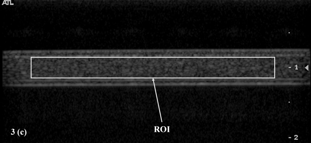
Echogenic liposomes (ELIP) are multifunctional ultrasound contrast agents (UCAs) with a lipid shell encapsulating both air and an aqueous core. ELIP are being developed for molecular imaging and image-guided therapeutic delivery. Stability of the echogenicity of ELIP in physiologic conditions is crucial to their successful translation to clinical use. In this study, we determined the effects of the surrounding media's dissolved air concentration, temperature transition and hydrodynamic pressure on the echogenicity of a chemically modified formulation of ELIP to promote stability and echogenicity. ELIP samples were diluted in porcine plasma or whole blood and pumped through a pulsatile flow system with adjustable hydrodynamic pressures and temperature. B-mode images were acquired using a clinical diagnostic scanner every 5 s for a total duration of 75 s. Echogenicity in porcine plasma was assessed as a function of total dissolved gas saturation. ELIP were added to plasma at room temperature (22 °C) or body temperature (37 °C) and pumped through a system maintained at 22 °C or 37 °C to study the effect of temperature transitions on ELIP echogenicity. Echogenicity at normotensive (120/80 mmHg) and hypertensive pressures (145/90 mmHg) was measured. ELIP were echogenic in plasma and whole blood at body temperature under normotensive to hypertensive pressures. Warming of samples from room temperature to body temperature did not alter echogenicity. However, in plasma cooled rapidly from body temperature to room temperature or in degassed plasma, ELIP lost echogenicity within 20 s at 120/80 mmHg. The stability of echogenicity of a modified ELIP formulation was determined in vitro at body temperature, physiologic gas concentration and throughout the physiologic pressure range. However, proper care should be taken to ensure that ELIP are not cooled rapidly from body temperature to room temperature as they will lose their echogenic properties. Further in vivo investigations will be needed to evaluate the optimal usage of ELIP as blood pool contrast agents.


Figures

Figure 1
Figure 1
Schematic of temperature controlled physiologic flow phantom with adjustable pulsatile pressure. Hydrodynamic pressure and temperature in the system were monitored throughout the experiment using inline sensors. L12–5 linear array transducer of the Philips HDI 5000 clinical scanner was used for B-mode imaging.
Figure 2
Figure 2
Example of measured pressure waveforms in the physiologic flow phantom: hypertensive pressure 145/90 mmHg (— —), normotensive pressure 120/80 mmHg (——), 60/20 mmHg (— · —) and atmospheric pressure 1 mmHg (⋯).
Figure 3
Figure 3
(a) Image of wire targets in tissue mimicking phantom used to calibrate mean digital intensity (MDI) based on mean gray scale value (MGSV). Region of interest, (ROI1) defined within the tissue mimicking speckle was used by Porter et al. (Porter et al. 2006). ROI2 defined in the hyperechoic region of uniform intensity is used in the current study. (b) Relationship between MDI and MGSV for a fixed grayscale map. (c) B-mode image of echogenic liposomes (ELIP) in plasma with the ROI defined within the tubing.
Figure 3
Figure 3
(a) Image of wire targets in tissue mimicking phantom used to calibrate mean digital intensity (MDI) based on mean gray scale value (MGSV). Region of interest, (ROI1) defined within the tissue mimicking speckle was used by Porter et al. (Porter et al. 2006). ROI2 defined in the hyperechoic region of uniform intensity is used in the current study. (b) Relationship between MDI and MGSV for a fixed grayscale map. (c) B-mode image of echogenic liposomes (ELIP) in plasma with the ROI defined within the tubing.
Figure 4
Figure 4
The echogenicity of ELIP was constant at pressures up to 120/80 mmHg for both arterial and venous dissolved gas levels. Only at physiologic pressures in degassed plasma did the ELIP lose echogenicity. (a) Echogenicity of ELIP in plasma at different dissolved gas concentrations and hydrodynamic overpressures. (b) Change in echogenicity of ELIP over 75 s in porcine plasma with arterial dissolved gas level (◊), venous dissolved gas level (♦), or degassed (▲) at 37 °C and 120/80 mmHg (N=5).
Figure 4
Figure 4
The echogenicity of ELIP was constant at pressures up to 120/80 mmHg for both arterial and venous dissolved gas levels. Only at physiologic pressures in degassed plasma did the ELIP lose echogenicity. (a) Echogenicity of ELIP in plasma at different dissolved gas concentrations and hydrodynamic overpressures. (b) Change in echogenicity of ELIP over 75 s in porcine plasma with arterial dissolved gas level (◊), venous dissolved gas level (♦), or degassed (▲) at 37 °C and 120/80 mmHg (N=5).
Figure 5
Figure 5
(a) B-mode images of ELIP in plasma at temperature transitions (ΔT) of −15 °C (37 °C to 22 °C), 0 °C (at 22 °C and at 37 °C), and 15 °C (22 °C to 37 °C) at 1 mmHg, 60/20 mmHg, and 120/80 mmHg pressures. (b) ELIP in plasma cooling from 37 °C to 22 °C showed a loss of echogenicity (p=0.002 (†)). Significant decreases in ELIP echogenicity at 120/80 mmHg were observed at room temperature (p<0.05 (*)) (N=5).
Figure 5
Figure 5
(a) B-mode images of ELIP in plasma at temperature transitions (ΔT) of −15 °C (37 °C to 22 °C), 0 °C (at 22 °C and at 37 °C), and 15 °C (22 °C to 37 °C) at 1 mmHg, 60/20 mmHg, and 120/80 mmHg pressures. (b) ELIP in plasma cooling from 37 °C to 22 °C showed a loss of echogenicity (p=0.002 (†)). Significant decreases in ELIP echogenicity at 120/80 mmHg were observed at room temperature (p<0.05 (*)) (N=5).
Figure 6
Figure 6
Effect of increased pulse pressure on echogenicity of ELIP in plasma at 37 °C. A marginal loss of ELIP echogenicity was observed (p=0.05 (*)) at 145/90 mmHg compared to 1 mmHg (N=6).
Figure 7
Figure 7
An insignificant decrease in echogenicity over time was observed at 120/80 mmHg in citrated whole blood and citrate-phosphate-dextrose (CPD) whole blood, but a significant decrease was observed in citrated whole blood with abnormal RBCs. Temporal changes in echogenicity of ELIP in CPD blood (♦) citrated blood (▲) and citrated blood with abnormal red blood cells (RBCs) (■) at 120/80 mmHg over 75 s is shown. Background echogenicity of CPD blood (◊) citrated blood (△) and citrated blood with abnormal RBCs (□) at 120/80 mmHg are also shown. (N=4).

Similar articles

Cited by

References

    1. Adam D, Sapunar M, Burla E. On the relationship between encapsulated ultrasound contrast agent and pressure. Ultrasound in Medicine and Biology. 2005;31:673–686. - PubMed
    1. Aksnes E, Rahn H. Measurement of total gas pressure in blood. J Appl Physiol. 1957;10:173–178. - PubMed
    1. Al-Jamal WT, Kostarelos K. Liposomes: From a clinically established drug delivery system to a nanoparticle platform for theranostic nanomedicine. Acc Chem Res. 2011;44:1094–1104. - PubMed
    1. Mines AH. Respiratory Physiology. New York: Raven Press; 1986.
    1. Alonso A, Reinz E, Jenne JW, Fatar M, Schmidt-Glenewinkel H, Hennerici MG, Meairs S. Reorganization of gap junctions after focused ultrasound blood-brain barrier opening in the rat brain. Journal of Cerebral Blood Flow and Metabolism. 2010;30:1394–1402. - PMC - PubMed

Publication types