

Intravenous mesenchymal stem cells prevented rejection of allogeneic corneal transplants by aborting the early inflammatory response
- PMID: 22929658
- PMCID: PMC3498800
- DOI: 10.1038/mt.2012.165
Intravenous mesenchymal stem cells prevented rejection of allogeneic corneal transplants by aborting the early inflammatory response
Abstract
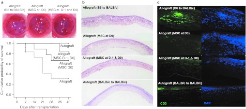
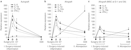
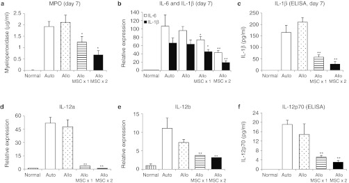
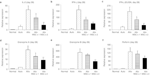
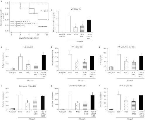
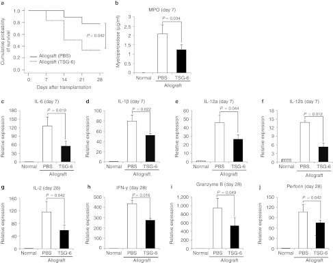
Mesenchymal stem/progenitor cells (MSCs) were reported to enhance the survival of cellular and organ transplants. However, their mode of action was not established. We here used a mouse model of corneal allotransplantation and demonstrated that peri-transplant intravenous (i.v.) infusion of human MSCs (hMSCs) decreased the early surgically induced inflammation and reduced the activation of antigen-presenting cells (APCs) in the cornea and draining lymph nodes (DLNs). Subsequently, immune rejection was decreased, and allograft survival was prolonged. Quantitative assays for human GAPDH revealed that <10 hMSCs out of 1 × 10(6) injected cells were recovered in the cornea 10 hours to 28 days after i.v. infusion. Most of hMSCs were trapped in lungs where they were activated to increase expression of the gene for a multifunctional anti-inflammatory protein tumor necrosis factor-α stimulated gene/protein 6 (TSG-6). i.v. hMSCs with a knockdown of TSG-6 did not suppress the early inflammation and failed to prolong the allograft survival. Also, i.v. infusion of recombinant TSG-6 reproduced the effects of hMSCs. Results suggest that hMSCs improve the survival of corneal allografts without engraftment and primarily by secreting TSG-6 that acts by aborting early inflammatory responses. The same mechanism may explain previous reports that MSCs decrease rejection of other organ transplants.
Figures
Comment in
-
A new vision of mesenchymal stromal cells.Mol Ther. 2012 Nov;20(11):2017-8. doi: 10.1038/mt.2012.221. Mol Ther. 2012. PMID: 23131854 Free PMC article. No abstract available.
Similar articles
-
Lentivirus-mediated IL-10-expressing Bone Marrow Mesenchymal Stem Cells promote corneal allograft survival via upregulating lncRNA 003946 in a rat model of corneal allograft rejection.Theranostics. 2020 Jul 9;10(18):8446-8467. doi: 10.7150/thno.31711. eCollection 2020. Theranostics. 2020. PMID: 32724480 Free PMC article.
-
Intravenous infusion of mesenchymal stem/stromal cells decreased CCR7(+) antigen presenting cells in mice with corneal allotransplantation.Curr Eye Res. 2014 Aug;39(8):780-9. doi: 10.3109/02713683.2013.877489. Epub 2014 Feb 6. Curr Eye Res. 2014. PMID: 24502523
-
Action at a distance: systemically administered adult stem/progenitor cells (MSCs) reduce inflammatory damage to the cornea without engraftment and primarily by secretion of TNF-α stimulated gene/protein 6.Stem Cells. 2011 Oct;29(10):1572-9. doi: 10.1002/stem.708. Stem Cells. 2011. PMID: 21837654
-
The mechanisms of corneal graft failure in the rat.Eye (Lond). 1995;9 ( Pt 2):197-207. doi: 10.1038/eye.1995.40. Eye (Lond). 1995. PMID: 7556718 Review.
-
Mesenchymal stem/stromal cells (MSCs): role as guardians of inflammation.Mol Ther. 2012 Jan;20(1):14-20. doi: 10.1038/mt.2011.211. Epub 2011 Oct 18. Mol Ther. 2012. PMID: 22008910 Free PMC article. Review.
Cited by
-
Mesenchymal Stem Cell-Based Therapy for Retinal Degenerative Diseases: Experimental Models and Clinical Trials.Cells. 2021 Mar 7;10(3):588. doi: 10.3390/cells10030588. Cells. 2021. PMID: 33799995 Free PMC article. Review.
-
Lentivirus-mediated IL-10-expressing Bone Marrow Mesenchymal Stem Cells promote corneal allograft survival via upregulating lncRNA 003946 in a rat model of corneal allograft rejection.Theranostics. 2020 Jul 9;10(18):8446-8467. doi: 10.7150/thno.31711. eCollection 2020. Theranostics. 2020. PMID: 32724480 Free PMC article.
-
Mesenchymal stem cells augment regulatory T cell function via CD80-mediated interactions and promote allograft survival.Am J Transplant. 2022 Jun;22(6):1564-1577. doi: 10.1111/ajt.17001. Epub 2022 Feb 26. Am J Transplant. 2022. PMID: 35170213 Free PMC article.
-
Mesenchymal Stem Cells Shift Mitochondrial Dynamics and Enhance Oxidative Phosphorylation in Recipient Cells.Front Physiol. 2018 Nov 13;9:1572. doi: 10.3389/fphys.2018.01572. eCollection 2018. Front Physiol. 2018. PMID: 30555336 Free PMC article.
-
Tumor Necrosis Factor (TNF)-α-Stimulated Gene 6 (TSG-6): A Promising Immunomodulatory Target in Acute Neurodegenerative Diseases.Int J Mol Sci. 2023 Jan 6;24(2):1162. doi: 10.3390/ijms24021162. Int J Mol Sci. 2023. PMID: 36674674 Free PMC article. Review.
References
-
- Hoogduijn MJ, Popp FC, Grohnert A, Crop MJ, van Rhijn M, Rowshani AT, MISOT Study Group et al. Advancement of mesenchymal stem cell therapy in solid organ transplantation (MISOT) Transplantation. 2010;90:124–126. - PubMed
-
- Le Blanc K, Frassoni F, Ball L, Locatelli F, Roelofs H, Lewis I, Developmental Committee of the European Group for Blood and Marrow Transplantation et al. Mesenchymal stem cells for treatment of steroid-resistant, severe, acute graft-versus-host disease: a phase II study. Lancet. 2008;371:1579–1586. - PubMed
-
- English K, French A., and, Wood KJ. Mesenchymal stromal cells: facilitators of successful transplantation. Cell Stem Cell. 2010;7:431–442. - PubMed
-
- Singer NG., and, Caplan AI. Mesenchymal stem cells: mechanisms of inflammation. Annu Rev Pathol. 2011;6:457–478. - PubMed
Publication types
MeSH terms
Substances
Grants and funding
LinkOut - more resources
Full Text Sources
Other Literature Sources
Research Materials
Miscellaneous
